Aug 14, 2024
Version 1
Exploring Glycoproteomic Profiles in Rectal Cancer: A Comparative N-Glycocapture Analysis of Tumor and Healthy Tissues Pre- and Post-Treatment V.1
- ABDULLAH BIN ZUBAIR1,
- David W. Larson1,
- Fatima Hamid2,
- Davide Ferrari1,
- K. L. Mathis1,
- Tommaso Violante1,
- Faisal Jehan3,
- RENEE HIRTE4
- 1Division of Colon and Rectal Surgery, Department of Surgery, Mayo Clinic, Rochester, Minnesota, United States;
- 2Department of Surgery, St. Agnes Hospital;
- 3Department of Surgical Oncology, Roswell Park Comprehensive Cancer Center, Buffalo, New York, United States;
- 4Department of Neurological Surgery, Mayo Clinic, Rochester, Minnisota, United States


Protocol Citation: ABDULLAH BIN ZUBAIR, David W. Larson, Fatima Hamid, Davide Ferrari, K. L. Mathis, Tommaso Violante, Faisal Jehan, RENEE HIRTE 2024. Exploring Glycoproteomic Profiles in Rectal Cancer: A Comparative N-Glycocapture Analysis of Tumor and Healthy Tissues Pre- and Post-Treatment. protocols.io https://dx.doi.org/10.17504/protocols.io.3byl49drogo5/v1Version created by ABDULLAH BIN ZUBAIR
License: This is an open access protocol distributed under the terms of the Creative Commons Attribution License, which permits unrestricted use, distribution, and reproduction in any medium, provided the original author and source are credited
Protocol status: In development
We are still developing and optimizing this protocol
Created: August 13, 2024
Last Modified: August 14, 2024
Protocol Integer ID: 105179
Keywords: Oncology, Immunotherapy, N-Glycocapture, KRAS, Chemotherapy , Immunotherapy , Mass Spec, Surgical Oncology, glycoproteomic profiles in rectal cancer, glycoproteins in rectal cancer, rectal cancer, glycoprotein profile, treatment strategies for rectal cancer, exploring glycoproteomic profile, rectal cancer management, glycocapture analysis of tumor, identification of specific glycoprotein, expressed glycoprotein, specific glycoprotein, recurrent tumor tissue, glycocapture analysis, biomarker, patients with stage iii crc, tumor
Disclaimer
This project is currently in the initial writing and development phases. Institutional Review Board (IRB) approval and funding have not yet been obtained. All proposed experiments and analyses described in this protocol are subject to approval and are not currently authorized for implementation. Further steps, including ethical review and securing financial support, are necessary before proceeding with the study.
Abstract
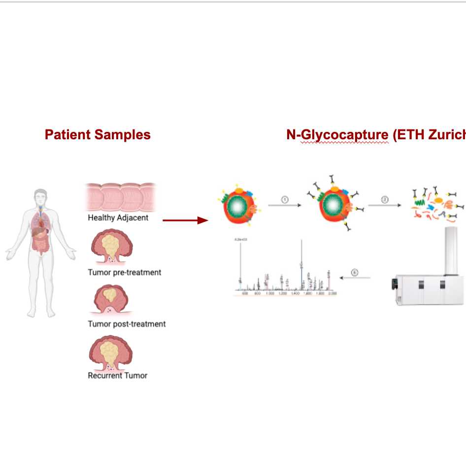
This protocol outlines the use of N-glycocapture analysis to identify differentially expressed glycoproteins in rectal cancer. We compare glycoprotein profiles from four tissue types: healthy adjacent tissue, tumor tissue pre-treatment, tumor tissue post-treatment, and recurrent tumor tissue, all derived from patients with Stage III CRC harboring the KRAS mutation. The aim is to uncover biomarkers associated with treatment response and recurrence, potentially leading to personalized treatment strategies for rectal cancer.
Expected results include the identification of specific glycoproteins and pathways dysregulated in CRC, which could serve as biomarkers for predicting treatment outcomes and guiding therapeutic decisions. This protocol is expected to contribute to the advancement of personalized medicine in rectal cancer management.
Guidelines
- Initial Development Stage: This protocol is currently in the early writing and development phase. No funding has been secured, and IRB approval has not yet been obtained.
- Ethical Considerations: Once the protocol is finalized, ensure that all ethical approvals are obtained before any research begins. This includes IRB approval for human subjects or IACUC approval for animal studies, if applicable.
- Sample Collection and Handling: Draft standardized procedures for the collection and handling of biological samples to maintain their integrity. These procedures will be finalized once the protocol is approved and funded.
- N-Glycocapture Protocol: Develop a detailed step-by-step N-glycocapture protocol, to be finalized after further consultation and once resources are available.
- Data Management Plan: Outline a preliminary plan for data management, including secure storage and documentation practices. The full plan will be implemented after funding is secured.
- Collaboration and Communication: Establish a clear communication plan with collaborators to ensure that everyone is aligned on the project's progress and next steps.
- Funding and Resources: Identify potential sources of funding and resources needed for the project. The protocol will be adjusted based on the resources that become available.
- Future Revisions: Be open to revising and updating the protocol as it develops, especially in response to feedback from potential reviewers or funders.
- Compliance with Regulations: Once the protocol is ready for implementation, ensure compliance with all relevant institutional, local, and international regulations.
- Reporting and Publication: Plan for the eventual publication of the study’s results, with the understanding that this will occur after the protocol is finalized, approved, and executed.
Safety warnings
- Pending Approvals: This protocol is still under development and has not yet received IRB approval or secured funding. The procedures and methodologies described are preliminary and may be subject to change based on ethical review and resource availability.
- Ethical Compliance: Ensure that all research activities comply with ethical guidelines and regulations once the protocol is finalized. No experiments should be conducted until IRB and, if applicable, IACUC approvals are obtained.
- Funding Requirements: Adequate funding must be secured before initiating the study. The current protocol does not include specific details about financial resources or budget considerations.
- Protocol Revisions: The protocol is a work in progress and may be revised as the project develops. Stay informed of any updates and ensure that you follow the most current version of the protocol.
- Data Handling: Proper data management and storage procedures will be established once the protocol is finalized. Do not begin data collection until the necessary systems and practices are in place to ensure data integrity and security.
- Sample Handling: Procedures for sample collection and handling are preliminary. Follow finalized guidelines to prevent contamination or degradation of samples once the protocol is approved and implemented.
Please review these warnings carefully and ensure that all required approvals and resources are in place before proceeding with any research activities.
Ethics statement
While this study does not involve any animal experiments, it’s important to note that if future research includes work with animals, it must adhere to the highest ethical standards. Approval from an Institutional Animal Care and Use Committee (IACUC) or a similar ethics board is required before any such experiments can be conducted. Researchers need to secure and document this approval, including any relevant permit numbers and the name of the overseeing committee, to ensure all ethical guidelines are followed.
Before start
This project is currently in the initial stages of protocol writing and development. Before any research can begin, the protocol will need to undergo further review, secure funding, and obtain IRB approval. These steps are essential to ensure that the study is ethically sound and adequately supported. The guidelines and procedures outlined in this document are preliminary and will be finalized once the necessary approvals and resources are in place.
Background and Rationale
Rectal cancer, a major contributor to global cancer mortality, poses a significant challenge in patient management, especially when complete surgical resection isn’t achieved. Even with advancements in treatment, many patients face disease recurrence, underscoring the urgent need for reliable biomarkers to guide personalized therapy. Biomarkers that can predict treatment response and recurrence could revolutionize how we approach rectal cancer, ultimately improving patient outcomes. N-glycocapture analysis has emerged as a groundbreaking technique for identifying glycoprotein biomarkers. This method targets glycoproteins, proteins with sugar chains attached, which play critical roles in cancer progression. Aberrant glycosylation, or the faulty addition of these sugar chains, is often seen in cancer, including colorectal cancer (CRC). By examining glycoproteins in tumor tissues, we can potentially uncover new biomarkers that indicate how well a patient might respond to treatment or if their cancer is likely to return. This study aims to leverage N-glycocapture analysis to compare glycoprotein profiles in four tissue types from patients with Stage III CRC, all harboring the KRAS mutation. By comparing healthy adjacent tissue, tumor tissue before treatment, tumor tissue after treatment, and recurrent tumor tissue, we hope to identify key glycoproteins and pathways linked to treatment response and recurrence. This research could open doors to more personalized treatment approaches for CRC patients.
Objective:
- Primary Objective: To identify and compare glycoproteins that are differentially expressed across healthy adjacent tissue, tumor tissue pre-treatment, tumor tissue post-treatment, and recurrent tumor tissue using N-glycocapture analysis.
- Secondary Objective: To assess the feasibility of using these glycoproteins as biomarkers for predicting treatment response, chemotherapy resistance, and recurrence in CRC.
Hypothesis
We hypothesize that N-glycocapture analysis will reveal specific glycoproteins that are differentially expressed between the four tissue types. These glycoproteins could serve as potential biomarkers for treatment response, resistance, and recurrence in rectal cancer, offering insights for personalized therapy.
Study Design
This is a comparative study involving 10 patients diagnosed with Stage III CRC, all with the KRAS mutation. Each patient will provide four tissue samples:
- Healthy Adjacent Tissue
- Tumor Tissue Pre-Treatment
- Tumor Tissue Post-Treatment
- Recurrent Tumor Tissue
Sample Collection: Frozen tissue samples will be collected during routine surgical procedures and stored at -80°C until further analysis. Each sample will be handled carefully to prevent degradation of proteins and ensure the integrity of glycoproteins for N-glycocapture analysis.
Materials
- Frozen Tissue Samples: Collected from 10 CRC patients.
- N-Glycocapture Kit: This kit will include lectins or other glycan-binding proteins that specifically capture N-glycosylated proteins from tissue lysates.
- Mass Spectrometry (MS) System: For analyzing the enriched glycoproteins and identifying differentially expressed ones.
- Lysis Buffer: To lyse tissues and extract proteins.
- Protease Inhibitors: To prevent protein degradation during extraction.
- Microcentrifuge Tubes: For sample processing.
- Centrifuge: For separating lysate components.
- LC-MS/MS (Liquid Chromatography-Mass Spectrometry): To separate and identify the captured glycoproteins.
Methodology
Sample Preparation
Tissue Lysis: Homogenize frozen tissue samples in lysis buffer supplemented with protease inhibitors to prevent protein degradation.
Protein Quantification: Measure the total protein concentration using a BCA assay to ensure equal loading of samples for N-glycocapture.
N-Glycocapture
Glycoprotein Enrichment: Apply the tissue lysates to the N-glycocapture kit. This kit contains lectins that specifically bind to N-glycans on glycoproteins, allowing selective capture of glycoproteins.
Washing Steps: Wash the bound glycoproteins to remove non-specifically bound proteins and other contaminants.
Elution: Elute the captured glycoproteins for further analysis.
Mass Spectrometry Analysis
Protein Digestion: Digest the eluted glycoproteins using trypsin to generate peptides.
LC-MS/MS Analysis: Inject the peptides into the LC-MS/MS system. The LC component separates peptides, while the MS/MS system identifies and quantifies the peptides, revealing the glycoproteins present in each sample.
Data Analysis
Differential Expression Analysis: Compare glycoprotein expression levels between the four tissue types to identify those that are differentially expressed.
Pathway Enrichment: Use bioinformatics tools to identify pathways and biological processes associated with the differentially expressed glycoproteins. This will help pinpoint potential mechanisms of treatment resistance or recurrence.
Validation:
Western Blotting/Immunohistochemistry: Validate key glycoproteins identified through N-glycocapture using independent techniques to ensure their relevance as biomarkers.
Inclusion and Exclusion Criteria
Inclusion Criteria:
- Patients diagnosed with Stage III CRC harboring the KRAS mutation.
- Patients who have undergone surgical resection of their tumors.
- Availability of frozen tissue samples for all four tissue types.
Exclusion Criteria:
- Patients who received adjuvant chemotherapy before tissue sample collection.
- Insufficient tissue sample quantity or quality.
- Co-morbid conditions that might interfere with the study outcomes.
Outcome Measures
- Primary Outcome: Identification of glycoproteins that are differentially expressed between healthy adjacent tissue, tumor tissue pre-treatment, tumor tissue post-treatment, and recurrent tumor tissue.
- Secondary Outcome: Evaluation of the potential of these glycoproteins as biomarkers for treatment response, chemotherapy resistance, and recurrence.
Statistical Analysis
- Comparative Analysis: Perform statistical tests (e.g., t-tests, ANOVA) to identify significant differences in glycoprotein expression between tissue types.
- Pathway Analysis: Use bioinformatics software to conduct pathway enrichment analysis, identifying dysregulated pathways in CRC.
Ethical Considerations
The study will adhere to ethical standards and will obtain approval from the Institutional Review Board (IRB). Informed consent will be obtained from all participants, ensuring transparency about the study's purpose, procedures, and potential risks.
Limitations
- Small Sample Size: The small cohort may limit the generalizability of the findings.
- Tumor Heterogeneity: Variability in tumor biology among patients could affect glycoprotein expression profiles.
- Technical Variability: Differences in sample handling, N-glycocapture efficiency, and MS analysis may introduce bias.
Expected Outcomes
This study expects to identify glycoproteins and pathways that are differentially expressed across the different tissue types in rectal cancer. These findings could lead to the identification of novel biomarkers for predicting treatment response, resistance, and recurrence, ultimately contributing to more personalized and effective treatment strategies for CRC patients.
Timeline
- Month 1-2: Patient recruitment and sample collection.
- Month 3-6: N-glycocapture analysis and mass spectrometry.
- Month 7-8: Data analysis and interpretation.
- Month 9: Validation of key findings.
- Month 10: Manuscript preparation and submission.
Protocol references
1. Le Rhun, E., Taillibert, S., Chamberlain, M. C., & Carpentier, A. F. (2017). Leptomeningeal metastases: a RANO proposal for response criteria. Neuro-oncology, 19(4), 484-492.
2. Boire, A., & Brastianos, P. K. (2019). Leptomeningeal metastasis: challenges in diagnosis and treatment. Current oncology reports, 21(12), 100.
3. Kudo, Y., Haymaker, C., Zhang, J., Reuben, A., Torres-Cabala, C., Quezada, S. A., ... & Tawbi, H. (2018). Suppressed immune microenvironment and repertoire in brain metastases from patients with resected melanoma. Journal of immunotherapy (Hagerstown, Md.: 1997), 41(9), 101-111.
5. Long, G. V., Grob, J. J., Nathan, P., Ribas, A., Robert, C., Schadendorf, D., ... & Hauschild, A. (2017). Factors predictive of response, disease progression, and overall survival after dabrafenib and trametinib combination treatment: a pooled analysis of individual patient data from randomised trials. The Lancet Oncology, 18(12), 1504-1516.
6. Omuro, A., DeAngelis, L. M., & Glioblastoma and Other Malignant Gliomas. (2013). Glioblastoma and other malignant gliomas: a clinical review. Jama, 310(17), 1842-1850.
7. Ballester, L. Y., Wang, Z., Shandilya, S., Muzaffar, M., Serrano, J., & Simeone, D. M. (2018). PD-L1 expression in leptomeningeal metastases: an exploratory analysis. Journal of neuro-oncology, 137(1), 95-101.
8. Cabel, L., Riva, F., Servois, V., Livartowski, J., Daniel, C., Rampanou, A., ... & Nicotra, C. (2018). Circulating tumor DNA changes for early monitoring of anti-PD1 immunotherapy: a proof-of-concept study. Annals of Oncology, 29(5), 1318-1324.
9. Farnia, B., & Allen, E. M. (2020). Leptomeningeal Metastasis in Solid Tumors. In Handbook of Brain Tumor Chemotherapy, Molecular Therapeutics, and Immunotherapy (pp. 357-375). Elsevier.
10. Gril, B., Paranjape, A. N., Woditschka, S., Hua, E., & Dolloff, N. G. (2017). Molecular pathways: the basis for rational combination using MEK inhibitors in KRAS-mutant cancers. Clinical Cancer Research, 23(16), 4172-4179.