Jun 02, 2026
Expansion and Mounting of Hydrogel Embedded Brain
- Rajvi Javeri1,
- Molly Logsdon1,
- Laura Roy1,
- Holly Myers1,
- Naveen Ouellette1,
- Andrew Recknagel2,
- Kevin Cao1,
- Jayaram Chandrashekar1
- 1Allen Institute / Neural Dynamics;
- 2Janelia Research Campus
- Allen Institute for Neural Dynamics


Protocol Citation: Rajvi Javeri, Molly Logsdon, Laura Roy, Holly Myers, Naveen Ouellette, Andrew Recknagel, Kevin Cao, Jayaram Chandrashekar 2026. Expansion and Mounting of Hydrogel Embedded Brain . protocols.io https://dx.doi.org/10.17504/protocols.io.5jyl8e996l2w/v1
License: This is an open access protocol distributed under the terms of the Creative Commons Attribution License, which permits unrestricted use, distribution, and reproduction in any medium, provided the original author and source are credited
Protocol status: Working
We use this protocol and it's working
Created: April 17, 2025
Last Modified: June 02, 2026
Protocol Integer ID: 126828
Keywords: Expansion Microscopy, Hydrogel, tissue mounting, mounting of hydrogel embedded brain, hydrogel embedded brain, expanded hydrogel, hydrogel, embedded brain, imaging on the exa, osmotic expansion, aqueous solutions due to osmotic expansion, gel, imaging, sheet imaging, charged polymer network, polymer network
Funders Acknowledgements:
Scalable technologies for brain-wide connectomics of transcriptomic cell types: focus on brainstem
Grant ID: RF1MH128841
Abstract
Hydrogel-embedded brains swell in aqueous solutions due to osmotic expansion of the charged polymer network. For imaging on the ExA-SPIM, samples are expanded in dilute saline-sodium citrate (SSC) buffer, so that gel swelling is limited to ~3× linear expansion. Expanded hydrogels are then mounted in a custom imaging chamber for light-sheet imaging.
Guidelines
Expanded hydrogels are extremely fragile. When exchanging 0.05x SSC or 10mM ascorbic acid, remove the hydrogel and strainer from the container before switching out the solutions. Pouring solutions directly on the hydrogels can break them.
Materials
20X SSCFisher ScientificCatalog #AM9770
Ascorbic AcidWard's Natural Science Establishment, Inc.Catalog #AA0425-100G
10N NaOHMerck MilliporeSigma (Sigma-Aldrich)Catalog #SX0607N-6
EthanolCatalog #64-17-5
1M HClMerck MilliporeSigma (Sigma-Aldrich)Catalog #1090571000
Equipment
Instrument Soaking Tray
NAME
Sklar
BRAND
10-3052
SKU
LINK
Equipment
Glass Cuvette Holder
NAME
N/A
BRAND
LINK
Equipment
Custom Cuvette
NAME
Azzota Scientific LLC
BRAND

Equipment
Lens Paper
NAME
Thomas Scientific
BRAND
6292F30
SKU
LINK
Equipment
1.5 mm Balldriver
NAME
Bondhus
BRAND
BD-1.5M
SKU
LINK
1.5mm
SPECIFICATIONS
Equipment
Pyrex® crystallizing dish
NAME
Pyrex
BRAND
CLS314080
SKU
LINK
Equipment
T-Handle Hex Key: 7/64, Hex End
NAME
Bondhus
BRAND
Mfr# 13206
SKU
LINK
Equipment
Single edge uncoated carbon steel blade
NAME
blade
TYPE
Pelco
BRAND
121-95
SKU
LINK
118mm long x 19mm wide x 0.229mm thick. (4.65 x 0.75 x 0.009")
SPECIFICATIONS
RECIPES
0.05X SSC: 20X SSC, Milli-Q Water
Combine the following reagents and stir at Room temperature until fully dissolved, store at Room temperature
| Reagent | Amount | |
| 20X SSC | 5 mL | |
| Milli-Q Water | 1995 mL | |
| Total | 2000 mL |
10mM Ascorbic Acid, pH 7.0
Combine the following reagents and stir at Room temperature until fully dissolved, adjust pH to 7.0 and then store at Room temperature . Make fresh before each use.
| Reagent | Amount | |
| Asorbic Acid | 3.52g | |
| Milli-Q Water | 2000 mL | |
| Total | 2000 mL |
Before start
Start with a brain that has been delipidated using the Tetrahydrofuran and Dichloromethane Delipidation of a Whole Mouse Brain and Aqueous (SBiP) Delipidation of a Whole Mouse Brain protocols. The sample may or may not be immunolabeled using the Immunolabeling of a Whole Mouse Brain protocol. The brain should be cleared following the Whole Mouse Brain Gelation and Digestion protocol.
Expansion of Hydrogel Embedded Brain
3d 1h
Submerge the hydrogel in 1L of 0.05X SSC.
- Replace 0.05X SSC once per day for at least 2 days at Room temperature .
As it expands, the hydrogel becomes more fragile. Use a gloved hand when handling the hydrogel and take care while handling and transferring to another container.
- The 0.05X SSC exchange is done using a 2L Instrument Soaking Tray with a strainer insert.
- The gel and strainer are lifted out of the solution, the solution is refreshed, and the strainer and gel are gently placed back in.
The hydrogel should expand to about 3 times its original size (size prior to digestion). If the gel is not fully expanded after 2d, change the SSC buffer again.
Transfer the expanded hydrogel to 1 L of a photoprotective solution of 10mM Ascorbic acid pH 7.0 at least 24:00:00 before the brain is to be imaged on the ExA-SPIM. Ensure that the expanded brain does not soak in the ascorbic acid solution for more than 6 days including the time taken to image the sample, as prolonged exposure to this solution can dissolve the hydrogel.
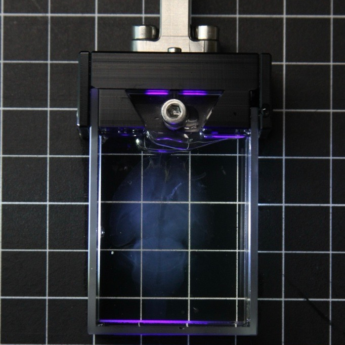
Mounting of Hydrogel in ExA-SPIM Chamber
The expanded brain is mounted in a custom chamber for imaging on the ExA-SPIM. The chamber is constructed from a detachable glass cuvette with an open face that holds the sample, and a holder that attaches the cuvette to the ExA-SPIM that has an adjustable face that secures the sample and also covers the cuvette. This chamber holds the specimen securely in place while allowing imaging access from three sides. Prior to mounting the brain, clean the chamber and cuvette with 70% ethanol and lens cleaning wipes to remove any dust.
Sample holder and cuvette used for imaging specimen
CAD visualization of glass cuvette holder (see materials for link to CAD file)
Carefully remove the sample from the soaking tray and gently place it into a large petri dish. Perform this slowly to prevent breaking the fragile hydrogel.
Using tissue slicer blades, trim the hydrogel on each side to size it to fit the cuvette. All surfaces of the hydrogel should be smooth after trimming to reduce the appearance of air bubbles while mounting.
- The trimmed dimensions of the hydrogel is ~3.9 cms - 4 cms medial-lateral axis and no more than 5cm along the rostral-caudal axis.
measurement of hydrogel after trimming along the medial-lateral axis
measurement of hydrogel after trimming along the rostral-caudal axis
Fill a large and deep petri dish with the same 10mM ascorbic acid solution used during expansion. Using the same ascorbic acid solution is essential to prevent unexpected expansion of the sample during imaging. Transfer the trimmed hydrogel to this petri dish.
Place the glass cuvette in the same petri dish and using a gloved hand, carefully guide the hydrogel into the cuvette. The sample should fit comfortably in the cuvette, and should be well pressed against all surfaces of the cuvette.
Once the sample has been placed in the cuvette, slide the cuvette through the slits in the holder. Slowly tighten the screws on either side using a 1.5mm ball driver screwdriver and stop once there is resistance. Check that the cuvette is well attached to the holder and does not move. If it is still loose, adjust the screws in very small increments (take care so as to not break the glass cuvette).
Next, tighten the top panel using a 7/64" T-Handle hex key until there is a little resistance and the panel is snug against the dorsal surface of the hydrogel.
Ensure that there are no air bubbles anywhere in the mounted sample. If there are bubbles, detach the cuvette to remove them and resume from step 2.3
Transfer the 10mM ascorbic acid solution to a container and submerge the mounted brain hydrogel chamber assembly into the container. Protect from light.
The sample is now ready to be imaged on the ExA-SPIM.
Protocol references
Citation
LINK
Citations
Glaser A, Chandrashekar J, Vasquez S, Arshadi C, Javeri R, Ouellette N, Jiang X, Baka J, Kovacs G, Woodard M, Seshamani S, Cao K, Clack N, Recknagel A, Grim A, Balaram P, Turschak E, Hooper M, Liddell A, Rohde J, Hellevik A, Takasaki K, Erion Barner L, Logsdon M, Chronopoulos C, de Vries SEJ, Ting JT, Perlmutter S, Kalmbach BE, Dembrow N, Tasic B, Reid RC, Feng D, Svoboda K. Expansion-assisted selective plane illumination microscopy for nanoscale imaging of centimeter-scale tissues.
https://doi.org/10.7554/eLife.91979