Jan 15, 2024
Evaluating GPNMB ACD mutants by Western Blotting and immunofluorescence.
- Erin Bogacki1,
- Patrick Lewis2,3,
- Susanne Herbst2,3
- 1NIH;
- 2Royal Veterinary College;
- 3The Michael J. Fox Foundation for Parkinson’s Research (MJFF) and the Aligning Science Across Parkinson’s (ASAP) Initiative


Protocol Citation: Erin Bogacki, Patrick Lewis, Susanne Herbst 2024. Evaluating GPNMB ACD mutants by Western Blotting and immunofluorescence.. protocols.io https://dx.doi.org/10.17504/protocols.io.4r3l22yz4l1y/v1
License: This is an open access protocol distributed under the terms of the Creative Commons Attribution License, which permits unrestricted use, distribution, and reproduction in any medium, provided the original author and source are credited
Protocol status: Working
We use this protocol and it's working
Created: November 13, 2023
Last Modified: June 01, 2024
Protocol Integer ID: 90843
Keywords: ASAPCRN, GPNMB, Western Blotting, Immunofluorescence, HEK293, gpnmb acd mutants by western blotting, gpnmb mutants by western blotting, evaluating gpnmb acd mutant, gpnmb mutant, immunofluorescence, immunofluorescent imaging, hek293 overexpression model, evaluation of cellular processing, cellular processing
Funders Acknowledgements:
The Michael J. Fox Foundation for Parkinson’s Research (MJFF) and the Aligning Science Across Parkinson’s (ASAP) Initiative
Grant ID: .
Abstract
This protocol describes the evaluation of cellular processing of GPNMB mutants by Western Blotting and Immunofluorescent imaging in a HEK293 overexpression model.
Materials
General
- HEK293T cells (ATCC CRL-3216)
- Fugene HD transfection reagent (E2311, Promega)
- pcDNA3.1-GPNMB-EGFP
 pcDNA3.1-GPNMB-EGFP.png104.5KB
 - PBS, pH 7.4: #14190250, ThermoFisher Scientific
Western Blotting
- Lysis buffer: 20 mM Tris-HCl (pH 7.5), 150 mM NaCl, 1 mM Na2EDTA, 1 mM EGTA, 1% (v/v) Triton X-100
NOTE: add protease and phosphatase inhibitors fresh each time (eg Halt Protease and Phosphatase Inhibitor Cocktail (100X), #78440, ThermoFisher Scientific)
- Loading buffer: NuPAGE LDS sample buffer (#NP0007, ThermoFisher Scientific)
- Sample reducing agent: NuPAGE sample reducing agent (#NP0009, ThermoFisher Scientific)
- 4-12% Bis-Tris NuPAGE gels (eg #NP0321BOX, ThermoFisher Scientific)
- SDS-PAGE running buffer: MES running buffer (#NP0002, ThermoFisher Scientific)
- Trans-Blot‱ Turbo‱ PVDF Transfer Packs: eg #1704157, BioRad
- TBS-T: 50 mM Tris–HCl, pH 7.5, 150 mM NaCl, 0.05% (v/v) Tween 20.
- Blocking and antibody dilution buffer: 5 % (w/v) non-fat milk powder in TBS-T
- Primary and secondary antibodies (see table 1 & 2 for antibody suggestions)
Immunofluorescence
- 4 % (v/v) PFA/PBS: Dilute 16 % Paraformaldehyde Aqueous Solution (#15710, Electron Microscopy Sciences) to 4 % in PBS
- Blocking and antibody dilution buffer: 0.3 % Triton X-100, 5 % (v/v) FCS in PBS
- DAPI staining solution: 300 nM DAPI in PBS (#D1306, ThermoFisher Scientific or similar)
- Mounting medium: DAKO Fluorescence Mounting medium, # S3023, Agilent or similar
- Coverslips #1.5 (eg 631-0150, VWR)
- Slides (eg SuperFrost Plus, J1800AMNZ, Epredia)
- Primary and secondary antibodies (see table 3 & 4 for antibody suggestions)
Seed cells
Seed HEK293 cells.
A) For Western Blotting, we recommend seeding 2.5 x 10^5 cells per well of a 12-well culture plate.
B) For Immunofluorescence, we recommend seeding 1.2 x 10^5 cells per well of a 24-well culture plate. Seed cells on Poly-D-Lysine coated coverslips.
Note: we routinely culture HEK293T cells in DMEM containing 10% FCS.
Incubate in a tissue culture incubator Overnight .
HEK 293 cell transfection
10m
This protocol uses a DNA: Fugene ratio of 1:3. Prepare the transfection complexes as follows:
| A | B | C | D | |
| DNA | Fugene | Serum-free DMEM | ||
| 12 well | 1000 ng | 3 ul | 100 ul | |
| 24 well | 500 ng | 1.5 ul | 50 ul |
Preparation of transfection complexes (quantities are per well)
Add the required amount of plasmid DNA to serum-free DMEM. Mix briefly.
Add the required amount of Fugene HD Transfection Reagent.
Vortex and incubate for 00:10:00 at Room temperature .
10m
In the meantime, change the medium on the cells to fresh DMEM containing 10 % FCS
Add 50 µL per 24-well or 100 µL per 12-well drop-wise to the cells and incubate Overnight
Western Blotting
25m
This section describes the sample preparation and analysis for Western Blotting.
Wash the cells gentle with PBS
Immediately add 100 µL per well of ice-cold cell lysis buffer and place cells on ice.
Scrape cells with a cell scraper and harvest cell lysate into a 1.5 ml Eppendorf tube.
Incubate the cells On ice for 00:10:00 , vortex occasionally
10m
Clear the cell lysate by spinning down at 16200 x g, 4°C for 00:15:00
15m
Transfer the post-nuclear supernatant to a fresh 1.5 ml Eppendorf tube and store at -20 °C .
Prepare the cell lysates for Western Blotting by adding LDS sample buffer and denaturing agent and denature the samples at 80 °C for 00:08:00 .
8m
Run samples on 4-12 % Bis-Tris SDS-page. (approx. 00:35:00 at 160 V const.)
35m
Transfer proteins onto a PVDF membrane using the Turbo transfer system (BioRad) or similar.
Block membranes in 5% milk/TBS-T for 01:00:00 at Room temperature .
1h
Incubate the membranes with primary antibodies at 4 °C Overnight .
| A | B | C | D | E | |
| Target | Cat # | Supplier | Raised in | Dilution | |
| GPNMB (N-terminal) | AF2550 | R&D Systems | Goat | 1:1000 | |
| GFP | MA5-15256 | ThermoFisher Scientific | Mouse | 1:1000 | |
| Actin | A1978 | Sigma | Mouse | 1:5000 |
Table 1: Primary antibodies for Western Blotting.
Wash the plots in TBS-T for 00:05:00 at Room temperature . Repeat this step twice for a total of three washes.
5m
Dilute the secondary antibody in 5% milk/TBS-T and incubate the membranes with secondary antibodies at Room temperature for 00:45:00 .
| A | B | C | |
| Antibody suggestion | Dilution | ||
| anti-mouse-Peroxidase | eg, A3682, Sigma | 1:10000 | |
| anti-goat-Peroxidase | eg, A5420, Sigma | 1:10000 | |
Table 2: Secondary antibodies for Western Blotting.
45m
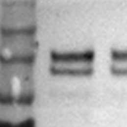
Develop the blots using an appropriate developer. Full-length GPNMB-EGFP is detected as a double band at ~125 kDa. A cleaved C-terminal GPNMB fragment can be detected with the anti-GFP antibody at ~35 kDa.
Immunofluorescence
2h 20m
Gently wash coverslips with PBS and fix in 4 % PFA/PBS for 00:15:00 min.
15m
Gently wash the cells with PBS. Replace the PBS and add the permeabilisation/blocking. Incubate coverslips for a minimum of 00:20:00 min at Room temperature .
20m
In the meantime, place a piece of Parafilm onto your bench and label if required. This will act as a flat surface to stain the coverslips on.
Prepare the antibody staining solution in Blocking and staining buffer. Find a suggestion of antibodies for counterstaining below:
| A | B | C | D | E | |
| Target | Cat # | Supplier | Raised in | Dilution | |
| LAMP-1 | H4A3 | DSHB | Mouse | 1:100 | |
| TGN46 | 13573-1-AP | Proteintech | Rabbit | 1:100 |
Table 3: Primary antibodies for immunofluorescence.
Pipette a 45 µL drop of the antibody staining solution onto the Parafilm and invert the coverslip onto the staining solution so that the cells face downwards.
Incubate for 01:00:00 hr in the dark.
1h
Wash the coverslips three times with PBS.
Prepare a staining solution containing the secondary antibody:
| A | B | |
| Antibody suggestion | Dilution | |
| anti-mouse-AF647 | 1:1000 | |
| anti-rabbit-AF586 | 1:1000 |
Table 4: Secondary antibodies for Immunofluorescence.
Pipette a 45 µL drop of the antibody staining solution onto the Parafilm and invert the coverslip onto the staining solution so that the cells face downwards.
Incubate for 00:45:00 min in the dark.
45m
Wash the coverslips once in PBS, stain with DAPI (or other nuclear stain), and mount onto glass slides.
GPNMB can be observed predominantly at the trans-Golgi network but can also be seen at lysosomal compartments.