May 30, 2026
Establishment of Symbiodiniaceae in vitro cultures from Anemonia viridis
- Elisabeth Beauson1,
- Clara Fricano1,
- Paola Furla1,
- Stéphanie Barnay-Verdier1,2
- 1Université Côte d'Azur, CNRS, INSERM, Institute for Research on Cancer and Aging, Nice (IRCAN), Nice - France;
- 2Sorbonne Université, UFR 927, Paris - France


Protocol Citation: Elisabeth Beauson, Clara Fricano, Paola Furla, Stéphanie Barnay-Verdier 2026. Establishment of Symbiodiniaceae in vitro cultures from Anemonia viridis. protocols.io https://dx.doi.org/10.17504/protocols.io.q26g7oje8vwz/v1
License: This is an open access protocol distributed under the terms of the Creative Commons Attribution License, which permits unrestricted use, distribution, and reproduction in any medium, provided the original author and source are credited
Protocol status: Working
We use this protocol and it's working.
Created: April 14, 2026
Last Modified: May 30, 2026
Protocol Integer ID: 314979
Keywords: Microalgae culture, Symbiosis, Symbiodiniaceae isolation, establishment of symbiodiniaceae, isolation of symbiodiniaceae, obtention of of symbiodiniaceae, symbiodiniaceae, cultures from anemonia, other symbiotic cnidarian, temperate sea anemone anemonia, anemonia, breviolum psygmophilum, proliferating symbiont, possessing big tentacle, big tentacle
Funders Acknowledgements:
Agence Nationale de la Recherche (ANR)
Grant ID: Coup de Foudre project number 257604
Gordon and Betty Moore Foundation
Grant ID: Symbiosis Model Systems Solicitation program; grant 9322
Abstract
This protocol allows the isolation of Symbiodiniaceae from the temperate sea anemone Anemonia viridis to establish viable and proliferating symbiont in vitro cultures. This is a detailed version of the methodology described by Beauson et al. 2025 (https://doi.org/10.1016/j.algal.2025.104410) which allowed the obtention of of Symbiodiniaceae in vitro cultures genetically characterized as Philozoon actiniarum and Breviolum psygmophilum. While this method has been develop on A. viridis, it could be extended to other symbiotic cnidarians possessing big tentacles/polyps.
Image Attribution
Photographs were taken by Keyla Plichon and Elisabeth Beauson.
Guidelines
Work in a sterile environment (biosafety cabinet).
Materials
Reagents :
- Artificial sea water (ASW) at 36–38 ‰
- Guillards (F/2) marine water enrichment solution (50x)Merck MilliporeSigma (Sigma-Aldrich)Catalog #G9903
- Antibiotic Antimycotic Solution (100X), StabilizedMerck MilliporeSigma (Sigma-Aldrich)Catalog #A5955
- Kanamycin solution from Streptomyces kanamyceticusMerck MilliporeSigma (Sigma-Aldrich)Catalog #K0254-20ML
Safety information
Kanamycin is a reprotoxic substance. Carefully read the Safety Data Sheet to handle the solution appropriately and safely.
- HCl solution
- NaOH solution
- EtOH 70%
Equipment :
- Biosafety cabinet
- Centrifuge
- Sterile filtration unit with a 0.22 µm PES filtration membrane
- Vacuum pump
- Fine dissection scissors
- Dissecting forceps
- Curved forceps
- Absorbant paper
- Digital clock
- Sterile 6-well plate
- Sterile Petri dish
- 2 mL microcentrifuge tubes
- 15 mL centrifuge tubes
- 22-gauge needle syringe
- 21-gauge needle syringe (optional)
- T25 tissue culture-treated flasks with vented caps
- Controlled climatic cabinet equipped with appropriate photoperiodic lightning
Safety warnings
Use of toxic reagents. Please read the SDS of every reagent carefully and take appropriate protection and waste disposals measures.
Ethics statement
This protocol involves invertebrate cnidarians and did not require any animal ethics approval under our institutional regulations. However one should verify the requirements in their own jurisdiction and institution beforehand.
Before start
Ensure clean aquarium conditions and that your symbiotic specimens are healthy.
Prepare Artificial Sea Water (ASW) at 36–38 ‰ using Prodibio Expert Reef salt (or equivalent) and MilliQ Water.
Autoclave dissection material (scissors and forceps) and absorbant paper.
Solutions preparation and material set up (Day 0)
1h 10m
Prepare the culture medium (1 L) :
- 20 mL of Guillards (F/2) marine water enrichment solution 50X
- 10 mL of Antibiotic Antimycotic Solution (AAS) 100X
- 10 mL of Kanamycin solution 10 mg/mL (reprotoxic solution, adopt appropriate safety measures after reading the Safety Data Sheets)
- 960 mL ASW
Adjust the pH to 7.80 using HCl or NaOH.
Filter the culture medium using a 0.22 µm PES filtration membrane and a vacuum pump to make it sterile.
30m
Filter the Artificial Sea Water (ASW) using a 0.22 µm PES filtration membrane and a vacuum pump to make it sterile.
10m
Prepare the 1 mM Glycine solution in sterile ASW.
10m
Prepare the 1% AAS solution in sterile ASW.
10m
Put the different solutions in a sterile 6-well plate (approximately 3 mL of solution per well) according the following plate layout :
| 1 | 2 | 3 | |
| A | Glycine 1 mM in sterile ASW | 1% AAS in sterile ASW | 1% AAS in sterile ASW |
| B | Sterile ASW |
10m
Symbiodiniaceae isolation (Day 0)
1h 50m
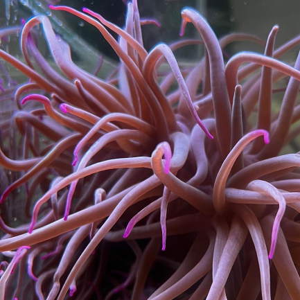
Collect ten A. viridis tentacles (approximately 5 cm each) using scissors and forceps disinfected with 70% ethanol beforehand and place them in sterile ASW.
10m
Place the tentacles in the 1 mM glycine solution and incubate for a 20 min incubation to allow cnidocytes discharge.
20m
Dry the tentacles on a sterile absorbant paper.
1m
Place the dried tentacles in the antibiotic-enriched ASW for a 2 min incubation at 200 rpm .
2m
Repeat this antibiotic washing step in another well of the six-wells plate 2 min at 200 rpm .
2m
Proceed to tentacles dissection one by one.
35m
Dry the tentacle on a sterile absorbant paper using curved forceps.
Place the tentacle on a sterile Petri dish lid and cut it lengthwise using fine scissors and forceps.
Open the tentacle with the gastrodermal layer upwards using curved forceps.
Gently scrape the tentacle to isolate the gastrodermal layer.
Collect the gastrodermis in 1 mL of sterile ASW in a 2 mL microcentrifuge tube.
5m
Homogenize the gastrodermis using a 22-gauge needle syringe. Optionally, if the tissue is too thick, start with a 21-gauge needle syringe.
10m
Transfer the homogenized tissue into a 15 mL centrifuge tube and add a chosen volume of ASW (3 to 5 mL).
5m
2000 x g, 00:05:00 and discard the supernatant and the animal gastrodermal cells (white layer on top of the Symbiodiniaceae pellet).
5m
Resuspend the Symbiodiniaceae pellet in a chosen volume of F/2 medium and collect a small volume of cell suspension for counting.
5m
Count Symbiodiniaceae using a Kova® Glasstic® slide or a hemocytometer.
5m
Seed Symbiodiniaceae at a final concentration of 1X10⁶ cells/mL in F/2 medium in a tissue culture-treated vented flask.
5m
Place your new culture in a controlled climatic cabinet with appropriate temperature and light conditions.
For instance our temperate Symbiodiniaceae cultures are maintained at 23 °C , under a standard 12h:12h photoperiod with an irradiance of 130 μmol.photons.m−2.s−1 (Beauson et al. 2025).
Symbiodiniaceae culture monitoring and care
5w
Check your culture daily under an inverted microscope to monitor cell health and detect any signs of contamination early.
Expected result
During the first weeks following Symbiodiniaceae isolation you may still observe cnidocytes and other types of animal cells in the culture, which will disappear afterwards.
Example of a Symbiodiniaceae culture 14 days post-isolation.
Refresh the culture weekly by replacing approximately 25% of the medium with fresh F/2 medium to maintain nutrient availability. Optionally, if bacterial contamination is detected, you may perform a gentle centrifugation step at 900 x g, 00:05:00 , discard the used medium and replace it completely by fresh medium while maintaining a similar final culture volume.
To track cell concentration overtime, you may count Symbiodiniaceae periodically by collecting a 100 µL culture aliquot and using a Kova® Glasstic® slide or a hemocytometer.
Expected result
Until approximately 4 weeks post-isolation Symbiodiniaceae are likely to undergo massive mortality. Afterwards they should start to actively divide and motile cells may be observed (see for reference Beauson et al. 2025).
When Symbiodiniaceae starts to actively divide you may propagate your newly established culture by seeding the cells in fresh F/2 medium in tissue-treated ventilated culture flasks at 1x10⁵ cells/mL. This step can be repeated monthly.
Protocol references
Beauson, E., Fricano, C., Porro, B., Sadoun, N., Pernice, R., Zamoum, T., Forcioli, D., Parisi, M.G., Furla, P., Barnay-Verdier, S., 2025. Expanding Symbiodiniaceae in vitro culture collections: Establishment and characterization of temperate Symbiodiniaceae cultures from the sea anemone Anemonia viridis. Algal Research 92, 104410. https://doi.org/10.1016/j.algal.2025.104410
Ventura, P., Toullec, G., Fricano, C., Chapron, L., Meunier, V., Röttinger, E., Furla, P., Barnay-Verdier, S., 2018. Cnidarian Primary Cell Culture as a Tool to Investigate the Effect of Thermal Stress at Cellular Level. Mar Biotechnol.
Acknowledgements
The authors thank Keyla Plichon for taking photographs of the dissection and tissue homogenization processes.