Jan 08, 2026
Endotracheal Wounding Using Derby Perry Excavator in Wistar Rats
- Josiah Irma1,2,
- Arief S. Kartasasmita1,3,
- Angga Kartiwa3,
- Irawati Irfani3,
- Ronny Lesmana4,5,
- Aziiz Mardanarian Rosdianto4,5,
- Anglita Yantisetiati6,
- Saraswati Anindita Rizki7
- 1Doctoral Program in Medical Sciences, Faculty of Medicine, Universitas Padjadjaran, Bandung, Indonesia;
- 2Ophthalmology Department, Universitas Pelita Harapan, Tangerang, Indonesia;
- 3Ophthalmology Department, Faculty of Medicine, Universitas Padjadjaran, Bandung, Indonesia;
- 4Department of Biomedical Sciences, Faculty of Medicine, Universitas Padjadjaran, Bandung, Indonesia;
- 5Biological Activity Division, Central Laboratory, Universitas Padjadjaran, Bandung, Indonesia;
- 6Pathology Anatomy Department, Faculty of Medicine, Universitas Padjadjaran, Bandung, Indonesia;
- 7Faculty of Medicine, Universitas Pelita Harapan, Tangerang, Indonesia


Protocol Citation: Josiah Irma, Arief S. Kartasasmita, Angga Kartiwa, Irawati Irfani, Ronny Lesmana, Aziiz Mardanarian Rosdianto, Anglita Yantisetiati, Saraswati Anindita Rizki 2026. Endotracheal Wounding Using Derby Perry Excavator in Wistar Rats. protocols.io https://dx.doi.org/10.17504/protocols.io.ewov1k41ygr2/v1
License: This is an open access protocol distributed under the terms of the Creative Commons Attribution License, which permits unrestricted use, distribution, and reproduction in any medium, provided the original author and source are credited
Protocol status: Working
We use this protocol and it's working
Created: January 06, 2026
Last Modified: January 08, 2026
Protocol Integer ID: 237121
Keywords: Endotracheal Wound, Rattus norvegicus, Wistar Rats, Trachea, Modified Derby Perry Excavator, vivo endotracheal wounding in wistar rat, vivo endotracheal wounding, derby perry excavator in wistar rat, endotracheal wounding, producing endotracheal injury, brush technique in mice, endotracheal injuries in wistar, thickness wound, derby perry dental excavator, brush technique, using derby perry excavator, dental excavator, acrylic attachment, wistar rat, glottis
Abstract
In vivo endotracheal wounding in Wistar rats presents notable technical challenges. Proper animal positioning and adequate illumination are necessary for clear visualization of the glottis and trachea, while selecting an appropriate instrument is critical to avoid perforation or insufficient injury. Although Hillel et al. previously described a wire-brush technique in mice, limited access to this tool motivated the development of alternative approaches. Utilization of a pen light with an acrylic attachment and modified Derby Perry dental excavator provided optimal visualization and generated consistent, controlled partial-thickness wounds. This approach proved to be the most effective technique for producing endotracheal injuries in Wistar
rats.

Materials
1. Male Wistar Rats (12 weeks old and weighing 200g)
2. Rat Intubation Table (Patented by Josiah Irma, Patent number EC00225045045)
3. Ketamine (PT. Ethica Industry Farmasi, Indonesia)
4. Xylazine
5. Modified Derby Perry Dental Excavator (Patented by Josiah Irma, Patent number EC00225117360; Original Derby Perry Dental Excavator by Bertamed, Indonesia)
6. Pen Light With Acrylic Attachment
7. Micropore Tape
8. Anatomical Forceps
9. Clamp
Methods
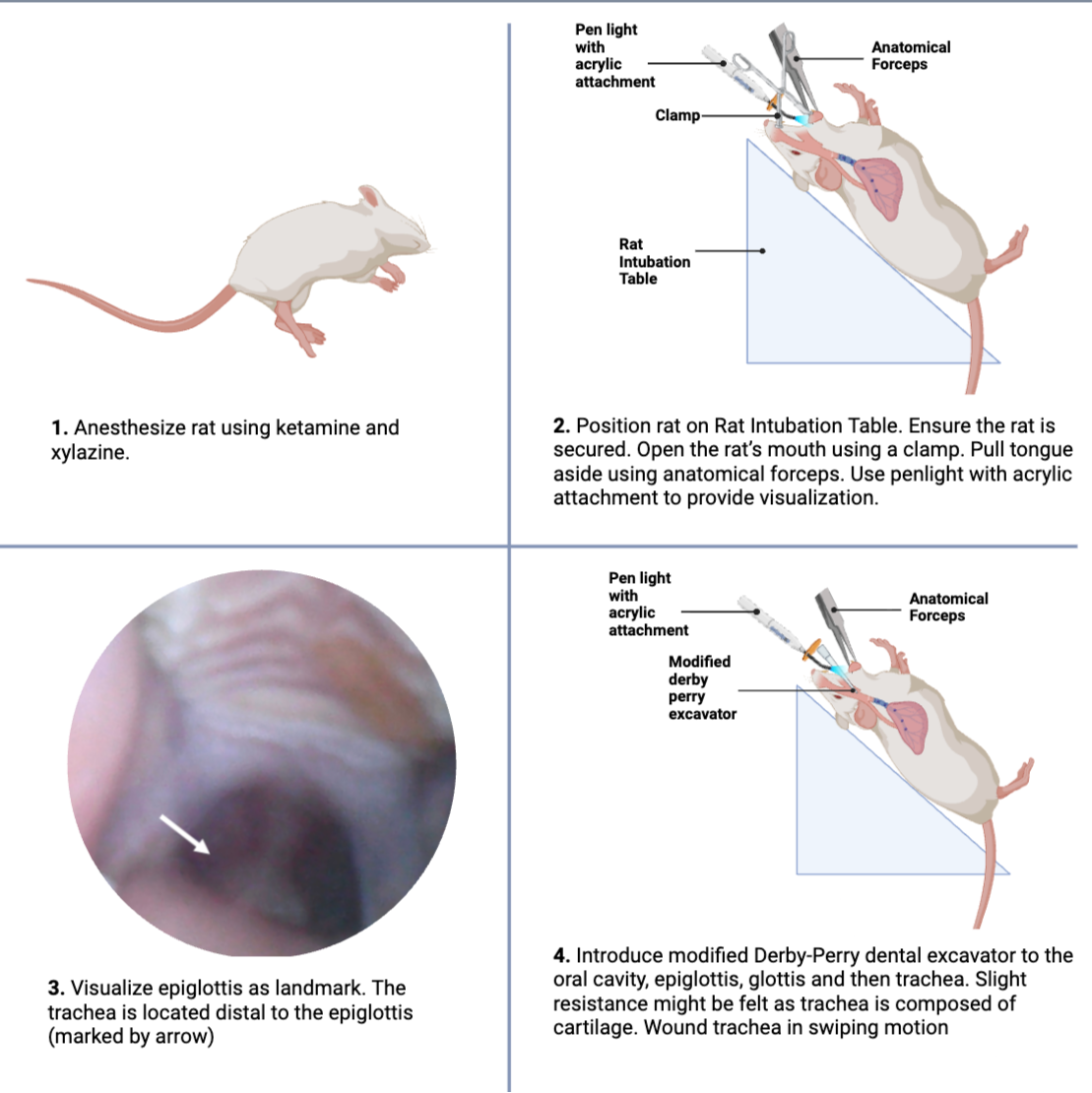
Anesthetize rat using Ketamine and Xylazine.
Position rat on Rat Intubation Table.
Ensure the rat’s sides are secured, and its body, rear, and front legs are taped using Micropore Tape.
Position table at 0°
Attach the rubber to the rat’s incisors.
Tilt tabletop to 60° angle
Open the rat’s mouth using a clamp.
Pull tongue aside using anatomical forceps.
Use penlight with acrylic attachment to provide visualization.
Visualize epiglottis as the landmark. The trachea is located distal to the epiglottis.
Introduce modified Derby-Perry dental excavator to the oral cavity, epiglottis, glottis and then trachea. Slight resistance might be felt as trachea is composed of cartilage.
Wound the trachea using a swiping motion.
Rat was then flipped to pronation position immediately after wounding.
Protocol references
Hillel AT, Namba D, Ding D, Pandian V, Elisseeff JH, Horton MR. An
in situ, in vivo murine model for the study of laryngotracheal stenosis. JAMA
Otolaryngol Head Neck Surg. 2014;140: 961–966. doi:10.1001/jamaoto.2014.1663