Jul 07, 2025
Embedding and Sectioning of Wheat Inflorescence Tissue
- Katie Long1,
- Ashleigh Lister2,
- Cristobal Uauy1
- 1John Innes Centre;
- 2Earlham Institute
- Uauy Lab - JIC


Protocol Citation: Katie Long, Ashleigh Lister, Cristobal Uauy 2025. Embedding and Sectioning of Wheat Inflorescence Tissue. protocols.io https://dx.doi.org/10.17504/protocols.io.rm7vzqwb4vx1/v1
Manuscript citation:
Spatial Transcriptomics Reveals Expression Gradients in Developing Wheat Inflorescences at Cellular Resolution
Katie A. Long, Ashleigh Lister, Maximillian R. W. Jones, Nikolai M. Adamski, Rob E. Ellis, Carole Chedid, Sophie J. Carpenter, Xuemei Liu, Anna E. Backhaus, Andrew Goldson, Vanda Knitlhoffer, Yuanrong Pei, Martin Vickers, Burkhard Steuernagel, Gemy G. Kaithakottil, Jun Xiao, Wilfried Haerty, Iain C. Macaulay, Cristobal Uauy
bioRxiv 2024.12.19.629411; doi: https://doi.org/10.1101/2024.12.19.629411
License: This is an open access protocol distributed under the terms of the Creative Commons Attribution License, which permits unrestricted use, distribution, and reproduction in any medium, provided the original author and source are credited
Protocol status: Working
We use this protocol and it's working
Created: June 24, 2025
Last Modified: July 07, 2025
Protocol Integer ID: 220750
Keywords: wheat, development, nuclei, DNA, cryosection, sectioning of wheat inflorescence tissue, wheat inflorescence tissue, multiplexed error robust fluorescence, fluorescence, situ hybridisation, tissue fixation, tissue
Funders Acknowledgements:
European Research Council
Grant ID: ERC-2019-COG-866328
UK Biotechnology and Biological Sciences Research Council (BBSRC)
Grant ID: BB/X011003/1
UK Biotechnology and Biological Sciences Research Council (BBSRC)
Grant ID: BB/X01102X/1
UK Biotechnology and Biological Sciences Research Council (BBSRC)
Grant ID: BB/X011070/1
UK Biotechnology and Biological Sciences Research Council (BBSRC)
Grant ID: BBS/E/23NB0006
UKRI Biotechnology and Biological Sciences Research Council Norwich Research Park Biosciences Doctoral Training Partnership
Grant ID: BB/T008717/1
Abstract
This protocol documents the steps for tissue fixation, embedding, and sectioning of wheat inflorescence tissue in preparation for a Multiplexed Error Robust Fluorescence In Situ Hybridisation (MERFISH) protocol; however, this protocol can be applied to other spatial transcriptomic and histological experiments. Accompanying information can be found at Long & Lister et al., 2024 doi: https://doi.org/10.1101/2024.12.19.629411
Guidelines
Safety warnings
- WARNING: Formaldehyde is a carcinogen. Use in a chemical fume hood.
- WARNING: Take care when using microtome blades. The cutting edge is extremely sharp. It is recommended to wear cut-resistant safety gloves. Before manipulating or moving a specimen, or taking a break, always lock the handwheel and cover the cutting edge with the knife guard.
- WARNING: Liquid nitrogen rapidly vaporizes to gas. Displacing air in a confined space such as a service lift may kill by asphyxiation. Cold and frostbite burns can occur from direct contact or contact with items cooled by liquid nitrogen.
Materials
Materials/Reagents
- 16% formaldehyde [w/v], methanol-free (Pierce, 28906M)
- Tissue-Tek mold, 25 x 20 x 5 mm (Thermo Fisher, AGG4580M)
- Blitz RNase Spray (Severn Biotech Ltd, 40173505M)
- MX35 Ultra Microtome Blade (Epredia, 3053835M)
- Tissue-Plus OCT compound (Agar Scientific, AGR1180M)
- Polysine Adhesion Slides (Epredia, J2800AMNZM)
- RNase-free 2ml Eppendorf Tubes
- Sucrose
- Plastic Petri Dishes
- 1x PBS in nuclease-free water
- Liquid Nitrogen
Equipment/Tools
- Liquid Nitrogen Carrier, 4 Litre (Agar Scientific, AGB7477M)
- Dissection Tools (GeeEdge ophthalmic slit knife model CJY01-2.2, GeeEdge MVR CJY04-20g)
- Stereo microscope with lighting & camera (Leica S9, HXCAM HiChrome HR4 Lite camera and a Photonic Optics light source)
- Cryostat (Leica CryoStar NX70M)
- Paintbrushes
Tissue Dissection & Fixation
Prepare 4% Paraformaldehyde solution in 1x PBS. Aliquot 1 mL 4% PFA solution into 2mL RNase-free Eppendorf Tube, store on ice.
Follow steps for tissue dissection documented in 'Wheat spike meristem microdissection' (dx.doi.org/10.17504/protocols.io.3byl49r2zgo5/v2). We modified this protocol by including the youngest leaves wrapped around the spike meristem.
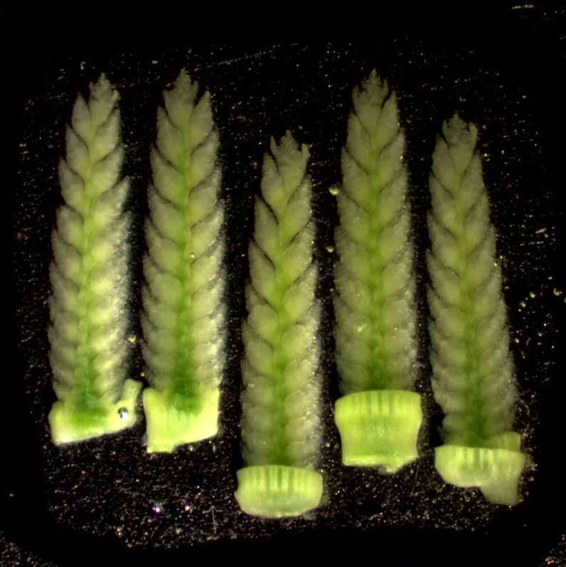
Dissected spike meristem prepared for MERFISH protocol, in four developmental stages
(Waddington stage W2.5, W3.25, W4, and W5). Taken on Leica Stereomicroscope.
Carefully transfer dissected spike meristems to 4% PFA solution with RNase-free pipette tip.
Vacuum infiltrate tissue submerged in 4% PFA solution for 00:10:00 , or until tissue sinks.
10m
Incubate tissues in 4% PFA solution overnight at 4 °C
Tissue Cryoprotection
Prepare 15% [w/v] sucrose solution with 1.5 g sucrose in 10 mL of 1x PBS.
Prepare 30% [w/v] sucrose solution with 3.0 g sucrose in 10 mL of 1x PBS.
Carefully aspirate 4% PFA solution from tissue.
Wash tissues with 1x PBS solution, repeat step 3x.
Immerse tissue in 15% sucrose solution for 06:00:00 at 4°C.
6h
Carefully aspirate 15% sucrose solution from tissues.
Immerse tissue in 30% sucrose solution, Overnight at 4 °C .
6h
Prior to embedding, clean all work surfaces and dissection tools with Blitz RNase Spray.
If protocol requires tissues be embedded within a limited area, proceed with the following step: With a lab marker, draw a 1 cm × 1 cm square on the back of a mold, centering the drawn square in the middle of the mold. Label the front of the mold with sample information.
Tissue-Tek mold (25 × 20 × 5 mm) with a 1cm2 square drawn on the back of the mold with lab marker
Tissue Embedding
Prepare a 60 mm Petri dish filled with O.C.T. compound, adding enough to entirely cover dissected tissues.
Collect a drop of O.C.T. compound on a dissection tool, and use to carefully pick up tissue from 30% sucrose solution.
With a dissection tool, carefully transfer tissue to the Petri dish, and submerge tissue into O.C.T. compound.
Examine tissue in O.C.T compound under a stereo microscope. With a dissection tool, gently move tissue within O.CT. for 1 minute, ensuring tissue is fully coated in O.C.T. compound. Remove any visible air bubbles from tissue, either by moving tissue gently around in O.C.T solution, or gently moving bubble with clean dissection tool.
Keeping tissue emerged in O.C.T. compound, trim excess tissue with dissection tools as required.
Using a dissection tool, move O.C.T. coated tissue into Tissue Tek mold. Push to the bottom of the mold and align tissue to desired orientation for sectioning.
Wheat Inflorescence embedded in O.C.T, placed within 1cm2 area of TissueTek mold
Freezing & Storage
Fill liquid nitrogen carrier with liquid nitrogen.
Carefully place a Petri dish lid upside down, floating on liquid nitrogen solution.
Place Tissue-Tek mold on top of Petri dish lid. Close liquid nitrogen carrier lid.
Once O.C.T is frozen (completely white & opaque), remove Tissue Tek mold.
Store Tissue Tek mold in sealed bag, parafilm, or aluminum foil at -70 °C
Cryosectioning
Clean the cryostat surface, forceps, and paintbrushes with Blitz RNase Spray before sectioning.
Set chamber temperature to -20 °C and blade temperature to-18 °C
Position a fresh MX35 Ultra Microtome Blade. Allow to cool completely before locking into place.
Warm cryomolds inside the cryostat for 00:30:00 at -20 °C .
30m
Trim O.C.T. blocks to remove excess O.C.T., ensuring a margin of O.C.T. left around tissues.
Frozen O.C.T block, trimmed to leave ~3 mm area around tissues
Mount O.C.T. block to chuck with room temperature O.C.T. Allow O.C.T. to solidify in cryostat.
Mount chuck and align tissues to blade.
Take 10 µm sections to trim down block. Throughout trimming, mount section to room temperature glass slides, and inspect sections to assess cutting angle and depth of tissue.
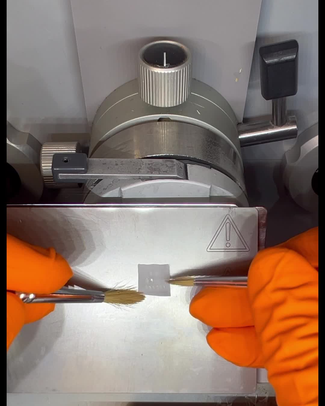
Once the region of interest is at the optimal depth and angle, take 10 µm sections, slowly and flatten with paintbrush as section is cut.
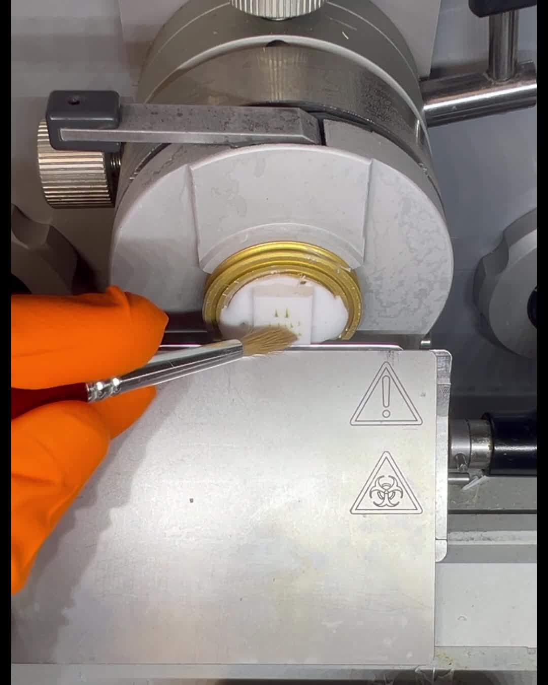
Detach section from blade gently, and continue to flatten edges of the section with paintbrush. To prevent further corners peeling or rolling up, flip the section with paintbrush.
Mount section to room temperature slides by quickly and precisely lowering slide into the cryostat chamber and pressing gently to section.
Move slide out of the chamber to room temperature. Visually inspect section has completely adhered to glass slide.
Example of wheat inflorescence cryosections adhered to glass slide- taken on Leica
Stereomicroscope.
Place the slides in 60 mm Petri dishes (or another Petri dish suited to the size of glass slide) and incubate in the cryostat chamber for00:30:00
30m
Wash slides 3x with 5 mL 1x PBS, incubate 00:05:00 each wash.
Aspirate 1xPBS. Add 5 mL 70% ethanol or until slide is completely submerged. Seal Petri dish with parafilm. Store at 4 °C for up to 1 month.
Next steps will vary depending further experimental design. For MERFISH experiments with the Vizgen MERSCOPE protocol, see MERSCOPE User Guide (Fresh and Fixed Frozen Tissue Sample Preparation, 91600002, Rev F).