Mar 27, 2026
Efficient insect DNA extraction protocol (using a tissue homogenizer)
Forked from Efficient insect DNA extraction protocol.
- Angelo José Rinaldi1,
- Juan José Quispe Haro2
- 1Universidade Federal de Viçosa;
- 2University of Helsinki
- Angelo José Rinaldi: Department of Biochemistry and Molecular Biology, Universidade Federal de Viçosa - UFV, BIOAGRO/INCT-IPP, Viçosa-MG, Brazil;
- Galleria protocols


Protocol Citation: Angelo José Rinaldi, Juan José Quispe Haro 2026. Efficient insect DNA extraction protocol (using a tissue homogenizer). protocols.io https://dx.doi.org/10.17504/protocols.io.dm6gp71e1gzp/v1
License: This is an open access protocol distributed under the terms of the Creative Commons Attribution License, which permits unrestricted use, distribution, and reproduction in any medium, provided the original author and source are credited
Protocol status: Working
We use this protocol and it's working
Created: March 26, 2026
Last Modified: March 27, 2026
Protocol Integer ID: 313937
Keywords: Extracting DNA, insects, DNA, CTAB, efficient insect dna extraction protocol, dna from insect, dna extraction technique, extracting dna, dna extraction, insect, extraction protocol, insect tissue, dna fragment, extraction process, quantity of the insect, dna, using different chemical, enzyme, galleria, mellonella, larvae, genomic, chloroform, isopropanol, efficient insect dna extraction protocol, dna from insect, extracting dna, dna extraction, dna extraction technique, genomic dna from galleria mellonella larvae, specific needs of the insect, dna fragment, insect tissue, dna, insect, many insect, genomic dna, extraction protocol, galleria mellonella larvae, extraction process, quantity of the insect, several enzyme, automated homogenization equipment, enzyme, presence of enzyme, molecular analysis, tissue homogenizer
Disclaimer
DISCLAIMER – FOR INFORMATIONAL PURPOSES ONLY; USE AT YOUR OWN RISK
The protocol content here is for informational purposes only and does not constitute legal, medical, clinical, or safety advice, or otherwise; content added to protocols.io is not peer reviewed and may not have undergone a formal approval of any kind. Information presented in this protocol should not substitute for independent professional judgment, advice, diagnosis, or treatment. Any action you take or refrain from taking using or relying upon the information presented here is strictly at your own risk. You agree that neither the Company nor any of the authors, contributors, administrators, or anyone else associated with protocols.io, can be held responsible for your use of the information contained in or linked to this protocol or any of our Sites/Apps and Services.
Abstract
This forked version modifies the original protocol by substituting manual maceration with a pestle for a tissue
homogenizer-based approach, making it more suitable for labs that have or wish to use automated homogenization equipment. In our lab, we use this protocol to extract genomic DNA from Galleria mellonella larvae.
Extracting DNA from insects can be a difficult process due to a number of factors. Some of these factors include the size and quantity of the insect, the presence of enzymes that degrade DNA, as well as the presence of chemical compounds that interfere with DNA extraction.
Some insects can be very small, making it difficult to collect enough tissue to extract DNA. In addition, insect tissues contain several enzymes that can rapidly degrade DNA, which can make it difficult to obtain DNA fragments long enough for molecular analysis.
Another factor that can make it difficult to extract DNA from insects is the presence of chemical compounds that interfere with the extraction process. For example, many insects produce chemical compounds to protect themselves from predators, which can interfere with DNA extraction techniques.
To overcome these challenges, researchers may need to optimize their DNA extraction techniques to meet the specific needs of the insect in question. This may involve using different chemicals and extraction protocols to remove chemical compounds and enzymes that interfere with DNA extraction.
Here is an example of an optimized and efficient insect DNA extraction protocol.
Guidelines
- For tough tissues such as trachea or cuticle, repeat the liquid nitrogen freezing and homogenization steps as needed to ensure complete tissue disruption.
- This protocol is optimized for Galleria mellonella larvae but can be adapted for other insect species or tissues.
- If Nanodrop measurements indicate low DNA quality (e.g., poor 260/230 or 260/280 ratios), consider using a DNA cleanup protocol to purify the sample further.
Materials
- Insects: Galleria mellonella larvae or other insect/tissue
- 70% ethanol, ice-cold
- Lysis buffer: 2% CTAB, 100 mM Tris-HCl pH 8.0, 1.4 M NaCl, 20 mM EDTA, 0.2% β-mercaptoethanol (add BME fresh before use)
- Chloroform: ice-cold, use in fume hood
- Isopropanol: ice-cold
- 70% ethanol: ice-cold
- 2 ml screw-cap microcentrifuge tubes with caps
- 1 mm glass beads
- Liquid nitrogen
- Tissue homogenizer (e.g., Precellys Evolution Touch)
- Water bath or heat block set to 65°C
- Ice bath
- Micropipettes and tips
- Refrigerated ultra centrifuge
- NanoDrop for DNA quantification and quality assessment
- Tweezers and scissors (clean, for handling insects)
Safety warnings
- Chloroform is toxic and volatile. Perform all steps involving chloroform in a fume hood and wear appropriate PPE (nitrile gloves, lab coat, safety goggles).
- Liquid nitrogen can cause severe cold burns. Handle with cryo-gloves and face protection.
Before start
- Preparing fresh lysis buffer for each extraction leads to optimal results, however, buffer without BME can be stored at room temperature for up to 6 months.
- Prepare an ice bath for ice-cold solvents (chloroform, isopropanol, 70% ethanol).
- Pre-heat a water bath or heat block to 65°C for lysis incubation.
- Use clean tweezers and scissors when handling insects to avoid cross-contamination.
- Ensure the tissue homogenizer is set up with the appropriate program before starting.
Buffer preparation
Lysis buffer solution
| A | B | C | D | E | |
| Component | Final Conc. | Stock Conc. | For 50 ml Buffer | For 10 ml Buffer | |
| CTAB | 2% (w/v) | Powder | 1.0 g | 0.2 g | |
| Tris-HCl pH 8.0 | 100 mM | 1 M | 5.0 ml | 1.0 ml | |
| NaCl | 1.4 M | 5 M | 14 ml | 2.8 ml | |
| EDTA | 20 mM | 0.5 M | 2.0 ml | 0.4 ml | |
| β-mercaptoethanol (BME) | 0.2% (v/v) | Liquid | 100 µl | 20 µl | |
| dH₂O | — | — | to 50 ml | to 10 ml |
Notes: Betamercaptoethanol (BME), added on day of use.
BME helps remove polyphenolic compounds, tannins, and proteins.
BME inhibits RNase activity. If using RNase to clean up RNA contamination of your DNA extraction, RNase will need to be added after your DNA has been precipitated and resuspended in the final storage buffer. RNase added to a CTAB solution containing BME will neutralize the RNase activity.
CTAB forms micelles in aqueous solutions and requires extensive vortexing or mixing to dissolve completely.
Cell lysis
1h 2m 30s
Collect the insects and wash them in distilled water to remove debris and residues.
Blot excess water from the insects with a paper towel.
Place the insects in a 2 ml microcentrifuge tube and add enough 70% ethanol to completely cover the insects.
Gently shake the tube and leave the insects in 70% ethanol for at least 30 minutes at room temperature. This dehydrates the insects and makes them more permeable to the lysis solution.
30m
Weigh 75 mg of insects/tissue and transfer to a 2 ml screw-cap microcentrifuge tube. For Galleria larvae, this represents about 25% of the body of a 7th instar larva.
Add 100 mg of 1 mm glass beads to each tube and close tightly.
Submerge tubes in liquid nitrogen for 30 seconds.
30s
Transfer tubes to a tissue homogenizer (e.g., Precellys Evolution Touch) and homogenize using the following program: 3 cycles of 20 seconds at 6000 rpm, with 15 seconds pause between cycles.
2m
OPTIONAL: Repeat steps 8 and 9 if necessary (e.g., for tracheal or cuticular tissue).
Incubate in a water bath at 65 °C for 30 minutes, gently shake the tubes halfway through incubation.
30m
Phase separation
10m
Prepare new 1.5 ml tubes with 650 µl of ice cold chloroform and add 750 µl from the incubated samples.
Gently mix each tube by inversion 4-5 times.
Centrifuge at 12 000 × g for 10 minutes at 4°C.
10m
Collect 600 µl of the supernatant and transfer to new 1.5 ml tubes in ice.
DNA precipitation
20m
Add 600 µl of ice-cold isopropanol, mix the tubes by inverting 4-5 times, and incubate for 30 minutes on ice.
Centrifuge at 12 000 × g for 10 minutes at 4°C, observe the formation of a pellet at the bottom of the tube.
10m
Discard the supernatant and gently wash the pellet with 1 ml of ice cold 70% ethanol.
Centrifuge again at 12 000 × g for 10 minutes at 4°C and discard the supernatant
10m
Resuspend DNA
10m
Leave the pellet to dry in the hood (∼10 minutes) and resuspend in 50 µl of deionized water.
10m
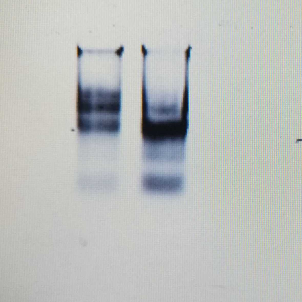
Measure the quality and quantity of eluted DNA using a NanoDrop.
Store at -20°C or -80°C until needed.
Protocol references
Asghar, U. , Malik, M. , Anwar, F. , Javed, A. and Raza, A. (2015) DNA Extraction from Insects by Using Different Techniques: A Review.Advances in Entomology,3, 132-138. doi:10.4236/ae.2015.34016.
HUNT, G. J. Insect DNA extraction protocol.Fingerprinting methods based on arbitrarily primed PCR, p. 21-24, 1997.
MOREAU, Corrie S. A practical guide to DNA extraction, PCR, and gene-based DNA sequencing in insects.Halteres, v. 5, p. 32-42, 2014.
OPPERT, Brenda et al. Optimized Extraction of Insect Genomic DNA for Long-Read Sequencing.Methods and protocols, v. 2, n. 4, p. 89, 2019.