Sep 01, 2025
Version 1
Efficient extraction of high molecular weight dsDNA from bacteriophage (QIAGEN silica-based membrane method) V.1
- Mitchell G Hedges1,
- Samuel Montgomery2
- 1The Kids Research Institute Australia;
- 2The Kids Research Institute Australia (formerly Telethon Kids Institute)


Protocol Citation: Mitchell G Hedges, Samuel Montgomery 2025. Efficient extraction of high molecular weight dsDNA from bacteriophage (QIAGEN silica-based membrane method). protocols.io https://dx.doi.org/10.17504/protocols.io.8epv5k2m6v1b/v1
License: This is an open access protocol distributed under the terms of the Creative Commons Attribution License, which permits unrestricted use, distribution, and reproduction in any medium, provided the original author and source are credited
Protocol status: Working
We use this protocol and it's working
Created: September 01, 2025
Last Modified: September 01, 2025
Protocol Integer ID: 226129
Keywords: dsDNA, DNA, bacteriophage, phage, DNEasy, Blood & Tissue, QIAGEN, silica-based membrane, silica, column, high, molecular, weight, extraction, DNA extraction, PhageWA, Oxford Nanopore, Nanopore, sequencing, whole genome sequencing, WGS, ONT, genome, fast, 2 hours, dsdna bacteriophage, efficient extraction of high molecular weight dsdna, phage dna concentration, phage dna, yield of phage dna, high molecular weight dsdna, high molecular weight dna, bacteriophage, impact from host cell dna contamination, host cell dna contamination, phage, read whole genome sequencing, phage lysate, oxford nanopore technology, whole genome sequencing, such as oxford nanopore technology, such as the qiagen dneasy blood, qiagen dneasy blood
Funders Acknowledgements:
Medical Research Future Fund
Grant ID: 2023559
Disclaimer
The digestion step (where exogenous bacterial nucleic acids are degraded) may require optimisation i.e. longer incubation times or additional volume of enzyme. The selected volume of 5uL of DNase and RNase has been tested and successfully applied in our phage extractions, and has been shown to minimise the amount of host DNA reporting via whole genome sequencing.
Sufficient removal of bacterial nucleic acids can be dependent on the growth media used for bacterial propagation, the growth characteristics of the host, the composition of the phage collection buffer and ultimately the yield of bacterial nucleic acid liberated during phage propagation.
Note that our group has detected nucleic acids within commercially available propagation broths ranging between ~2 - 10 ng/uL of DNA and ~25 - 100 ng/uL of RNA with fragment sizes from ~50 bp to 200 bp, which are consistently removed following our pre-treatment steps. We note this to alert users that sources of nucleic acid within your sample could be from either host, phage or reagent(s). We recommend the testing of all reagents for endogenous nucleic acids prior to use in DNA extractions.
Abstract
High molecular weight DNA can be efficiently extracted from dsDNA bacteriophages (phage) using a silica-based membrane method, such as the QIAGEN DNeasy Blood & Tissue Kit, with some modifications to significantly reduce the impact from host cell DNA contamination. Users can expect phage DNA concentrations of 2 to 100 ng/uL in 40 uL of elution buffer (median of approximately 15 ng/uL) if extracted from 450 uL of phage lysate containing > 1 x 10⁹ PFU/mL. The concentration and yield of phage DNA is appropriate for long read whole genome sequencing, such as Oxford Nanopore Technology.
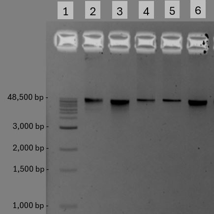
Image Attribution
Mitchell G Hedges 2025 (20250429_MH_EXPMH143)
Guidelines
- Expected input: It is ideal to extract DNA from phages which have a titre of 1 x 10⁹ to 1 x 10¹¹ PFU/mL, however lower titres may be extracted successfully.
- Expected output: Users can expect phage DNA concentrations in the range of 2 to 100 ng/uL in 40 uL of elution buffer (median of approximately 15 ng/uL), if extracting from 450 uL of phage lysate containing > 1 x 10⁹ PFU/mL.
- Intended End Use: Phage DNA extracted using this protocol is appropriate for whole genome sequencing such as Oxford Nanopore Technology long read sequencing, or PCR based applications.
Materials
- DNeasy Blood & Tissue Kit (QIAGEN #69504 or 69506)
- 100% Ethyl alcohol Molecular Biology Grade (Sigma Merck #E7023-1L)
- RNase A, DNase & Protease-Free (Thermo Fisher #EN0531)
- DNAse I [2000U/mL] (New England Biolabs #M0303 S/L)
- DNAse I 10x reaction buffer (New England Biolabs #B0303SVIAL)
- UltraPure 0.5M EDTA, pH 8.0 (Thermo Fisher #15575020)
- 1.5 ml Eppendorf DNA LoBind tubes (ThermoFisher #30108.051)
- Thermoshaker heating block (Thermo Fisher #0006286)
- Microcentrifuge (Thermo Fisher #43231565)
Before start
This protocol extracts DNA from a 450 μL aliquot of bacteriophage lysate, however the starting lysate volume can be scaled up. If scaling up, adjust the reagent volumes in all steps until the lysate mixture has been passed through the silica-membrane, ensuring consistent ratios throughout the protocol to this point. Resume the protocol volumes used at the Buffer AW1 step.
Preparation of reagents
5m
DNase & RNase Digestion Master Mix
- Calculate the required volume of Digestion Mix for the number of samples you wish to process
- Each 450 µL sample of bacteriophage lysate requires 50 µL of Digestion Mix
- Remove DNase I, DNase 10x buffer and RNase A from storage and place On ice to thaw
- Gently pipette mix each tube 5 to 10 times. Do not vortex DNase I as it is sensitive to physical denaturation
- To a clean microcentrifuge tube, add 5 µL of DNAse I and 5 µL of RNAse A to 40 µL of 10x DNAse I reaction buffer, store On ice until required
5m
DNEasy Blood & Tissue Kit Buffer Preparation
Prepare kits buffers Buffer AW1 and Buffer AW2 with 100% molecular grade ethanol as per the manufacturer's manual
Phage DNA extraction
1h 41m
Add 50 µL of Digestion Master Mix to 450 µL of syringe filtered 0.22 µm bacteriophage lysate in a 1.5mL LoBind microtube and mix via gentle pipetting
1m
Incubate for 00:30:00 at 37 °C using a Thermomixer without shaking
Note: longer incubation times may be necessary to achieve complete degradation of bacterial DNA and RNA. It is advisable to quantify the amount of bacterial DNA and RNA pre- and post-digestion to ensure bacterial nucleic acids do not contaminate phage DNA. The volume of enzyme prescribed significantly degrades host DNA and RNA and has been demonstrated to facilitate whole genome sequencing without host DNA contamination. Assessing the DNA quality of both untreated lysate and treated lysate using agarose gel electrophoresis can be useful to determine the host nucleic acid sizes in your samples, and whether a post-extraction SPRI bead clean up may be required (described elsewhere).
30m
Add 25 µL of 0.5 Mass Percent EDTA (final concentration >20 mM) to inactivate the DNase and incubate for 00:01:00 at 37 °C after gently pipette mixing
2m
Incubate for 80 °C for 00:15:00 to heat inactivate DNase. Chill the tube at 4 °C for 00:01:00 and spin down tubes briefly to collect condensation. Hot tubes may pop open, so take care to avoid cross contamination
18m
Add 20 µL proteinase K and 180 µL Buffer ATL to the sample, and mix by vortexing, incubate for 00:15:00 at 56 °C . Total volume of sample is now 720 µL
15m
Vortex the tube for 15 seconds and spin down tubes
1m
Aliquot 360 µL of the mixture into a second microcentrifuge tube (ensure it is labelled) to split the sample which allows processing of the volumes in subsequent steps
1m
To each tube containing the lysis mixture, add 360 µL of Buffer AL and mix thoroughly by vortexing
1m
Add 360 µL of ethanol (96 – 100%) and mix again thoroughly by vortexing
It is essential that the sample, Buffer AL, and ethanol are mixed immediately and thoroughly by vortexing or pipetting to yield a homogeneous solution
1m
Incubate at 56 °C for 00:05:00
5m
Pipette 600 µL of the phage lysate mixture into a labelled silica-membrane spin column
1m
Centrifuge the viral spin column in the collection tube at 6000 x g for 00:01:00 , discarding the throughflow into a waste container. The throughflow is not compatible with bleach. Discard the tube to avoid liquid cross contamination
2m
Repeat the above two steps until all phage sample has passed through the column
6m
Place the silica-membrane spin column in a new collection tube, discard any used collection tubes and any throughflow
1m
Add 500 µL of ethanol Wash Buffer 1 (AW1) onto the spin column
1m
Centrifuge the spin column in the collection tube at 6000 x g for 00:01:00
1m
Place the spin column in a new and clean collection tube. Discard the throughflow and do not combine with bleach. Discard the used collection tube
1m
Add 500 µL of Wash Buffer 2 (AW2) into the spin column
1m
Centrifuge the spin column in the collection tube at maximum speed 16000 x g to 20000 x g for 00:01:00
2m
Discard the throughflow (this is compatible with bleach if required by laboratory processes) and replace the column back into the collection tube and spin at maximum speed 16000 x g to 20000 x g for 00:03:00 to dry the column
4m
Place the spin column in a labelled, clean 1.5 ml LoBind tube (which will become the final sample tube for storage) and pipette 40 µL of warmed (56 °C ) Buffer AE directly onto the membrane. Incubate at 56 °C for 00:01:00 and then centrifuge for 00:01:00 at ≥ 6000 x g to elute
3m
Re-elute the sample by pipetting the eluate back onto the silica membrane and incubate at 56 °C for 00:01:00 then centrifuge for 00:01:00 at ≥ 6000 x g to re-elute
2m
Store the eluate containing the phage DNA at 2 - 8 ℃ in a labelled tube and take forward to DNA QA/QC processes / whole genome sequencing
1m
Optional (recommended): Quantify the extracted DNA using a 1x dsDNA High Sensitivity Kit and a sample volume of 2 µL . Following favourable quantification, load between 5 ng to 20 ng of phage DNA with a loading dye onto a 1% TAE agarose gel prepared with 1X SYBR Safe Stain and run at 100V for 01:00:00 to assess DNA quality. Ensure a ladder such as 1kb Extend DNA Ladder (NEB) is used and loaded at the same mass as the phage DNA sample for visualisation on a trans-UV system such as ChemiDoc
Protocol references
QIAGEN DNeasy Blood & Tissue Handbook (June 2023)
Jakočiūnė D, Moodley A. A Rapid Bacteriophage DNA Extraction Method. Methods and Protocols. 2018; 1(3):27. https://doi.org/10.3390/mps1030027