Feb 23, 2023
Version 2
Effective and efficient cytoskeleton (actin and microtubules) fluorescence staining of adherent eukaryotic cells V.2
- 1Department of Zoology, University of Sao Paulo;
- 2Department of Biological Sciences, Mississippi State University, USA;
- 3Misssissippi State University


External link: http://amoeba.msstate.edu
Protocol Citation: Alfredo Leonardo Porfirio-Sousa, Matthew Brown, Tristan Henderson 2023. Effective and efficient cytoskeleton (actin and microtubules) fluorescence staining of adherent eukaryotic cells. protocols.io https://dx.doi.org/10.17504/protocols.io.kxygxeerzv8j/v2Version created by Alfredo Leonardo Porfirio-Sousa
License: This is an open access protocol distributed under the terms of the Creative Commons Attribution License, which permits unrestricted use, distribution, and reproduction in any medium, provided the original author and source are credited
Protocol status: Working
We use this protocol and it's working
Created: February 22, 2023
Last Modified: February 23, 2023
Protocol Integer ID: 77454
Keywords: immunofluorescence, protist, microscopy, confocal, tubulin, microtubules, actin, nucleus, Phalloidin, antibody, Hoechst, DNA, visualizing actin microfilament, microtubule, actin microfilament, diverse organisms with complex cytoskeletal element, amoeboid protist, adherent eukaryotic cells eukaryotic microbe, efficient cytoskeleton, other adherent eukaryotic cell, cytoskeletal elements researcher, alpha tubulin antibody, complex cytoskeletal element, cell, phalloidin, immunocytochemistry, microscopical approach, fluorescence
Funders Acknowledgements:
National Science Foundation
Grant ID: 2100888
Abstract
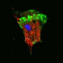
Eukaryotic microbes, protists, are highly diverse organisms with complex cytoskeletal elements used for movement consisting mostly of actin-myosin and microtubules. In order to visualize the cytoskeletal elements researchers may take a microscopical approach based on immunocytochemistry. Presented here is an efficient and effective for staining and visualizing actin microfilaments stained with phalloidin, nuclei stained with Hoechst 33342, and microtubules labeled using an alpha tubulin antibody. This protocol was developed for amoeboid protists, but will likely work on other adherent eukaryotic cells.
Protocol is adapted from the following citations.
Citation
LINK
Citation
LINK
Citation
Guidelines
Follow and adhere to all manufacturer's guidelines and warnings. Users must understand the MSDS data for each reagent before proceeding.
If staining only for Actin and nucleus, and not for microtubules, skip steps 9, 10, 11, 12, and 16.
Materials
MATERIALS
Bovine Serum AlbuminMerck MilliporeSigma (Sigma-Aldrich)Catalog #A2153
Triton X-100 Merck MilliporeSigma (Sigma-Aldrich)Catalog #X100
ParaformaldehydeMerck MilliporeSigma (Sigma-Aldrich)Catalog #158127
Fluoromount-G™Thermo FisherCatalog #00-4958-02
Nunc™ Lab-Tek™ II Chamber Slide™ System, 2 wellThermo FisherCatalog #154461PK
Normal Goat SerumThermo FisherCatalog #PCN5000
ActinGreen™ 488 ReadyProbes™ ReagentThermo FisherCatalog #R37110
NucBlue™ Live ReadyProbes™ ReagentThermo Fisher ScientificCatalog #R37605
Alpha-Tubulin Monoclonal Antibody (B-5-1-2) Thermo Fisher ScientificCatalog #32-2500
Goat anti-Mouse IgG (H L) Highly Cross-Adsorbed Secondary AntibodyThermo Fisher ScientificCatalog #A-11032
Nest Scientific 230122 Cell Culture Chamber Slides 2 Well with Glass Slide 4.55 cm2 1.2-2.5 mL 2Nest ScienficCatalog #230122
10X - Phosphate Buffered Saline (PBS):
For PBS recipe please see http://cshprotocols.cshlp.org/content/2006/1/pdb.rec8247
1L - 10X Stock Solution Recipe:
NaCl, 80 g
KCl, 2 g
Na2HPO4, 14.4 g
KH2PO4, 2.4 g
Dissolve the chemicals listed above in 800 mL of H2O. Adjust the pH to 7.4 (or 7.2, if required) with HCl, and then add H2O to 1 L. Sterilize via autoclave.
1X - Phosphate Buffered Saline (PBS):
To a 50mL of 1X PBS solution dilute 5 mL of 10X PBS (above) in 45 mL H2O. Filter sterilize 0.22µm into a 50 mL conical tube.
1X Serum Blocking Buffer (Preferred Blocking Reagent):
Serum Blocking Buffer (1X PBS [recipe above] / 5% normal serum [Thermo Fisher #: PCN5000] / 0.3% Triton™ X-100 [Sigma-Aldrich # X100-5ML]): To prepare 10 ml, add 0.5 ml normal goat serum (i.e., from the same species as the secondary antibody - GOAT) to 9.5 ml 1X PBS) and mix well. While stirring, add 30 µl Triton™ X-100. – FILTER STERILIZE 0.22µm, store in 4C.
1X BSA Blocking Buffer (Can be used in replacement of above):
To make a 500mL Blocking buffer: Weigh 0.5 g BSA (Bovine serum albumin, Sigma-Aldrich A2153) [1X = 0.5g in 500ml PBS] and add to 500 ml PBS in a 600-ml beaker. – FILTER STERILIZE 0.22µm, store in 4C.
Paraformaldehyde (PFA) Solution (8%):
For PFA recipe please see https://www.aatbio.com/resources/buffer-preparations-and-recipes/paraformaldehyde-solution-8
To make a 40mL Paraformaldehyde (8%): Prepare 32 mL of distilled water in a glass bottle containing a stir bar. Heat to 60º C on a magnetic heating plate and add 2-3 drops of 1 N NaoH. Add distilled water until the volume is 40 mL. - store at 4º C.
0.5% Triton X-100 in PBS:
To make a 2mL 0.5% Triton™ X-100 in PBS: Make a 10% Triton™ X-100 stock solution, add 50 µl Triton™ X-100 to 950 µl 1X PBS stirring well; this 10% stock solution is easier to handle. Make 2 mL 0.5% Triton™ X-100, add 0.2 µl 10% Triton™ X-100 to 1.8 µl 1X PBS.
Primary Alpha-Tubulin Antibody Dilution:
Prepare fresh PRIMARY antibody (1:500) dilution in PBS:
For 1000µL, add 2µL of primary antibody (Alpha-Tubulin Monoclonal Antibody (B-5-1-2) = Thermo Fisher Scientific | Catalog # 32-2500 | 100 µg | Antibody is at 0.5 mg/mL) in 998µL of PBS. Store at 4C in the dark.
Secondary Antibody Dilution:
Prepare fresh SECONDARY antibody (1:1000) dilution in PBS:
For 1000uL, add 1µL of secondary antibody (Goat anti-Mouse IgG (H+L) Highly Cross-Adsorbed Secondary Antibody, Alexa Fluor 594 = Thermo Fisher Scientific | Catalog # A-11032 | 1mg | Antibody is at 2 mg/mL) in 999µL of PBS
. Store at 4C in the dark
Safety warnings
Paraformaldehyde is hazardous and should be HANDLED WITH CAUTION. Before start review Paraformaldehyde and all other reagents manufacture’s Safety Data Sheet.
Move cells onto a chamber culture slide (Lab-Tek™ II Chamber Slide - Thermo Fisher Scientific - 154461) according to how the cells are being grown, see below.
If cells are growing on agar plates, cut block where there is dense area of cells. Place upside down on chamber culture slide (Lab-Tek™ II Chamber Slide - Thermo Fisher Scientific - 154461 or Nest Scientific 2 well slide 230122). Add 500µl of liquid media (same media as agar is made) and allow to sit for 00:15:00 to Overnight under normal incubation conditions. Check on the inverted microscope to see if your cells have adhered.
If cells are growing in liquid media in a tissue culture flask, scrap cells with a cell scraper to dislodge cells from tissue culture flask. Move 1 mL to each chamber of the chamber culture slide (Lab-Tek™ II Chamber Slide - Thermo Fisher Scientific - 154461). Allow to sit for 00:15:00 to Overnight under normal incubation conditions. Check on the inverted microscope to see if your cells have adhered
Ensure Paraformaldehyde (8%) is at Room temperature .
Prepare all reagents as listed in the materials section. Prepare FRESH primary and secondary antibody dilutions before you proceed. The blocking buffer may be made in bulk beforehand.
If cells were grown on agar, remove agar block gently. Be sure to remove all agar chunks.
Aspirate liquid VERY gently and discard to bleach solution. Add 500 µL of liquid media (same media the cells were growing or same media as agar is made) to chamber's side and allowing the liquid to gently flow down onto glass surface. Cells should still be attached to the chamber slide.
Fix cells by gently adding 500 µL of Paraformaldehyde (8%) at Room temperature to chamber's side and allowing the liquid to gently flow down onto glass surface. This will bring the solution to 4% Paraformaldehyde.
Incubate at Room temperature for 00:10:00 .
10m
Gently aspirate liquid with a 1mL pipette and discard.
Rinse gently by adding 500 µL PBS to chamber's side and allowing the liquid to gently flow down onto glass surface. Wash a total of three times for 00:03:00 each.
Gently aspirate liquid with a 1mL pipette and discard. Permeabilize cells by adding 500 µL of 0.5% Triton™ X-100 and incubate for 00:05:00 at Room temperature .
5m
Rinse gently by adding 500 µL PBS to chamber's side and allowing the liquid to gently flow down onto glass surface. Wash a total of three times for 00:03:00 each.
3m
Gently aspirate liquid with a 1mL pipette and discard. Add 500 µL of Serum Blocking Buffer per chamber (this is the blocking agent) and incubate for 00:10:00 at Room temperature .
If Serum Blocking Buffer is not available, you may use 1X BSA Blocking Buffer as above.
Add 500 µL of 1:500 primary antibody [monoclonal Anti-α-Tubulin antibody produced in mouse clone B-5-1-2] to the chamber slide and incubate 00:30:00 at Room temperature . This will bring your entire volume up to 1000µL.
For a negative control, add 500 µL of PBS to the other chamber slide and incubate 00:30:00 at Room temperature . This will bring your entire volume up to 1000µL.
Add 2 drops of ActinGreen 488nm ReadyProbes Reagent (Thermo Fisher Scientific | R37110) to each chamber slide and incubate 00:30:00 at Room temperature .
Gently aspirate liquid with a 1mL pipette and discard.
Rinse gently by adding 500 µL PBS to chamber's side and allowing the liquid to gently flow down onto glass surface. Wash a total of four times for00:05:00 each. Aspirate liquid completely after final wash.
5m
Add 500 µL of 1:1000 secondary antibody [Goat anti-Mouse IgG (H L) Secondary Antibody, Alexa 594] to each chamber and incubate for a 00:15:00 at Room temperature . Keep dark by covering with a box.
Add 2 drops of ActinGreen 488nm ReadyProbes Reagent (Thermo Fisher Scientific | R37110) to each chamber slide and incubate 00:10:00 at Room temperature . Keep dark by covering with a box.
Add 2 drops of NucBlue ReadyProbes (Thermo Fisher Scientific | R37605) to each chamber. Continue to incubate at Room temperature for 00:10:00 .
Rinse gently by adding 500 µL PBS to chamber's side and allowing the liquid to gently flow down onto glass surface. Wash a total of three times for00:05:00 each. Aspirate liquid completely after final wash.
5m
Remove culture slide chamber sides with removal tool included with Lab-Tek™ II Chamber Slide (Thermo Fisher Scientific - 154461) kit.
Mount your sample using a drop of Fluoromount-G (Thermo Fisher Scientific | 00-4958-02) (~100µL) and place a clean 1.5H cover slip (22x22mm) on one side of the chamber area and allow the coverslip to gently set down to avoid air bubbles. Allow to incubate at Room temperature for 00:15:00 . Keep dark by covering with a box.
Seal the edges of the cover slip with transparent nail lacquer. Let the nail lacquer dry for00:15:00 at Room temperature . Keep dark by covering with a box.
Visualize slide on a fluorescence microscope with DAPI, GFP, and TexasRed cubes or on a confocal microscope with 405, 488, 532/561 nm excitation lasers. Store slides horizontally in 4 °C in the dark.
Citations
Shadwick LL, Brown MW, Tice AK, Spiegel FW.. A new amoeba with protosteloid fruiting: Luapeleamoeba hula n. g. n. sp.
10.4467/16890027AP.16.012.5744Garajová M, Mrva M, Vaškovicová N, Martinka M, Melicherová J, Valigurová A. Cellulose fibrils formation and organisation of cytoskeleton during encystment are essential for Acanthamoeba cyst wall architecture.
https://doi.org/10.1038/s41598-019-41084-6Tekle YI, Williams JR. Cytoskeletal architecture and its evolutionary significance in amoeboid eukaryotes and their mode of locomotion.