May 01, 2025
Eco-Friendly Efficient DNA Staining Using Modified Methylene Blue Solution: A Frugal and Safer Alternative to Ethidium Bromide in Low-Resource Biotechnology Laboratories
- Joseph shenekji1,
- Kamar Shayah1,
- Sara Shanta1,
- Ladi Serawan1,
- Elaf Bobaky1,
- Ahmad Mouzayek1
- 1Department of Biotechnology Engineering, Faculty of Technical Engineering, University of Aleppo
- Joseph shenekji: PhD in Biotechnology Engineering - [email protected]
- Kamar Shayah: PhD in Biotechnology Engineering
- Sara Shanta: Graduate student in Biotechnology Engineering
- Ladi Serawan: Graduate student in Biotechnology Engineering
- Elaf Bobaky: Graduate student in Biotechnology Engineering
- Ahmad Mouzayek: Graduate student in Biotechnology Engineering
- Reclone.org (The Reagent Collaboration Network)Tech. support email: [email protected]


Protocol Citation: Joseph shenekji, Kamar Shayah, Sara Shanta, Ladi Serawan, Elaf Bobaky, Ahmad Mouzayek 2025. Eco-Friendly Efficient DNA Staining Using Modified Methylene Blue Solution: A Frugal and Safer Alternative to Ethidium Bromide in Low-Resource Biotechnology Laboratories. protocols.io https://dx.doi.org/10.17504/protocols.io.dm6gp27p8lzp/v1
License: This is an open access protocol distributed under the terms of the Creative Commons Attribution License, which permits unrestricted use, distribution, and reproduction in any medium, provided the original author and source are credited
Protocol status: Working
We use this protocol and it's working
Created: May 01, 2025
Last Modified: May 01, 2025
Protocol Integer ID: 206713
Keywords: DNA, staining, methylene blue, low-cost, eco-friendly, friendly efficient dna staining, need for carcinogenic dye, safe dyes like sybr, safe dye, carcinogenic dye, using modified methylene blue solution, visualizing dna, modified methylene blue solution, safer alternative to ethidium bromide, ethidium bromide, resource biotechnology laboratories this protocol, research biotechnology laboratory, resource biotechnology laboratory, biotechnology, agarose gel electrophoresi, using methylene, ethidium, laboratory, dna
Abstract
This protocol presents a validated, low-cost, and environmentally sustainable method for visualizing DNA using methylene blue staining after agarose gel electrophoresis. It eliminates the need for carcinogenic dyes like ethidium bromide and does not require UV transilluminators, knowing that safe dyes like SYBR safe are considered expensive for low-budget labs, making this method especially suitable for educational and research biotechnology laboratories in developing regions, in Aleppo in our case.
Materials
Materials and Reagents
| A | B | |
| Reagent | Purpose | |
| Methylene blue (powder) | Intercalating dye that binds to nucleic acids | |
| Sodium acetate (CH₃COONa) | Enhances DNA-dye retention via ionic stabilization and pH control | |
| Glacial acetic acid | Adjusts pH for optimal dye-DNA interaction (acidic range) | |
| Sodium chloride (NaCl) | Increases ionic strength, promoting stable dye binding to DNA | |
| Ethanol (99%) or Acetic acid 5% | Fixation of stained DNA to prevent fading | |
| Distilled water (dH₂O) | Solvent and diluent | |
| Loading dye | Adds density and tracking dyes to DNA samples for gel loading | |
| Agarose | Medium for DNA electrophoresis | |
| TAE or TBE buffer | Electrophoresis running buffer | |
| DNA ladder/marker | Reference for molecular weight visualization |
Equipment
- Electrophoresis apparatus with power supply
- Gel casting tray and combs
- Light box or white-light transilluminator
- pH meter or pH paper
- Balance (precision: ±0.001 g)
- Pipettes and microcentrifuge tubes
- Beakers, measuring cylinders
- Heating source (optional for increasing stain uptake)
Introduction
Ethidium bromide (EtBr), though widely used for DNA staining, poses significant health and environmental risks due to its mutagenicity and the need for UV light exposure. Methylene blue offers a non-toxic, UV-free alternative. This protocol describes an optimized approach for using methylene blue to visualize DNA ladder bands, focusing on the molecular roles of each component for improved clarity and reproducibility.
Preparation of Enhanced Methylene Blue Staining Solution
Prepare 0.5 M Sodium Acetate (pH 5.5):
To prepare a robust, low-toxicity staining solution that maximizes DNA interaction.
- Weigh4.1 g sodium acetate and dissolve in ~80 mL distilled water.
- Adjust pH to5.5 using glacial acetic acid (kitchen vinegar could do just fine).
- Bring volume to 100 mL .
Molecular Role: Sodium acetate maintains an acidic environment that enhances the electrostatic binding between methylene blue and DNA phosphate backbones.
0.25 gr methylene bluePreparation of Methylene Blue Stock Solution:
- Weigh 0.25 g 0.25 g methylene blue and dissolve in 1 L 1 L distilled water.
0.25 gr methylene blue
dissolving methylene blue in 1 L of distilled water
- Add 100 mL 100 mL of0.5 Mass Percent 0.5 Mass Percent sodium acetate (final concentration ~0.05 Mass Percent 0.05 Mass Percent ).
- Optional: Add 0.1 Mass Percent 0.1 Mass Percent NaCl (0.580.58 g 0.58 g g/100 mL) to stabilize staining.
Molecular Role: Methylene blue intercalates into the minor groove of DNA, especially under acidic conditions where the positive charge on the dye is stabilized.
Optional Fixative Preparation:
- 10% Ethanol: Mix 10 mL 10 mL ethanol + 90 mL 90 mL water.
- 5% Acetic Acid: Mix5 mL 5 mL glacial acetic acid + 95 mL 95 mL water.
DNA Electrophoresis
1h 57m
Prepare 1% Agarose Gel:
- Dissolve 1 g agarose in 100 mL TAE buffer.
- Heat until fully melted.
- Cool slightly, then pour into casting tray with comb.
loading ladder markerLoad DNA Ladder:
- Mix 5 µL loading dye with DNA ladder. (1kb Gene Ruler Fermentas)
- Load samples into wells and run gel at:
- 50 V for00:05:00 , then
- 100 V for 00:30:00 .
loading ladder marker
Function: Electrophoresis separates DNA based on molecular weight.
5m
- Staining the Gel
- Post-Electrophoresis Staining:
- Immerse gel in prepared methylene blue staining solution.
- Stain at room temperature for 00:20:00 , or for 01:00:00 for enhanced sensitivity in lower temperatures in the winter.
staining solution
Optional Fixation (Recommended):
Soak stained gel in 10 % (v/v) ethanol or5 % (v/v) acetic acid for00:10:00 to fix bands, you could put 10 mL of ethanol or 5 mL acetic acid to the same solution of staining containing methylene blue.
1h 30m
Destaining:
- Rinse gel in dH₂O or 1X TAE for 00:15:00 .
- Change wash solution every 00:05:00 until background is sufficiently clear.
- use 30 °C c water to have faster results.
20m
Visualization and Interpretation
- Visualize DNA bands under white light or a light box. (we used a white plexiglass and a fixed a light under it).
- Bands appear as dark blue lines against a lighter background.
- Faint bands may require extended destaining or longer staining time.
- in case you rinsed too much and the bands disappeared, you can simply put the gel back into staining solution for 5-10 minutes and rinse for 00:02:00 , and the bands will reappear.
- you could use smartphone image editing apps to enhance contrast.
2m
Troubleshooting and Optimization
| A | B | C | |
| Issue | Cause | Solution | |
| Faint bands | Insufficient staining time or low DNA amount | Increase staining time or DNA concentration | |
| High background | Incomplete rinsing or too high dye concentration | Increase washing steps, optimize dye concentration | |
| Bands fade quickly | No fixation step applied | Use 10% ethanol or 5% acetic acid to fix dye | |
| Uneven staining | Poor mixing or uneven gel immersion | Ensure uniform staining solution coverage |
Troubleshooting several problems
Comparison Table: Methylene Blue vs. Ethidium Bromide
| A | B | C | |
| Feature | Methylene Blue | Ethidium Bromide | |
| Cost | Low | High | |
| Safety | Non-toxic | Carcinogenic | |
| UV Light Required | No (white light suffices) | Yes | |
| Environmental Impact | Eco-friendly | Hazardous waste | |
| Staining Time | 20 - 30 minutes | 15–30 min | |
| Band Persistence | Long-lasting (if fixed) | Fades quickly under UV | |
| Sensitivity | Moderate | High | |
| Ease of Disposal | Simple | Requires hazardous disposal |
comparison between EtBr and Methylene Blue
Results
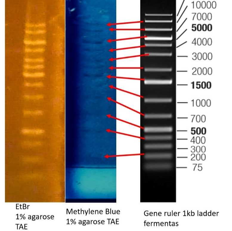
Interpretation of Gel Electrophoresis Results Using Ethidium Bromide and Methylene Blue Compared to a Commercial DNA Ladder
comparing DNA staining using EtBr, Methylene blue, and Commercial standard.
This image presents a comparative analysis of DNA band visualization using two common staining methods-ethidium bromide (EtBr) and methylene blue-on a 1% agarose gel in TAE buffer, alongside a commercial 1 kb DNA ladder (GeneRuler, Fermentas) as a molecular weight reference.
Band Patterns and Sensitivity
The EtBr-stained lane displays 14 distinct bands, visualized as bright orange-yellow under UV illumination. Ethidium bromide intercalates between DNA bases, providing high sensitivity and clear band definition, especially for fragments of medium and lower molecular weight.
The methylene blue-stained lane reveals 12 visible bands, appearing as dark blue bands on a lighter blue background. Methylene blue enables DNA visualization under normal white light, offering a safer alternative to EtBr, though with slightly reduced sensitivity, particularly for smaller DNA fragments.
The reference DNA ladder lane contains 15 bands, corresponding to fragment sizes ranging from 75 to 20,000 base pairs, with key marker bands (5000, 1500, 500 bp) highlighted for orientation. Red arrows in the image indicate the correspondence between visible bands in the methylene blue lane and their respective sizes in the ladder.
Comparative Analysis
Ethidium bromide demonstrates higher sensitivity, allowing detection of more bands and lower DNA quantities compared to methylene blue. This is evident from the 14 bands visualized with EtBr versus 12 with methylene blue, while the original ladder contains 15 bands.
Methylene blue, while less sensitive, still provides adequate resolution for most DNA fragments, and its bands align accurately with the expected sizes in the commercial ladder, as indicated by the red arrows.
Both staining methods allow for reliable estimation of DNA fragment sizes when compared to the reference ladder, but EtBr is superior for detecting low-abundance or small fragments.
Practical Implications
Ethidium bromide is recommended when maximum sensitivity is required, such as for detecting low-concentration DNA samples. However, due to its mutagenic properties, appropriate safety measures must be implemented.
Methylene blue offers a safer, non-mutagenic alternative suitable for routine applications and for downstream recovery of DNA, albeit with lower sensitivity for small or faint bands.
The choice of stain should be guided by experimental requirements: EtBr for maximal sensitivity, methylene blue for user safety and DNA integrity.
Conclusion
The comparison demonstrates that while both stains are effective for DNA visualization in agarose gels, ethidium bromide provides greater sensitivity and band resolution, closely approaching the full band pattern of the original commercial ladder. Methylene blue, though less sensitive, remains a practical and safer option for many standard Biotechnology laboratory applications, with low-cost and high availability, it is the best candidate for students labs, in cases where safe DNA stains are not available or expensive.
Protocol references
- Hu, Z., & Tong, C. (2007). Synchronous fluorescence determination of DNA based on the interaction between methylene blue and DNA.. Analytica chimica acta, 587 2, 187-93 . https://doi.org/10.1016/J.ACA.2007.01.050.
- Vardevanyan PO, Antonyan AP, Parsadanyan MA, Shahinyan MA, Hambardzumyan LA. Mechanisms for binding between methylene blue and DNA. Journal of Applied Spectroscopy. 2013 Sep;80:595-9.
- Merdekawati F, Gustira Rahayu I, Khoirul Abror Y. Check Similarity: METHYLENE BLUE AS ALTERNATIVE DNA STAINING IN ELECTROPHORESIS. InProceeding of The 5th International Conference on Interprofessional Health Collaboration and Community Empowerment 2022 (Vol. 4, No. 1, pp. 87-92). PPM Poltekkes Kemenkes Bandung M Poltekkes Kemenkes Bandung.