Apr 28, 2026
DNA Extraction Protocol and 3RAD Library Construction for Metabolite-Rich Greyia Leaf Samples
- Iné Botha1,2,
- Simo Njabulo Maduna3,
- Snorre B. Hagen3,
- Dave K. Berger1,2
- 1Department of Plant and Soil Sciences, University of Pretoria, Hatfield, 0002, South Africa;
- 2Forestry and Agricultural Biotechnology Institute (FABI), University of Pretoria, Hatfield, 0002, South Africa;
- 3Department of Ecosystems in the Barents Region, Svanhovd Research Station, Norwegian Institute of Bioeconomy Research (NIBIO), Svanvik, 9925, Norway
- Iné Botha: 0000-0003-2805-3887;
- Simo Njabulo Maduna: 0000-0002-9372-4360;
- Snorre B. Hagen: 0000-0001-8289-7752
- Dave K. Berger: 0000-0003-0634-1407


Protocol Citation: Iné Botha, Simo Njabulo Maduna, Snorre B. Hagen, Dave K. Berger 2026. DNA Extraction Protocol and 3RAD Library Construction for Metabolite-Rich Greyia Leaf Samples. protocols.io https://dx.doi.org/10.17504/protocols.io.dm6gp7mrpgzp/v1
License: This is an open access protocol distributed under the terms of the Creative Commons Attribution License, which permits unrestricted use, distribution, and reproduction in any medium, provided the original author and source are credited
Protocol status: Working
We use this protocol and it's working
Created: March 17, 2026
Last Modified: April 28, 2026
Protocol Integer ID: 313413
Keywords: Species identification, medicinal plant, genotyping, genetic diversity, phytomedicine, molecular systematics, BioMark™ HD, rich greyia leaf samples this protocol, rich greyia leaf sample, greyia tree, greyia leaf tissue, dna extraction protocol, medicinal southern african tree, dna extraction method, 3rad library construction for metabolite, greyia hook, genomic dna, dna quality, 3rad library construction method, robust snp discovery for subsequent genotyping
Funders Acknowledgements:
National Research Foundation South Africa (NRF) Foundational Biodiversity Information Programme grant
Grant ID: FBIS2204041924
NRF SARChI grant
Grant ID: SARCI150227114490
DSI Funding
Grant ID: DST/CON 0024-2015
Disclaimer
DISCLAIMER – FOR INFORMATIONAL PURPOSES ONLY; USE AT YOUR OWN RISK
The protocol content here is for informational purposes only and does not constitute legal, medical, clinical, or safety advice, or otherwise; content added to protocols.io is not peer reviewed and may not have undergone a formal approval of any kind. Information presented in this protocol should not substitute for independent professional judgment, advice, diagnosis, or treatment. Any action you take or refrain from taking using or relying upon the information presented here is strictly at your own risk. You agree that neither the Company nor any of the authors, contributors, administrators, or anyone else associated with protocols.io, can be held responsible for your use of the information contained in or linked to this protocol or any of our Sites/Apps and Services.
Abstract
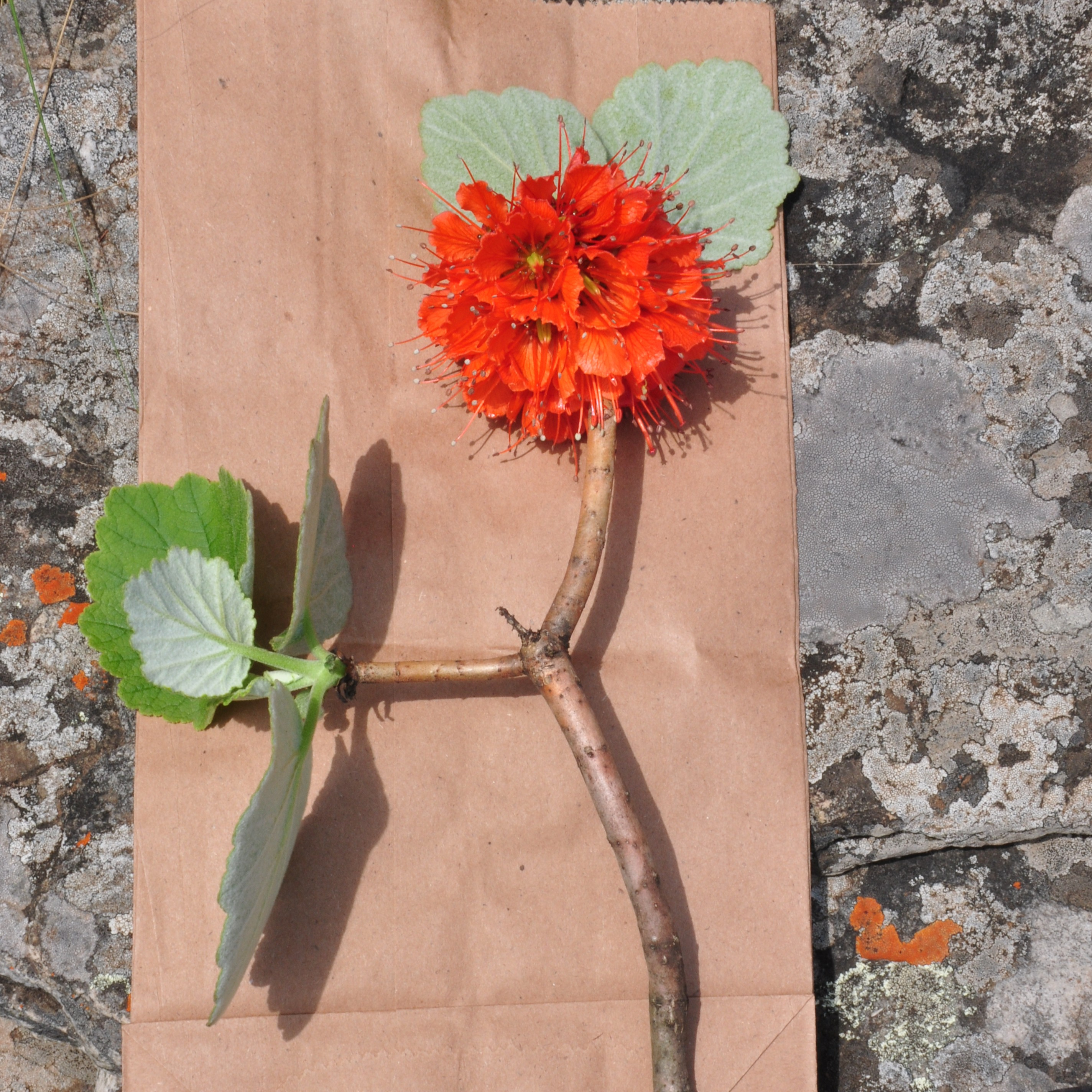
This protocol describes a DNA extraction method and 3RAD library construction method developed for the medicinal southern African tree genus Greyia Hook & Harv. Due to substantial sample-to-sample variation in secondary metabolites in Greyia leaf tissue, which frequently co-purify with genomic DNA and inhibit PCR, the protocol has been iteratively refined to improve DNA quality and reliability. Together, these protocols enable robust SNP discovery for subsequent genotyping of Greyia trees using field-collected, silica-dried material.
Image Attribution
D.K. Berger
Materials
Genomic DNA extraction protocol (for 3RAD library construction)
- Silica-dried Greyia leaf material (preferably at leaf flush stage, 1.5–2.5 cm diameter, 2g)
- Airtight zip-lock bags
- Silica gel beads (2–5 mm, Sigma-Aldrich, catalogue# 1.07735)
- Liquid nitrogen
- Mortar and pestle
- 50 mL Falcon polypropylene tubes
- CTAB buffer (see composition below)
- Polyvinylpyrrolidone (PVP-10)
- β-mercaptoethanol
- Chloroform:isoamyl alcohol (24:1)
- Phenol:chloroform:isoamyl alcohol (25:24:1)
- DNase-free RNase
- Isopropanol (propan-2-ol)
- Centrifuge capable of 6,000 × g and 10,000 × g
- Vortex
- Water bath or incubator at 37 °C and 65 °C
Safety warnings
Note: DNA quality and yield is highest if young leaf material (newly emerged flush) is used. This protocol did not completely remove secondary metabolites and other potential PCR inhibitors, so dilutions of 1/10 to 1/100 were required for successful PCR amplification in some cases.
Before start
Greyia leaf material was sampled in the field by collecting three leaves, preferably at leaf flush stage (1.5-2.5 cm diameter each), and placing them in airtight zip-lock bags containing silica gel beads, desiccant (2-5 mm) (catalogue# 1.07735, Sigma-Aldrich) for at least one week. (Note: To date, PCR-amplifiable gDNA has been isolated with this protocol from dried Greyia leaf material in silica desiccant or dried herbarium material that has been stored for up to four years.)
DNA Extraction protocol (for 3RAD library construction)
Prepare the CTAB Buffer Lysis Solution (500 mL)
Reagents needed:
- CTAB (Cetyltrimethylammonium bromide)
- Tris-HCl (pH 8)
- EDTA (disodium salt, pH 8)
- NaCl
- Sodium sulfite (Na₂SO₃)
- Distilled water
Exact amounts for 500 mL:
- CTAB: 10 g (2% w/v)
- Tris-HCl: 6.06 g (100 mM)
- EDTA: 0.372 g (20 mM)
- NaCl: 40.9 g (1.4 M)
- Na₂SO₃: 3.16 g (80 mM)
Procedure:
- Dissolve CTAB: In a 1 L beaker, add ~400 mL of distilled water and slowly dissolve 10 g of CTAB with gentle stirring.
- Add Tris-HCl: Add 6.06 g of Tris-HCl and stir until completely dissolved. Ensure the pH is around 8.0.
- Add EDTA: Add 0.372 g of EDTA (pH 8) and mix thoroughly.
- Add NaCl: Dissolve 40.9 g of NaCl into the solution with continuous stirring.
- Add Sodium Sulfite: Add 3.16 g of Na₂SO₃ and mix until fully dissolved.
- Adjust Volume: Bring the total volume up to 500 mL with distilled water.
- Mix and Store: Stir the solution until completely homogeneous. Store the buffer at room temperature or 4°C as appropriate.
NOTE: Use freshly activated buffer for experiments by adding 5% polyvinylpyrrolidone (PVP-10) and 2% v/v β-mercaptoethanol. Pre-warm at 65 °C before use.
Sample Preparation:
- Weigh out 2g of leaf material.
- Grind the leaf material to a fine powder in liquid nitrogen using a mortar and pestle.
- Transfer the ground plant material to a 50 ml Falcon® (polypropylene) conical tube.
Tips:
- Highest DNA quality and yield is obtained from young leaf flush.
- Field-collected leaves should be immediately placed in ziplock bags with silica gel beads.
- Alternatively, DNA may be extracted from fresh tissue or material stored at –20 °C.
Lysis:
- Add 8 mL of pre-warmed, activated CTAB buffer to the ground tissue.
- Vortex thoroughly to mix.
- Incubate at 65 °C for 30 minutes.
- Mix by inversion and incubate for an additional 30 minutes at 65 °C.
Chloroform-based DNA extraction:
- Add an equal volume (8 mL) of chloroform:isoamyl alcohol (24:1) to the lysate.
- Mix thoroughly by inversion.
- Centrifuge at 6,000 × g for 12 minutes at room temperature.
- Carefully transfer the supernatant to a new 50 mL Falcon® tube.
RNA removal:
- Add 5 µg DNase-free RNase to the supernatant.
- Incubate at 37 °C for 30 minutes.
Phenol-chloroform purification:
- Add an equal volume of phenol:chloroform:isoamyl alcohol (25:24:1).
- Mix thoroughly by vortexing.
- Centrifuge at 6,000 × g for 12 minutes.
- Transfer the supernatant to a clean tube.
DNA Precipitation:
- Add an equal volume of isopropanol (propan-2-ol) to the supernatant.
- Incubate at –20 °C for 1 hour.
- Centrifuge at 10,000 × g for 30 minutes.
- Carefully discard the supernatant.
- Perform an ethanol wash with two volumes of 70% ethanol.
- Repeat the wash step.
- Resuspend the DNA in DNase/RNase-free deionized water or low TE buffer.
Solid-Phase Reversible Immobilization (SPRI) bead-based inhibitor removal (following Koskinen
et al., 2018) :
- Add 100 µL of Sera-Mag SpeedBeads/ AMPure XP Beads to 50 µL of DNA extract (2X bead-based cleanup).
- Mix thoroughly by pipetting up and down five times.
- Incubate at room temperature for 10 minutes. This allows the DNA to bind to the SPRI beads.
- Spin down briefly and place on magnet for 3 minutes or until a pellet is fully formed.
- While keeping the tube in the magnet, remove and discard the supernatant. During this phase, the pellet is usually well attached to the side of the well.
- Perform an ethanol wash: keeping the tube on the magnet, add 100 µL of 80% ethanol Let stand for 1 minute and remove the ethanol.
- Repeat the wash once.
- After removing the second washing ethanol, keeping the samples on the magnet rack, let dry with the lids/caps open at room temperature for 3 minutes, or until nearly all the ethanol has evaporated, but not overdry. Caution: over-drying the beads may result in reduced yield.
- Remove the samples from the magnet stand.
- Add 30 µL (or equal amount of original extract) of purified and distilled water to sample. To concentrate the sample, add a smaller volume of water. Cover the plate carefully, vortex and spin down.
- Incubate the tube at room temperature for 5 minutes to elute DNA.
- Place the samples back on the magnet and incubate until beads separate fully from the solution (the liquid should be clear).
- Carefully transfer 30 µL of the clear supernatant to a new tube and proceed with DNA qualification and quantification.
- Carryover of small amounts of SPRI beads will not interfere with subsequent steps. Store purified DNA at 2°C to 8°C for 1 – 2 weeks, or at -15°C to -25°C.
Protocol references
Botha I, De Canha MN, Oberlander K, Botes J, Lall N, Berger DK (2026) DNA barcoding and anti-tyrosinase activities of three species-representative populations of the genus Greyia Hook & Harv. South African Journal of Botany 189:55-67.