Jun 11, 2026
Dissection-Based Method for Identifying Sex in Anthopleura elegantissima
- Elaina Berdyck1,
- Nathan Kirk2,
- Rowan McLachlan2
- 1University of Oregon;
- 2Oregon State University
- Anemones at War Protocols


Protocol Citation: Elaina Berdyck, Nathan Kirk, Rowan McLachlan 2026. Dissection-Based Method for Identifying Sex in Anthopleura elegantissima. protocols.io https://dx.doi.org/10.17504/protocols.io.6qpvrb5o2lmk/v1
License: This is an open access protocol distributed under the terms of the Creative Commons Attribution License, which permits unrestricted use, distribution, and reproduction in any medium, provided the original author and source are credited
Protocol status: Working
We use this protocol and it's working.
Created: April 24, 2026
Last Modified: June 11, 2026
Protocol Integer ID: 315710
Keywords: Anthopleura elegantissima, sea anemone, sex determination, gonad development, gonad index, reproductive biology, mesentery filaments, gonad assessment, anatomical dissection, marine invertebrate, Cnidaria, dissecting microscopy, compound microscopy, sexual maturity, polyp anatomy, asexual reproduction via longitudinal fission, asexual reproduction, sex in anthopleura elegantissima, assessing gonad development, sexual reproduction, gonad index, aggregating sea anemone anthopleura elegantissima, white spermary, sea anemone anthopleura elegantissima, gonad, determining sex, gonad staging, gonad index of bingham et al, identifying sex, common intertidal cnidarian along the pacific coast, dissection, anthopleura elegantissima, pink to brown ovary, longitudinal fission, brown ovary, examination of mesenterial tissue, peripheral polyp, central polyp
Abstract
The aggregating sea anemone Anthopleura elegantissima is a common intertidal cnidarian along the Pacific coast of North America that reproduces both sexually and asexually. Asexual reproduction via longitudinal fission produces dense, genetically uniform clonal aggregations, while sexual reproduction occurs annually through broadcast spawning, with clones typically exhibiting gonochorism. Within colonies, central polyps are often more reproductively active than peripheral polyps, reflecting functional specialization.
Gonads develop along mesenteries within the gastrovascular cavity and are seasonally and size-dependent, with males producing yellowish-white spermaries and females producing pink to brown ovaries.
This protocol describes a dissection-based approach for determining sex and assessing gonad development in A. elegantissima through examination of mesenterial tissue under dissecting and compound microscopy. Gonad staging follows the gonad index of Bingham et al. (2014). Although invasive, this method can be minimally destructive, as individuals may recover from careful pedal disc incisions that preserve viability.
Guidelines
Before beginning dissection, ensure all necessary tools and equipment are prepared in advance to streamline the procedure and allow adequate time for polyp recovery. Photograph each polyp’s gonads to provide a reference for later verification if needed.
Materials
Disposable Materials:
- Nitrile gloves
- Glass cover slips
Reusable Materials:
- Scalpel with No. 22 blade
- 4 ½” dissecting scissors
- Bent teasing needle
- Straight teasing needle
- 4 ½” straight forceps
- Small, wax dissection tray
- Fine dissection pins
- Glass microscope slides
- 200 mL volume glass beaker
Equipment:
- Compound microscope (this protocol used a ZeissPrimo Star with 10x/0.25 numerical aperture and 40x/0.65 numerical aperture objectives with brightfield illumination.)
- Dissecting microscope and light source (this protocol used an Olympus SZ61 with zoom range 6.7x to 45x with a built-in light source).
Chemicals:
- Filtered seawater (natural or artificial)
- Magnesium chloride
Software:
- Microsoft Excel(R)
Safety warnings
Electrical Hazards Warnings: Scientific scales and other laboratory equipment are powered by high-voltage outlets. Keep all liquids away from cords, plugs, and outlets to prevent electrical shock or short circuits. Always ensure hands and work surfaces are dry before handling electrical equipment.
Broken Glass and Sharps Warning: Glassware is fragile and may break easily. Handle with care to avoid cuts. Never dispose of broken glass in regular trash — always place it in a designated sharps or glass disposal container.
Biological Hazard Warning: While nematocysts of Anthopleura spp. are typically unable to penetrate human skin, repeated or prolonged handling of live specimens has been reported to cause sensitization and subsequent allergic responses in some individuals.
Ethics statement
All Anthopleura elegantissima specimens used in this protocol were collected in compliance with state regulations under the Oregon Department of Fish and Wildlife’s Scientific Taking Permit (Permit number 28795, Permit Applicant: Maya Watts, Organization: Oregon Institute of Marine Biology). Collection and handling procedures followed institutional guidelines for the ethical use of invertebrates in research and teaching. All efforts were made to minimize stress and potential harm to the animals, and only the number of specimens necessary to achieve the educational and methodological objectives was collected.
Before start
For the aggregating anemone, Anthopleura elegantissima, sex is most easily determined in late spring and summer due to peak gonad development during this period.
Preparation of equipment and tools
Set up the compound microscope. Note: This protocol utilized a ZeissPrimo Star with 10x/0.25 numerical aperture and 40x/0.65 numerical aperture objectives with brightfield illumination (Fig. 1). Place the compound microscope on a stable laboratory bench and connect it to a power source. Turn on the illumination system and set the light intensity to a moderate level. Rotate the objective turret so that the lowest-power objective lens is in position. Place a prepared test slide on the stage and secure it with the stage clips. While viewing from the side, raise the stage close to the objective lens without making contact. Looking through the eyepieces, use the coarse focus knob to bring the specimen into view. Use the fine focus knob to sharpen the image and adjust the condenser and iris diaphragm for optimal contrast. Increase magnification as needed by rotating to higher-power objectives and refocusing with the fine focus knob only.
Figure 1. Major components of the ZEISS Primostar 3 compound microscope used for gonad examination. Adapted from the ZEISS Primostar 3 User Manual (Carl Zeiss Microscopy GmbH.)
Set up the dissecting microscope. Note: this protocol utilized an Olympus SZ61 stereomicroscope with a magnification range of 6.7×–45× and an integrated illumination system (Fig. 2). Place the dissecting microscope on a stable laboratory bench and connect it to a power source. Turn on the light source as appropriate for specimen observation. Adjust the zoom magnification to the lowest setting. Place a test object on the stage. Focus using the coarse focus knob while viewing through the eyepieces. Adjust the interpupillary distance and diopter settings, if necessary, to obtain a clear image. Increase magnification as needed and refocus before beginning the procedure.
Figure 2. Major components of the Olympus SZ61 stereomicroscope used for gonad examination. Adapted from the Olympus SZ61 User Manual (Olympus Corporation).
Prepare your workspace. Gather, clean, and lay out the tools you will use during the dissection (Fig. 3).
Figure 3. Dissection tools used for Anthopleura elegantissima sex determination. Tools shown (excluding dissection pins) are from the Carolina Intermediate Dissecting Set I. From left to right: scalpel with No. 22 blade, 4½-inch dissecting scissors, bent teasing needle, 4½-inch straight forceps, straight teasing needle, and a wax dissecting tray containing an anemone polyp. (Photo credit: Elaina Berdyck.)
Dissection to expose mesentery filaments
Pin the anemone polyp. Place a single Anthopleura elegantissima polyp on a wax dissecting tray with the oral surface facing downward to maximize exposure of the pedal disc (Fig. 4A). Secure the specimen using fine dissection pins (Fig. 4B). If necessary, gently remove debris from the pedal disc using forceps and teasing needles prior to dissection.
Figure 4. Pinning and preparation of Anthopleura elegantissima prior to dissection. (A) Partially pinned anemone with visible debris on the pedal disc. (B) Fully pinned specimen following debris removal and positioning for dissection. (Photo credit: Elaina Berdyck.)
Open the pedal disc. Using a scalpel, make a small incision at the edge of the pedal disc adjacent to the top-most pin (Fig. 5A). Insert the tips of the dissection scissors into the incision and perform a single, shallow longitudinal cut along the center of the pedal disc. Ensure that all cuts remain superficial, targeting only the pedal disc tissue and avoiding damage to the underlying mesentery filaments. If needed, make additional small perpendicular cuts to facilitate spreading and pinning of the tissue (Fig. 5B). Use additional dissecting pins to fully open and expose the mesentery filaments (Fig. 5C).
Figure 5. Dissection of the pedal disc of Anthopleura elegantissima for internal anatomy exposure. (A) Initial incision of the pedal disc using a No. 22 blade scalpel. (B) Pedal disc immediately following longitudinal incision. (C) Fully opened and pinned specimen with internal structures exposed. (Photo credit: Elaina Berdyck.)
Determine the sex of the sea anemone
Explore the internal anatomy. Using a bent teasing needle and straight forceps, carefully examine the mesentery tissues and distinguish between (A) mesentery filaments, (B) gonads, and (C) the siphonoglyph (Fig. 6). Begin observations with the unaided eye, then transfer the dissection tray to the stage of the dissecting microscope for higher-magnification examination of internal structures.
Figure 6. Internal anatomy of Anthopleura elegantissima showing key mesenterial structures. (A) Mesenteries, (B) siphonoglyph, and (C) gonads (male spermaries, shown in this specimen). (Photo credit: Elaina Berdyck.)
Determine sex and gonad development level. Sex assignment (male or female) and gonad development stage may be determined by visual inspection or may require examination using a dissecting microscope (10×) or compound microscope (100×). The size, morphology, and prominence of gonadal tissues on the mesenteries vary with the sexual maturity of the polyp. Gonad development levels are assigned according to the gonad index described in Bingham et al. (2014) (Table 1).
Table 1. Gonad index scale for Anthopleura elegantissima, adapted from Bingham et al. (2014).
Level 0 gonads (Fig. 7). Assigned when no visible gonadal protrusions/bumps are present on the mesentery filaments under compound microscopy at 100× magnification; sex cannot be determined at this stage.
To prepare a slide, excise a small section of mesentery using a scalpel or dissecting scissors and place it on a clean glass microscope slide. Gently apply a glass coverslip, taking care to avoid excessive compression of the tissue. Position the slide on the compound microscope stage for observation. Multiple sections of mesentery should be sampled by cutting several pieces per specimen and mounting 2–3 pieces per slide, or by preparing multiple slides as needed, to ensure a representative assessment of gonadal tissue distribution.
Figure 7. Level 0 gonadal development in Anthopleura elegantissima. Mesenteries without detectable gonadal structures, shown under (A) a dissecting microscope at 10× magnification and (B) a compound microscope at 100× magnification. (Photo credit: Elaina Berdyck.)
Level 1 gonads. Characterized by small, discrete protrusions/bumps on the mesentery filaments that require examination under a compound microscope at 100× magnification for detection and sex differentiation. No specimens exhibiting Level 1 gonadal development were observed at the time of this study; accordingly, no representative image is included. Future iterations/forks of this protocol may incorporate visual documentation of this stage when available.
Level 2 gonads (Fig. 8). Characterized by swollen mesenteries that are visible under a dissecting microscope; however, definitive sex determination requires examination of mesenterial tissues at 100× magnification using a compound microscope.
Figure 8. Level 2 gonadal development in Anthopleura elegantissima. Swollen mesenteries are shown for (A) a male specimen and (B) a female specimen, visualized under a compound microscope at 100× magnification. (Photo credit: Nathan Kirk.)
Level 3 gonads (Fig. 9). Characterized by single-lobed gonadal masses that are visible under a dissecting microscope at 10× magnification; sex can be distinguished at this stage based on gonadal morphology.
Figure 9. Level 3 gonadal development in Anthopleura elegantissima. Single-lobed gonadal masses are shown in (A) a male specimen and (B) a female specimen, observed under a compound microscope at 10× magnification. (Photo credit: Nathan Kirk.)
Level 4 gonads (Fig. 10). Characterized by multi-lobed gonadal masses that are visible to the unaided eye and less turgid than Level 5 gonads. However, confident sex determination requires examination under a dissecting microscope at 10× magnification. Gonadal tissue may not be present on all mesenteries.
Figure 10. Level 4 gonadal development in Anthopleura elegantissima. Multilobed gonadal masses are shown in (A) a male specimen and (B) a female specimen, observed under a dissecting microscope at 10× magnification. (Photo credits: A, Brian Bingham; B, Elaina Berdyck.)
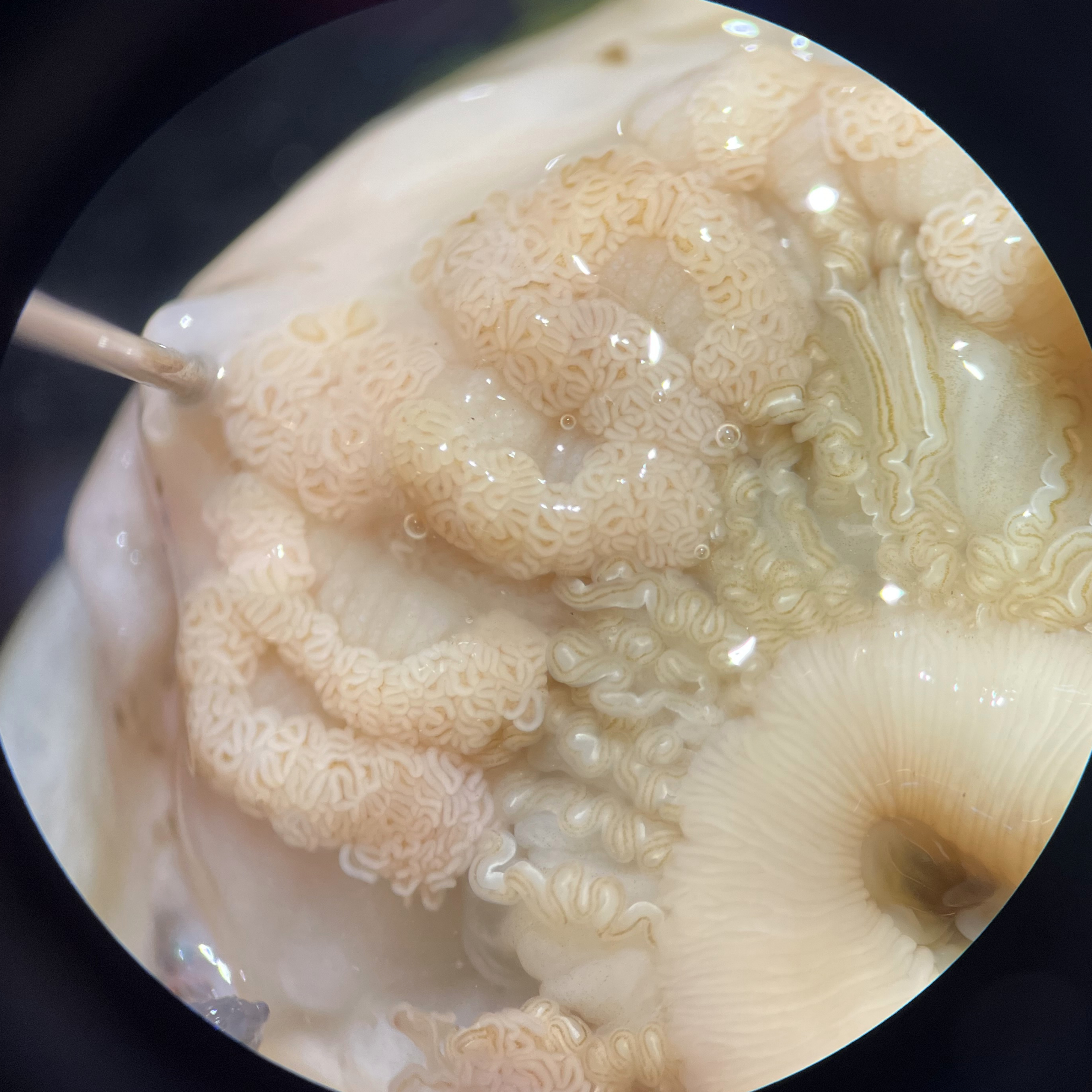
Level 5 gonads (Fig. 11). Characterized by abundant, highly turgid, multi-lobed gonadal masses that are readily visible to the unaided eye. Gonads are present on most to all mesenteries.
Figure 11. Level 5 gonadal development in Anthopleura elegantissima. Highly developed, multi-lobed gonadal masses are shown in (A) a male specimen and (B) a female specimen, observed under a dissecting microscope at 10× magnification. (Photo credit: Elaina Berdyck.)
Protocol references
Bingham, B. L., Dimond, J. L., & Muller-Parker, G. (2014). Symbiotic state influences life-history strategy of a clonal cnidarian. Proceedings of the Royal Society B: Biological Sciences, 281(1789).
Acknowledgements
We gratefully acknowledge the University of Oregon’s Oregon Institute of Marine Biology for providing access to teaching facilities, equipment, and support from faculty and staff. We thank the administrative staff, Laura Screen and Shawna Johnston, for their assistance in ordering supplies and materials. Special thanks are extended to Ian Washington, Education Support Specialist, for his support in setting up the teaching laboratory space and equipment, as well as for his help in locating additional supplies. Finally, we thank all students enrolled in BI457 Anemones at War for their contributions in refining and troubleshooting the methods described in this protocol.
Funders Acknowledgements: All supplies and materials not available through the Oregon Institute of Marine Biology’s teaching inventory were purchased using student class fees.