May 16, 2022
Differentiation of iPSCs with the hNIL construct into motor neurons protocol
- Maria Sckaff1,
- Kenneth Wu2,
- Hana Ghanim2,
- Aradhana Sachdev2,
- Gokul N Ramadoss2,
- Carissa M. Feliciano2,
- Luke M. Judge2,
- Bruce Conklin2,
- Claire D Clelland3
- 1University of California, San Francisco;
- 2Gladstone Institutes, San Francisco, CA, United States;
- 3University of California, San Francisco, Weill Institute for Neurosciences, San Francisco, CA, United States
- Claire D Clelland: [email protected];


Protocol Citation: Maria Sckaff, Kenneth Wu, Hana Ghanim, Aradhana Sachdev, Gokul N Ramadoss, Carissa M. Feliciano, Luke M. Judge, Bruce Conklin, Claire D Clelland 2022. Differentiation of iPSCs with the hNIL construct into motor neurons protocol. protocols.io https://dx.doi.org/10.17504/protocols.io.14egn76kqv5d/v1
License: This is an open access protocol distributed under the terms of the Creative Commons Attribution License, which permits unrestricted use, distribution, and reproduction in any medium, provided the original author and source are credited
Protocol status: Working
We use this protocol and it’s working.
Created: March 24, 2022
Last Modified: May 16, 2022
Protocol Integer ID: 59827
Keywords: hNIL differentiation, Maintaining iPSCs, Passaging iPSCs, motor neurons, iPSC, stem cells, hnil construct into motor neurons protocol, induced pluripotent stem cell, pluripotent stem cell, ipscs with the hnil, motor neurons protocol, differentiation of ipsc, motor neuron, hnil transgenic factor, ipsc, hnil construct, hnil, stem
Abstract
This protocol describes the differentiation of induced pluripotent stem cells (iPSCs) into motor neurons using the hNIL transgenic factors in a CLYBL safe harbor site.
Attachments

Guidelines
This protocol is based on hNIL differentiation protocol by Fernandopulle et al. from Michael Ward’s
lab1.
References
[1] Michael S Fernandopulle, Ryan Prestil, Christopher Grunseich, Chao Wang, Li Gan, Michael E Ward. Transcription Factor-Mediated Differentiation of Human iPSCs into Neurons. Curr Protoc Cell Biol. 2018 Jun;79(1): e51. doi: 10.1002/cpcb.51. Epub 2018 May 18. PMID: 29924488; PMCID: PMC6993937.
[2] Erika Lara Flores (2021). Maintenance protocol of iPSCs. Protocols.io
[3] Merissa Chen*, Nina Draeger*, Martin Kampmann*, Kun Leng*, Emmy Li*, Connor Ludwig*, Greg Mohl*, Avi Samelson*, Syd Sattler*, Ruilin Tian* (2019). Kampmann Lab iNeuron pre-differentiation & differentiation protocol. Protocols.io
Materials
Items needed for the creation of the media and reagents used in the differentiation of hNIL iPSCs into motor neurons.
| A | B | C | |
| Item | Manufacturer | Catalog Number | |
| KnockOut DMEM | ThermoFisher Scientific | 10829018 | |
| Growth Factor Reduced Matrigel | Corning | 356231 | |
| mTeSR™ Plus and supplement | Stemcell Technologies | 1000276 | |
| ReLeSR™ | Stemcell Technologies | 05872 | |
| Gibco™ DPBS, no Ca, no Mg | ThermoFisher Scientific | 14-190-235 | |
| Gibco™ DPBS, Ca, Mg | ThermoFisher Scientific | 14-040-117 | |
| Accutase | Stemcell Technologies | 07920 | |
| DMEM/F-12, HEPES | ThermoFisher Scientific | 11330032 | |
| N-2 Supplement | ThermoFisher Scientific | 17502048 | |
| MEM Non-Essential Amino Acids Solution (NEAA) | ThermoFisher Scientific | 11140050 | |
| GlutaMAX Supplement | ThermoFisher Scientific | 35050061 | |
| Culture One Supplement | ThermoFisher Scientific | A3320201 | |
| B-27 Supplement, serum free | ThermoFisher Scientific | 17504044 | |
| y-Secretase Inhibitor XXI, Compound E | Millipore Sigma | 565790 | |
| Poly-D-Lysine (PDL) | ThermoFisher Scientific | A3890401 | |
| Laminin Mouse Protein, Natural | ThermoFisher Scientific | 23017015 | |
| Doxycycline Hyclate (reconstituted in water) | Millipore Sigma | D3447 | |
| 5-Bromo-2ʹ-deoxyuridine (BrdU) | Millipore Sigma | B9285 | |
| ROCK1 Inhibitor (Y-27632 2HCl) | Selleckchem | S1049 | |
| Neurobasal | Life Technologies | 21103-049 | |
| HyClone Characterized Fetal Bovine Serum (FBS) | Cytiva | SH30071.03HI | |
| Recombinant Human BDNF | Peprotech | 450-10 | |
| Recombinant Human GDNF | Peprotech | 450-02 | |
| Recombinant Human NT3 | Peprotech | 450-03 | |
| Aphidicolin | Millipore Sigma | 89458 |
Note
Notes on some items that need to be reconstituted
Compound E: 1 mg is reconstituted in 255 µL of ethanol and 255 µL of DMSO to make a 10,000 stock, then aliquoted and stored at-20 °C for up to 6 months, minimizing exposure to light. Doxycycline: diluted in cell culture grade water to 2 mg/mL and stored at-20 °C (long-term storage) or 4 °C (short-term storage), minimizing exposure to light.
BrdU: reconstituted in water to a stock of 40 millimolar (mM) (12.284 mg/mL ).
BDNF, GDNF, and NT3: 50 µg reconstituted in filtered 1X DPBS with 0.1% BSA, then aliquoted and stored at -20 °C for up to 3 months.
KnockOut™ DMEMThermo Fisher ScientificCatalog #10829018
Corning® Matrigel® Growth Factor Reduced (GFR) Basement Membrane MatrixCorningCatalog #356231
mTeSR™ PlusSTEMCELL Technologies Inc.Catalog #1000276
ReLeSR™ 100 mL
STEMCELL Technologies Inc.Catalog #5872
Gibco™ DPBS no calcium no magnesiumThermo Fisher ScientificCatalog #14190235
Gibco™ DPBS calcium magnesiumThermo Fisher ScientificCatalog #14040117
ACCUTASE™STEMCELL Technologies Inc.Catalog #07920
DMEM/F-12, HEPESThermo Fisher ScientificCatalog #11330032
N2 supplement (100x supplement)Gibco - Thermo Fisher ScientificCatalog #17502048
MEAA (MEM Non-Essential Amino Acids)Gibco - Thermo Fisher ScientificCatalog #11140050
GlutaMAX™ SupplementThermo Fisher ScientificCatalog #35050061
CultureOne™ Supplement (100X)Thermo FisherCatalog #A3320201
B-27™ Supplement (50X), serum freeGibco - Thermo Fisher ScientificCatalog #17504044
γ-Secretase Inhibitor XXI Compound EMerck Millipore (EMD Millipore)Catalog #565790
Poly-D-LysineThermo Fisher ScientificCatalog #A3890401
Laminin Mouse Protein, NaturalThermo FisherCatalog #23017015
5-Bromo-2′-deoxyuridineMerck MilliporeSigma (Sigma-Aldrich)Catalog #B9285
Y-27632SelleckchemCatalog #S1049
Gibco™ Neurobasal™ MediumThermo Fisher ScientificCatalog #21103049
HyClone Characterized Fetal Bovine Serum (FBS)CytivaCatalog #SH30071.03HI
Recombinant human GDNFpeprotechCatalog #450-10
Recombinant human BDNF peprotechCatalog #450-02
Recombinant Human NT-3peprotechCatalog #450-03
Equipment usedin the differentiationof hNIL iPSCs intomotor neurons.
| A | B | C | |
| Equipment | Manufacturer | Catalog Number | |
| Falcon® 96-well Black/Clear Flat Bottom TC-treated Imaging Microplate with Lid | Corning | 353219 | |
| Eppendorf® Centrifuge 5810/5810R | Millipore Sigma | EP022628168 | |
| Invitrogen Countess™ II automated cell counter | ThermoFisher Scientific | AMQAX1000 |
Equipment
6-well Black/Clear Flat Bottom TC-treated Imaging Microplate with Lid
NAME
Microplate
TYPE
Falcon®
BRAND
353219
SKU
LINK
Equipment
Centrifuge 5810/5810R
NAME
Centrifuge
TYPE
Eppendorf®
BRAND
EP022628168-1EA
SKU
LINK
Equipment
LIFE TECHNOLOGIES COUNTESS II
NAME
Automated Cell Counter
TYPE
Invitrogen Countess™
BRAND
AMQAX1000
SKU
LINK
Maintenance and Preparation of the iPSCs for the hNIL differentiation into motor neurons: Maintaining iPSCs
20m
Culture iPSCs for at least 2-3 passages after initial hNIL transfection or from frozen stocks before starting a motor neuron differentiation.
Note
iPSCs that have been recently thawed or are otherwise stressed (e.g., recently nucleofected) can result in phenotypically abnormal motor neurons and poor differentiation. See Flores et al. for complete iPSC culture methods2.
Briefly, we maintain iPSCs on Matrigel-coated plastic culture dishes (Growth Factor Reduced Matrigel diluted in 50 mL of KnockOut DMEM to a concentration of 80 µL ) with mTeSR™ Plus medium.
Change media every other day, and passage every 4-5 days with ReLeSR™ (see below).
Add ROCK1 inhibitor at 10 micromolar (µM) in media to freshly passaged iPSCs for 1 day to limit spontaneous differentiation.
Maintenance and Preparation of the iPSCs for the hNIL differentiation into motor neurons: Passaging iPSCs
20m
Passage iPSCs as clumps (for routine expansion of iPSC cultures) or single cells (for differentiation).
For routine expansion, wash the well with DPBS -Ca/-Mg (1-2 mL/well*) then add 1 mL per well* of ReLeSR™ for 00:01:00 .
1m
Aspirate most of the ReLeSR™, but do not overdry, and incubate the wells at Room temperature for 3-4 minutes.
4m
Add fresh mTeSR™ Plus media to the well (1 mL /well*) and pipette gently to detach the iPSCs and break colonies into small clumps.
Note
Clumps should be visible by eye.
A confluent well can be split 1:6 to 1:20 depending on the desired confluency and rate of growth of the iPSC line.
If desired, before replating, cells can be centrifuged at 800 rpm, 00:00:15 to pellet clumps and remove any single cells (which remain in suspension).
Note
ROCK1 inhibitor is not necessary when clump passaging, but can be added at 10 micromolar (µM) (final concentration in well solution) for 1 day to limit spontaneous differentiation.
15s
For differentiation (or anytime cell counting is needed), wash the well with DPBS -Ca/-Mg (1-2 mL/well*) then add 0.5 mL * of Accutase®.
Note
* These volumes are based on experiments run on a 6-well plate and are scalable to other cell culture vessels.
Incubate the plate for 00:05:00 at 37 °C .
5m
After the incubation, tap the plate to fully detach the iPSCs, then add 0.5 mL * of KnockOut DMEM with 20% FBS (alternately, 2.5 mL/well* of DPBS +Ca/+Mg can be used in place of DMEM/FBS).
Note
* These volumes are based on experiments run on a 6-well plate and are scalable to other cell culture vessels.
Transfer the cells and solution to a conical vial and centrifuge for 800 rpm, 00:03:00 to pellet the cells.
3m
Aspirate the supernatant carefully as to not disturb the cell pellet, and resuspend the cells in mTeSR™ Plus with ROCK1 inhibitor at 10 micromolar (µM) .
Note
ROCK1 inhibitor is highly recommended when passaging iPSCs as single cells to prevent spontaneous differentiation.
hNIL differentiation into motor neurons: Day 0
1h
Coat the receiving vessel with Matrigel at least 00:30:00 before starting Day 0 (but no more than 24-36 hours prior to starting) and incubate at 37 °C .
30m
Bring the mTeSR™ Plus with ROCK1 inhibitor (final concentration of 10 micromolar (µM) in well, 1000X dilution from 10 millimolar (mM) stock) and the Accutase® to Room temperature .
Aspirate the spent media and wash the wells with cells once with 1X DPBS -Ca/-Mg.
Add appropriate volume of Accutase® per well.
Incubate the plate for 00:05:00 at 37 °C .
5m
Remove plate from the incubator and tap the plate to release the cells.
Quench the Accutase® using five times the volume DPBS +Ca/+Mg (if you used 0.5 mL of Accutase®, quench with 2.5 mL of DPBS).
Transfer the cells to a conical vial.
Centrifuge the vial at 800 rpm, 00:03:00 .
3m
Aspirate the Accutase® with DPBS, being careful not to disturb the cell pellet at the bottom of the vial.
Resuspend the cells with an appropriate volume of mTeSR™ Plus with ROCK1 inhibitor (final concentration of 10 micromolar (µM) in well, 1000X dilution from 10 millimolar (mM) stock). Aim for roughly 1 mL of mTeSR™ Plus with ROCK1 inhibitor per 1 million cells.
Pipette up and down to mix well and produce an evenly distributed solution.
Count the number of cells using a Countess II (any preferred equivalent way of counting cells is also appropriate).
Note
Countess II count parameters:
- Size 0 to 30
- Brightness 0 to 255
- Circularity 80
- Auto Lighting selected
5m
Aspirate the Matrigel from the receiving vessel.
Add appropriate volume of media (the mTeSR™ Plus with ROCK1 inhibitor at 10 millimolar (mM) ) to the receiving vessel.
Add volume of cell mixture appropriate for the number of cells desired in the well. For a 10cm dish, we have found 1-2 million iPSCs at day 0 produces 3-5 million cells at day 3.
Place the receiving vessels (which now has your cells) back in the incubator at 37 °C .
5m
Shake the plate in all four directions to ensure the cells are evenly distributed in the well.
hNIL differentiation into motor neurons: Day 1
30m
Prepare the neural induction medium (NIM) as follows:
| A | B | |
| DMEM/F12 | 97 mL | |
| N-2 | 1 mL | |
| NEAA | 1 mL | |
| GlutaMAX | 1 mL |
Calculate the volume of freshly prepared NIM needed to perform a full media change (see Table 2) and aliquot in a vial of appropriate size.
| A | B | C | D | E | |
| Cell culture vessel | Surface area (cm2) | Media volume per well | Day 3 Seeding density for ICC applications | Day 3 Seeding density for protein, RNA, DNA extraction | |
| 96-well plate | 0.32 | 150 µL | 20-25k cells | - | |
| 24-well plate | 1.9 | 500 µL | 80-100k cells | - | |
| 12-well plate | 3.5 | 1 mL | 2 x 10⁵ cells | - | |
| 6-well plate | 9.6 | 2 mL | 0.5 x 10⁶ cells | 1 x 10⁶ cells | |
| 10-cm dish | 56.7 | 10 mL | - | 5 x 10⁶ cells | |
| 15-cm dish | 145 | 30 mL | - | 15 x 106 cells |
Table 2: Number of cells and volume of media matrix needed on the day-3 replating in the differentiation of hNIL iPSCs into motor neurons based on the size and format of the cell culture vessel used, as well as the application. Modification from the Kampmann’s Lab protocol3.
Add the appropriate factors for day 1 to the NIM aliquot you aliquoted in step 35:
| A | B | C | D | |
| Factor | Final Concentration | Stock | Dilution | |
| ROCK1 inhibitor | 10 μM | 10 mM | 1:1,000 | |
| Doxycycline | 2 μg/mL | 2 mg/mL | 1:1,000 | |
| Compound E | 0.2 μM | 2 mM | 1:10,000 |
Aspirate the spent media from the wells with cells.
Add the appropriate volume of media to each well.
Place the plate with cells in fresh media back in the incubator at 37 °C .
hNIL differentiation into motor neurons: Day 2
10m
Select the receiving cell-culture vessel. This is the plate on which your neurons will grow permanently (see recommendations below).
Note
Recommendations for protein or RNA extraction: A minimum surface area equivalent to three 6-wells is recommended per RNA or protein sample to be extracted. Regular cell-culture plates (plastic, flat bottom) are well-suited for this application.
Recommendations for imaging purposes: We recommend high quality plastic plates, such as the Falcon® 96-well Black/Clear Flat Bottom TC-treated Imaging Microplate (see Table 4) due to improved long-term neuron attachment. Glass-bottom plates or coverslips may also be used, but in our hands, about 20-50% of the wells experience lifting as the neurons age past day 14 in culture. Glass-bottom plates are recommended when the neurons will be fixed for imaging at an earlier stage, such as between day 7 and day 14 in culture. Premature detachment can be minimized by very gentle handling, slow pipetting, and never completely removing the media from the wells (including when PFA fixing, see below).
Note on PDL pre-coated plates: If using a PDL pre-coated plate, you will only need to rehydrate it on day 3 in DPBS once prior to coating receiving wells with laminin and the seeding of the cells.
Coat the wells which will be receiving cells on day 3 with the appropriate volume of 1X PDL at 0.1 mg/mL (see Table 1).
| A | B | C | D | |
| Cell culture vessel | Matrigel, PDL, and laminin in KO DMEM volume per well | DPBS volume for washes per well | Accutase® volume per well | |
| 96-well plate | 120 µL | 150 µL | 50 µL | |
| 24-well plate | 250 µL | 400 µL | 125 µL | |
| 12-well plate | 500 µL | 1 mL | 250 µL | |
| 6-well plate | 1 mL | 2 mL | 500 µL | |
| 10-cm dish | 4 mL | 6 mL | 3 mL | |
| 15-cm dish | 12 mL | 18 mL | 9 mL |
Table 1: Volumes of coating solutions (Matrigel, PDL, and laminin in KO DMEM), DPBS, and Accutase® for each cell culture vessel.
Place the coated plate back in the incubator at 37 °C .
hNIL differentiation into motor neurons: Day 3; Part 1
40m
Note
If using PDL pre-coated plates, rinse once with DPBS and skip to step 46.
Aspirate out the 1X PDL from the receiving cell-culture vessel.
Wash the receiving wells.
Wash the receiving wells with 1X DPBS. (1/2)
Wash the receiving wells with 1X DPBS. (2/2)
Let receiving wells dry in the laminar flow hood for 20-30 minutes.
30m
Coat receiving cell-culture vessel with the appropriate volume of NIM with laminin mouse protein (at a concentration of 15 µL ) for 2-5 hours at 37 °C (see Table 1 for volume)
| A | B | C | D | |
| Cell culture vessel | Matrigel, PDL, and laminin in KO DMEM volume per well | DPBS volume for washes per well | Accutase® volume per well | |
| 96-well plate | 120 µL | 150 µL | 50 µL | |
| 24-well plate | 250 µL | 400 µL | 125 µL | |
| 12-well plate | 500 µL | 1 mL | 250 µL | |
| 6-well plate | 1 mL | 2 mL | 500 µL | |
| 10-cm dish | 4 mL | 6 mL | 3 mL | |
| 15-cm dish | 12 mL | 18 mL | 9 mL |
hNIL differentiation into motor neurons: Day 3; Part 2
1h 30m
Prepare quenching media DMEM/F12 with 20% FBS at an appropriate volume. Per well in a 6- well plate, 0.5 mL of quenching media will be needed.
Calculating the volume of day 3 NIM media needed
Note
- When calculating the volume of day 3 NIM media needed, consider that you will need the volumes in Table 2, as well as 1 mL per 1 million cells resuspended before seeding.
- That is, if you have about 3 million cells that you will seed on 2 wells of a 6-well plate and 72 wells of a 96-well plate, you will need roughly 3 mL + 4 mL + 10.8 mL = 17.8 mL of day 3 media. However, you should always prepare about 10% more to account for loss and round up, which in this case would be 20 mL of day 3 NIM media.
| A | B | C | D | E | |
| Cell culture vessel | Surface area (cm2) | Media volume per well | Day 3 Seeding density for ICC applications | Day 3 Seeding density for protein, RNA, DNA extraction | |
| 96-well plate | 0.32 | 150 µL | 20-25k cells | - | |
| 24-well plate | 1.9 | 500 µL | 80-100k cells | - | |
| 12-well plate | 3.5 | 1 mL | 2 x 10⁵ cells | - | |
| 6-well plate | 9.6 | 2 mL | 0.5 x 10⁶ cells | 1 x 10⁶ cells | |
| 10-cm dish | 56.7 | 10 mL | - | 5 x 10⁶ cells | |
| 15-cm dish | 145 | 30 mL | - | 15 x 106 cells |
Table 2: Number of cells and volume of media matrix needed on the day-3 replating in the differentiation of hNIL iPSCs into motor neurons based on the size and format of the cell culture vessel used, as well as the application. Modification from the Kampmann’s Lab protocol3.
Preparing day 3 NIM media:
| A | B | C | D | |
| Factor | Final Concentration | Stock | Dilution | |
| ROCK1 inhibitor | 10 μM | 10 mM | 1:1,000 | |
| Doxycycline | 2 μg/mL | 2 mg/mL | 1:1,000 | |
| Compound E | 0.2 μM | 2 mM | 1:10,000 | |
| Laminin | 1 μg/mL | 1 mg/mL | 1:1,000 | |
| BrdU | 40 μM | 40 mM | 1:1,000 |
Note
Recommendation to minimize the chances of mitotically-active cells overwhelming the culture: Include Aphidicolin (a reversible polB inhibitor) at a final concentration of 5 μM in the well at every media change starting at day 3. This is particularly useful if maintaining the motor neurons for more than four weeks and for imaging purposes.
Aspirate the spent media and wash the wells with cells once with 1X DPBS -Ca/-Mg.
Add appropriate volume of Accutase® per well (0.5 mL /well in a 6-well plate).
Incubate the plate for 00:05:00 at 37 °C .
5m
Remove plate from the incubator and tap the plate to release the cells.
Quench the Accutase® with DMEM/F12 + 20% FBS, at a volume equal to the volume of Accutase® per well (if you used 0.5 mL of Accutase®, quench Accutase® with 0.5 mL of DMEM/F12 + 20% FBS). DPBS +Ca/+Mg is also suitable.
Pipette the solution gently around the well thoroughly to ensure cells have dissociated from the well bottom.
Move the cells from their wells to a 15-mL vial (or vial of appropriate volume depending on how much volume of cells you have).
Centrifuge the vial at 800 rpm, 00:03:00 .
3m
Aspirate the Accutase® with DMEM/F12 + 20% FBS, being careful not to disturb the cell pellet at the bottom of the vial.
Resuspend the cells with an appropriate volume of day 3 NIM media (about 1 mL per 1 million cells in the vial).
Count the cells using a Countess™ (or preferred counting method) using the same parameters from Day 0.
Aspirate the NIM with 15 µL laminin from each receiving well.
Plate appropriate number of cells per well using the appropriate media volume per well depending on your receiving cell culture vessel.
Sample calculation and coating advice for uniform seeding of cells onto receiving vessel:
Calculate the volume of day 3 NIM media needed per well.
Multiply that volume by the desired concentration of cells per well (e.g., 20k cells/well or 1 x 10⁶ cells/well).
Divide that number by the concentration found by counting using the Countess™ (e.g. 2 x 10⁶ cells/well).
The resulting number is the volume you need from the cell solution you resuspended in day 3 NIM media in step 59.
Add that volume to your final desired volume of day 3 NIM media (e.g., 4 mL if seeding onto 2 wells of a 6-well plate).
Mix well to ensure a uniform distribution of cells.
Seed appropriate media volume (which now has cells as well) onto the receiving cell culture vessel.
Note
This is particularly important when seeding cells onto small wells (e.g., 96-well plates), where a uniform distribution of cells can improve survival rates.
Carefully place the plate with cells back in the incubator at 37 °C , rocking the plate briefly after placing the plate back, such that the cells are uniformly distributed onto the well.
hNIL differentiation into motor neurons: Day 4
30m
Prepare the day 4 NIM media for performing a full-media change
| A | B | C | D | |
| Factor | Final Concentration | Stock | Dilution | |
| B-27 Supplement | 50x | 01:50 | ||
| Culture One Supplement | 100x | 1:100 | ||
| Laminin | 1 μg/mL | 1 mg/mL | 1:1,000 | |
| BDNF | 20 ng/mL | 20 μg/mL | 1:1,000 | |
| GDNF | 20 ng/mL | 20 μg/mL | 1:1,000 | |
| NT3 | 20 ng/mL | 20 μg/mL | 1:1,000 |
Note
In addition, consider including Aphidicolin (a reversible polB inhibitor) at a final concentration of 5 μM in the well, if appropriate for your application.
Aspirate the media slowly from the wells, using a pipette, discarding into a reservoir.
Add the fresh media slowly at a direction normal to the well side wall.
Note
Never add the fresh media directly onto the well bottom, which will produce a shear force capable of lifting (and consequently killing) the neurons.
Place the plate back in the incubator at 37 °C .
hNIL differentiation into motor neurons: Day 7
30m
The risk of lifting only increases as the motor neurons age and mature on the well bottom.
Note
- Proper care is needed to ensure the neurons neither dry out nor lift; consider increasing the volume of media per well, starting on day 7 by up to 50%.
- That is, if you had 150 µL per well in a 96-well plate, then consider maintaining 200 µL per well from now on, by aspirating 50 µL per well on day 7, and adding back 100 µL per well of fresh day 7 media.
Prepare the day 7 NIM media for performing a half-media change.
Note
Note on growth factors BDNF, GDNF, and NT3 concentrations: although this is a half-media feed, we provide the full amount of growth factors, so their dilution doubled. This doubling of the growth factor dilution is maintained during all half-media changes from here.
| A | B | C | D | |
| Factor | Final Concentration | Stock | Dilution | |
| B-27 Supplement | 50x | 1:50 | ||
| Culture One Supplement | 100x | 1:100 | ||
| Laminin | 500 ng/mL | 1 mg/mL | 1:1,000 | |
| BDNF | 20 ng/mL | 20 μg/mL | 1:500 | |
| GDNF | 20 ng/mL | 20 μg/mL | 1:500 | |
| NT3 | 20 ng/mL | 20 μg/mL | 1:500 |
Note
In addition, consider including Aphidicolin (a reversible polB inhibitor) at a final concentration of 5 μM in the well, if appropriate for your application.
Aspirate the media slowly from the wells, using a pipette, discarding into a reservoir.
Add the fresh media slowly in at a direction normal to the well side wall (which is easier by tilting the plate about 60-degrees towards you).
Note
Never add the fresh media directly onto the well bottom, which will produce a shear force capable of lifting (and consequently killing) the motor neurons.
Place the plate of motor neurons back in the incubator at 37 °C .
hNIL differentiation into motor neurons: Day 10 on
30m
Prepare Neurobasal Medium (NMM) as follows:
| A | B | |
| Neurobasal | 97 mL | |
| N-2 | 1 mL | |
| NEAA | 1 mL | |
| GlutaMAX | 1 mL |
Prepare the day 10 NMM media for performing a half-media change.
| A | B | C | D | |
| Factor | Final Concentration | Stock | Dilution | |
| B-27 Supplement | 50x | 1:50 | ||
| Culture One Supplement | 100x | 1:100 | ||
| Laminin | 500 ng/mL | 1 mg/mL | 1:1,000 | |
| BDNF | 20 ng/mL | 20 μg/mL | 1:500 | |
| GDNF | 20 ng/mL | 20 μg/mL | 1:500 | |
| NT3 | 20 ng/mL | 20 μg/mL | 1:500 |
Note
In addition, consider including Aphidicolin (a reversible polB inhibitor) at a final concentration of 5 μM in the well, if appropriate for your application.
Aspirate the media slowly from the wells, using a pipette, discarding into a reservoir.
Add the fresh media slowly in at a direction normal to the well side wall (which is easier by tilting the plate about 60-degrees towards you).
Place the plate of motor neurons back in the incubator at 37 °C .
Perform a half-media change with NMM and factors every 7 days for up to 9 weeks or more (if using the plate for imaging) or every 4 days (if using the plate for protein, RNA, or DNA extraction).
Note
The frequency of half-media changes was optimized for our genes of interest and applications and is recommended to be optimized for different genes and applications.
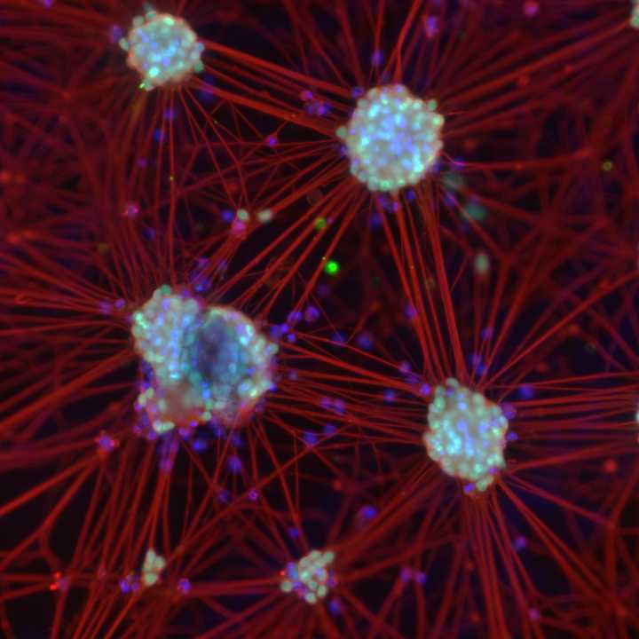
Figure: hNIL differentiation of iPSCs into motor neurons of WTC line initially on a 6-well plate (10X), then transferred to a 6-well plate and a 96-well plate (20X) after day-3 differentiation.