Oct 06, 2025
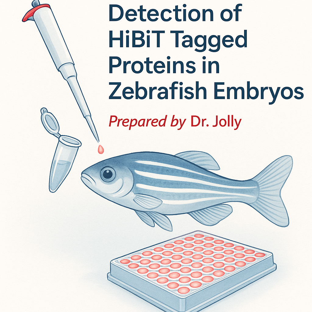
Detection of HiBit Tagged Proteins in Zebrafish Embryos (V1 10.05.25)
- 1University of Utah
- Jolly Protocols


Protocol Citation: Jeffery Jolly 2025. Detection of HiBit Tagged Proteins in Zebrafish Embryos (V1 10.05.25). protocols.io https://dx.doi.org/10.17504/protocols.io.rm7vz9165gx1/v1
License: This is an open access protocol distributed under the terms of the Creative Commons Attribution License, which permits unrestricted use, distribution, and reproduction in any medium, provided the original author and source are credited
Protocol status: Working
We use this protocol and it's working
Created: October 02, 2025
Last Modified: October 06, 2025
Protocol Integer ID: 228898
Keywords: Zebrafish, HiBit, Embryo, Detection, Luciferase , Protein, detection of hibit tagged protein, hibit tagged protein, proteins in zebrafish embryo, zebrafish embryo, microinjection buffer for optimal mrna delivery, zebrafish application, mrna microinjection, reagent ratios for zebrafish application, tagged protein, following mrna microinjection, optimal mrna delivery, glo hibit system, stage embryo, expressed protein, embryo, hibit activity, defined microinjection buffer, lytic reagent, zebrafish, protein
Disclaimer
Always double-check institutional guidelines for the sacrifice of zebrafish embryos.
Abstract
This protocol describes a rapid method for detecting HiBiT-tagged proteins in zebrafish embryos following mRNA microinjection. It outlines construct design, in vitro transcription with capping and polyadenylation, and preparation of a defined microinjection buffer for optimal mRNA delivery. Embryos are lysed by freeze–thaw in E3 medium, followed by the addition of HiBiT lytic reagent to enable quantitative luminescent detection of tagged proteins using the Nano-Glo HiBiT system.
The method yields reliable detection of exogenously expressed proteins within hours post-fertilization, requiring only minimal sample processing. Compared with enzymatic or mechanical lysis methods, this approach improves reproducibility, reduces processing time, and preserves HiBiT activity. Version 1 (10.05.25) standardizes freeze–thaw parameters and reagent ratios for zebrafish applications, ensuring compatibility with both early- and late-stage embryos.
Image Attribution
Made in conjunction with ChatGPT-5
Guidelines
This protocol describes a rapid method for detecting HiBiT-tagged proteins in zebrafish embryos following mRNA microinjection.
It is designed for efficiency, reproducibility, and compatibility with standard zebrafish workflows.
Key guidelines:
- Perform all steps using RNase-free reagents and consumables to prevent RNA degradation.
- Maintain embryos in E3 medium without additives (no methylene blue or HEPES) prior to lysis.
- Use consistent embryo numbers (20–30 per sample) to allow accurate comparison of luminescence values.
- For detection, use Promega Nano-Glo HiBiT Lytic Reagent or a comparable luciferase-compatible lysis buffer.
- Keep samples on ice whenever possible and minimize unnecessary freeze–thaw cycles after lysis.
Materials
1: 96-well white wall plates
2: Nano-Glo HiBiT Lytic Detection System (Promega N3030)
3: Centrifuge and luminescence plate reader
Safety warnings
- Do not use SDS or high-salt buffers, as these can inhibit NanoLuc/HiBiT luminescence reactions.
- Avoid vigorous homogenization or vortexing, which can shear DNA and increase background signal.
- Handle embryos and lysates in RNase-free conditions to prevent mRNA degradation before translation.
- Do not incubate lysis reactions above room temperature; elevated temperatures reduce luciferase activity.
- Dispose of all zebrafish embryos and lysates following institutional IACUC and biosafety guidelines.
Ethics statement
Experiments involving animals must be conducted according to internationally-accepted standards and should always have prior approval from an Institutional Animal Care and Use Committee (IACUC) or equivalent ethics committee(s).
Before start
Before beginning the assay, ensure that HiBiT fusion constructs and mRNA preparations are fully validated and free of contaminants.
- Prepare capped and polyadenylated mRNA following the “Jolly T7 IVT with Capping and Poly(A) Reaction” protocol.
- Verify mRNA concentration and purity (A260/280 ~2.0) before microinjection.
- Prepare the microinjection buffer (150 mM KCl, 20 mM HEPES pH 7.3) and pre-load needles to deliver 50 pg mRNA per embryo (1 nL at 50 ng/µL).
- Label all sample tubes and prepare ice for embryo collection and processing.
- If using embryos older than 1 day post-fertilization, plan for two freeze–thaw cycles to achieve complete lysis.
Construct Design, IVT, and Microinjection
Before you begin, practical notes:
Design constructs with C-terminal or internal HiBit fusion. These fusions are preferred over N-terminal tags as the N-terminal HiBit could still be expressed despite possible failure of transcription/translation of the downstream construct.
For the expression of proteins in zebrafish from mRNA:
1- Ensure your IVT template includes a Kozak sequence, along with both a start and a stop codon.
2- Example of Kozak sequence: GCCACC.ATG (Start Codon)
3- Ensure mRNA products are 5' capped. Cap-0 is sufficient for zebrafish microinjection
4- A Poly(A) reaction with your IVT products greatly increases stability and expression, but is not explicitly required for most applications.
5- See "Jolly T7 IVT with Capping and Poly(A) reaction Protocol" for more details regarding the preparation of mRNA for microinjection
Optimal Zebrafish Microinjection Buffer:
- 150 mM KCl
- 20 mM HEPES (pH 7.3)
Prepare under sterile conditions with RNAse-free water and aliquot for future use.
Preparation of mRNA for microinjection.
Resuspend mRNA in the buffer described above using extreme caution to avoid RNAse contamination.
Prepare mixes at 50 ng/uL. Aliquot RNA and injection buffer, and prepare the mixes fresh the morning of injections.
Keep the injection mixes on ice and minimize exposure to open air.
Inject 1 nL per embryo to deliver 50 pg of RNA per embryo. Can adjust to deliver 25 or 100 pg as needed.
Lyse Zebrafish Embryos and Detect HiBit Protein
47m
Collect x20 zebrafish embryos in 100 µL E3 media (without Methylene Blue or HEPES) in an Eppendorf tube. Fill the tube with the required volume to collect the embryos, then carefully remove the excess media, leaving behind a final volume of 100 µL.
Flash-freeze the embryos to enhance cell lysis. The preferred method for this is to flash freeze with liquid nitrogen for a few seconds. Alternatively, place the 100 µL samples into the -80 °C freezer for 20 minutes.
* Samples can be stored in this state without loss of signal for future analysis.
5m
Thaw samples at room temperature (~10 minutes).
10m
Prepare the needed amount of 2x Nano-Glo HiBit Lytic Reagent:
Adapted from Nano-Glo HiBiT Lytic Detection System
Promega Catalog #N3030
Recipe for 1 mL Nano-Glo HiBit Lytic Reagent:
- 970 µL Lysis Buffer
- 20 µL Substrate Solution
- 10 µL LgBit Protein Solution
Prepare a fresh solution for each experiment. Keep the reagents on ice and return to the -20 °C as soon as possible.
Add 100 µL of Nano-Glo HiBit Lytic Reagent (1:1 ratio). Vortex and gently spin down. Incubate at room temperature for 15 minutes.
15m
Spin down the mixtures at max speed (16,000-21,000 RCF) for 2 minutes.
2m
Use a disposable pestle to homogenize the solution, being careful to minimize volume loss.
Incubate the homogenized mixtures for an additional 15 minutes at room temperature.
15m
Clarify the solution by centrifugation at max speed (16,000-21,000 RCF) for 5 minutes.
Transfer 150 µL of the cleared solution to white 96-well plates, being careful to avoid bubbles.
Tips:
- Use a white-walled and white-bottom 96-well plate to maximize signal detection.
- If possible, plate the assay by skipping adjacent wells for sample reads to minimize bleed between wells. *Especially if utilizing a positive control sample.
- If bubbles persist, spin down the plate in the centrifuge before reading.
- Assay is adaptable to 100 µL per well if sample volumes are variable, with a reduction in signal.
Also, inject a buffer-only group to use for survival/norphology comparisons, as well as a negative control for HiBit detect.
Read luminescence signal on a compatible plate reader.
Detection Notes:
Protein expression and stability can vary significantly from that ofinjected mRNA. Protein detection can occur as soon as 3 hours, but starts to decline dramatically after 24 hours for most constructs.
GFP signal seems to peak at 24 HPF, while integrase protein expression appears to peak between 6 and 9 HPF and rapidly declines by 24 HPF. Therefore, it is best to detect samples over a time course when expressing novel proteins in the system.