Jun 23, 2025
Detection of alpha-synuclein aggregates in floating sections using antibody 5G4
- Bryan Killinger1,
- atousa bahrami1
- 1Rush University
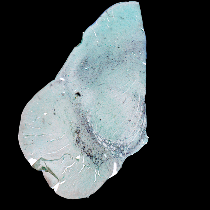


Protocol Citation: Bryan Killinger, atousa bahrami 2025. Detection of alpha-synuclein aggregates in floating sections using antibody 5G4. protocols.io https://dx.doi.org/10.17504/protocols.io.bp2l6yo15vqe/v1
License: This is an open access protocol distributed under the terms of the Creative Commons Attribution License, which permits unrestricted use, distribution, and reproduction in any medium, provided the original author and source are credited
Protocol status: Working
We use this protocol and it's working
Created: June 15, 2025
Last Modified: June 23, 2025
Protocol Integer ID: 220724
Keywords: Rehydration, Antigen, Peroxidase quenching , synuclein aggregate, synuclein protein, micron human brain section, disease aggregate, antibody with an epitope, using antibody, disease aggregates from oligomer, including thicker peripheral tissue biopsy, antibody, biomarker, thicker peripheral tissue biopsy, thicker tissue format, use of this antibody, human brain section
Funders Acknowledgements:
Co-Pathologies Drive Neuroinflammation and Progression in PD
Grant ID: ASAP-021030
PROTEOMIC MAPPING OF PATHOLOGICAL INTERACTIONS ACROSS THE SYNUCLEINOPATHY BRAIN
Grant ID: 5R01NS128467
Abstract
5G4 is an antibody with an epitope in the region of aa44-57 of human alpha-synuclein protein, with a high specificity for aggregates. 5G4 reacts with a wide variety of disease aggregates from oligomers to fibrillar type, and shows very little reactivity for physiologic monomeric asyn, making it an excellent generalized “marker” for misfolded pathogenic asyn species. Use of this antibody has been limited to paraffin-embedded sections. Here, we developed a working protocol for fixed floating 40-micron human brain sections. Using the thicker tissue format may help apply 5G4 to a wide variety of specimens, including thicker peripheral tissue biopsies, which are the preferred format for use as a biomarker.
Materials
5G4: Anitbody can be purchased from millipore-sigma (Millipore Cat# MABN389, RRID:AB_2716647)
Anti-α-Synuclein (SNCA) AntibodyMerck MilliporeSigma (Sigma-Aldrich)Catalog #MABN389
Before start
Mount tissue on slide:
Mount tissue on superfrost plus adhesion slides and dry overnight in oven at 37 (make sure it’s very dry).
Day 1: Dehydrate tissues
41m
Di water for 00:02:00 .
2m
50% ethanol for 00:03:00 .
3m
70% ethanol for 00:03:00 .
3m
95% ethanol for 00:03:00 .
3m
100% ethanol 2x5 min each in a chemical hood. (100% ethanol is denature) (absolute ethanol is the anhydrous.
100% ethanol 00:05:00 each in a chemical hood. (100% ethanol is denature) (absolute ethanol is the anhydrous. (1/2)
5m
100% ethanol 00:05:00 each in a chemical hood. (100% ethanol is denature) (absolute ethanol is the anhydrous. (2/2)
5m
Xylenes 2x10 min in the chemical hood.
Xylenes 00:10:00 in the chemical hood. (1/2)
10m
Xylenes 00:10:00 in the chemical hood. (2/2)
10m
Day 1: Rehydration
41m
Xylenes 2x10 min in the chemical hood.
Xylenes 00:10:00 in the chemical hood. (1/2)
10m
Xylenes 00:10:00 in the chemical hood. (2/2)
10m
100% ethanol 2x5 min each in a chemical hood. (100% ethanol is denature) (absolute ethanol is the anhydrous.
100% ethanol 00:05:00 each in a chemical hood. (100% ethanol is denature) (absolute ethanol is the anhydrous. (1/2)
5m
100% ethanol 00:05:00 each in a chemical hood. (100% ethanol is denature) (absolute ethanol is the anhydrous. (2/2)
5m
95% ethanol for 00:03:00 .
3m
70% ethanol for 00:03:00 .
3m
50% ethanol for 00:03:00 .
3m
Di water for 00:02:00 .
2m
Day 1: Antigen Retrieval (~45min)
1h 15m
Wash sections with sodium citrate buffer for 00:05:00 .
5m
Microwaving: 00:10:00 in citrate buffer (pH 6).
- 1000 L H2O
- 2.94 g sodium citrate (dihydrate)
- PH to 6.0
- 0.5 mL Tween -20
10m
Let citrate acid to cool ~00:30:00 .
30m
00:30:00 in 88% formic acid.
30m
Quick wash with DM.
DM preparation:
| A | B | |
| Tris-HCl | 50mM | |
| NaCl | 150mM | |
| Triton X-100 | 0.05% |
Day 1: Blocking and peroxidase quenching (1h 40 min)
1h 40m
Make blocking buffer: DM + 3% BSA + 3% serum+ Triton.
- For 100 mL:
| A | B | |
| DM | 100 mL | |
| BSA | 3 g | |
| Serum | 3 mL | |
| Triton | 0.5 mL |
2. Serum must be from host species of secondary antibody
3. Take 50 mL out for blocking buffer and leave the other 50 for primary antibody
4. Add peroxidase quenching to the blocking buffer solution
- For 50ml:
| A | B | |
| 30%H2O2 | 0.5 mL | |
| Sodium azide (stock 10%) | 0.5 mL |
Dip slides in blocking buffer for 01:30:00 at Room temperature .
1h 30m
Dip in TBS 2x5 min each.
Note
This removes excess DM which interferes with the hydrophobic barrier.
Dip in TBS 00:05:00 each. (1/2)
5m
Dip in TBS 00:05:00 each. (2/2)
5m
Day 1: Primary antibody
5G4 should be diluted 1:5000.
Cover the tissue section in primary antibody solution. Overnight at 4 °C .
Day 2: Secondary antibody (1h 50 min)
1h 50m
Dip in DM 3x5 min.
Dip in DM 00:05:00 . (1/3)
5m
Dip in DM 00:05:00 . (2/3)
5m
Dip in DM 00:05:00 . (3/3)
5m
Dip in TBS 2x5 min.
Dip in TBS 00:05:00 . (1/2)
5m
Dip in TBS 00:05:00 . (2/2)
5m
Make secondary blocking buffer:
- For 50 mL working solution:
| A | B | |
| DM | 50 mL | |
| BSA | 1 g | |
| Serum | 2 mL |
Dilute secondary antibody (Horse Anti-Mouse IgG Antibody (H+L), Biotinylated (BA-2000-1.5) from VectorLabs) in secondary blocking buffer (for biotinylated secondary 1:200).
Cover sections in the secondary antibody for 01:00:00 at Room temperature .
1h
Dip in DM 3x5 min.
Dip in DM 00:05:00 . (1/3)
5m
Dip in DM 00:05:00 . (2/3)
5m
Dip in DM 00:05:00 . (3/3)
5m
Dip in TBS 2x5 min.
Dip in TBS 00:05:00 . (1/2)
5m
Dip in TBS 00:05:00 . (2/2)
5m
Day 2: ABC (35 min)
1h 20m
Prepare ABC reagent. Must be prepared 00:30:00 prior to use. Stable for 24:00:00 after prep.
- For 25mL:
- 2.5 mL blocking buffer + 1 drop A + 1 drop B
- After 30 minutes, fill to 25 mL
Cover sections with prepared ABC reagent for 01:15:00 at Room temperature .
1h 15m
Dip in DM 00:05:00 .
5m
Day 2: DAB (~1 hr)
1h 7m
Make imidazole buffer (IB).
- For 1 L : 6.8 sodium acetate + 0.68 imidazole.
- Once IB is homogeneous, separate 100 mL into a separate beaker with stirbar.
- For 900 mL of IB, pH to 7.2-7.4.
- For 100 mL of non-pH’d IB, add 50 mg DAB followed by 2.5 g nickel. Dissolve completely.
Dip slides in pH’d IB (i.e. pH 7.2-7.4) 3x10 min each.
Dip slides in pH’d IB (i.e. pH 7.2-7.4) 00:10:00 each. (1/3)
10m
Dip slides in pH’d IB (i.e. pH 7.2-7.4) 00:10:00 each. (2/3)
10m
Dip slides in pH’d IB (i.e. pH 7.2-7.4) 00:10:00 each. (3/3)
10m
Immediately before use add hydrogen peroxide to DAB solution.
- For 100mL DAB solution add 16 µL of 30% H202.
Dip sections in DAB solution for development.
- Strong signals 00:00:30 -00:02:00
- Medium signals, 00:02:00 -00:05:00
- Weaker signals 00:05:00 -00:10:00 .
- Development up to 10 min is typically free of background. Over 10 min and background may develop.
17m
Monitor color development. (Nickel DAB = jet black, DAB = brown).
- To check color development slides can be placed in a coplin jar with pH’d IB and then checked under microscope.
- If color intensity is too weak, place sections back into DAB solution for additional time.
- Can repeat this procedure until desired color intensity is achieved.
Once done developing wash sections in PBS 2x10min.
Once done developing wash sections in PBS 00:10:00 . (1/2)
10m
Once done developing wash sections in PBS 00:10:00 . (2/2)
10m
Deactivate DAB solution with bleach and dispose in DAB waste container.