Nov 26, 2024
Cyclic Heat Induced Epitope Retrieval (CHIER)
- Victor Dieriks1,2
- 1Department Anatomy and Medical Imaging, Faculty of Medical and Heath Sciences, The University of Auckland;
- 2Centre for Brain Research, The University of Auckland


Protocol Citation: Victor Dieriks 2024. Cyclic Heat Induced Epitope Retrieval (CHIER). protocols.io https://dx.doi.org/10.17504/protocols.io.kxygx3284g8j/v1
License: This is an open access protocol distributed under the terms of the Creative Commons Attribution License, which permits unrestricted use, distribution, and reproduction in any medium, provided the original author and source are credited
Protocol status: Working
We use this protocol and it's working
Created: November 26, 2023
Last Modified: November 26, 2024
Protocol Integer ID: 91432
Keywords: antigen retrieval, immunohistochemistry, IHC, immunofluorescence, IFC, formalin-fixed, paraffin-embedded (FFPE) tissue, cyclic heat induced epitope retrieval, novel antigen retrieval protocol, efficiency of antigen retrieval, traditional antigen retrieval method, precise antigen detection, effective unmasking of epitope, epitope preservation, persistent challenges in immunolabeling, detection of the protein, specificity of antibody, epitope, antibody, aggregated protein, essential for accurate protein localisation, accurate protein localisation, involving protein, immunolabeling, protein localisation, critical insights into protein localisation, subtle epitope, protein, introducing chier
Funders Acknowledgements:
Health Research Council Hercus
Grant ID: 21/034
he School of Medical Science, University of Auckland
The Neurological Foundation of New Zealand
Grant ID: 2026 PRG
Royal Society of New Zealand Te Apārangi
Grant ID: 22-UOA-049-CSG
Abstract
Immunolabeling is a cornerstone technique in molecular biology and pathology, providing critical insights into protein localisation and function within biological tissues. However, one of the persistent challenges in immunolabeling is the effective unmasking of epitopes, which are often concealed in formalin-fixed, paraffin-embedded (FFPE) tissue samples. While effective to an extent, traditional antigen retrieval methods have limitations in consistency, epitope preservation, and overall labelling efficiency. We introduce a novel antigen retrieval protocol, Cyclic Heat Induced Epitope Retrieval (CHIER) to address these issues.
The development of CHIER is particularly crucial for studies involving proteins that are notoriously difficult to label due to dense cross-linking or subtle epitope masking. By improving the efficiency of antigen retrieval, CHIER enhances the sensitivity and specificity of antibody binding in immunohistochemical (IHC) and immunofluorescence (IF) assays. This results in more robust staining and can facilitate the detection of densely aggregated proteins, which is essential for accurate protein localisation and quantification studies.
Significantly, CHIER demonstrated a remarkable ability to increase the detection of the protein of interest (SWI/SNF related, matrix-associated, actin-dependent regulator of chromatin subfamily c member 2, SMARCC2), with results showing a 3x increase in detection compared to traditional methods. This enhancement in detection efficiency makes CHIER a valuable tool for researchers in diverse fields, including neurobiology, cancer research, and tissue pathology, where precise antigen detection is paramount. Introducing CHIER into these fields is expected to significantly advance our understanding of complex protein dynamics and interactions in various biological and disease contexts.
Guidelines
Heat block reaches 70° Celcius and can cause burns when skin comes into contact with the hot surface.
Materials
Tris-EDTA antigen retrieval buffer in dH20
- 10mM Tris Base
- 1 mM EDTA Solution
Adjust to pH 9.0
- 0.05% Tween 20
Troubleshooting
Safety warnings
Heat block reaches 70° Celcius and can cause burns when skin comes into contact with the hot surface.
Make sure chemical waste is disposed of according to on-site regulations.
Ethics statement
The human post-mortem brain tissue used in this project was sourced from the Neurological Foundation Human Brain Bank (Centre for Brain Research, University of Auckland, New Zealand). All brain tissue was donated upon written, informed consent from the individual(s) and/or next of kin before brain removal.
Please ensure consent is obtained, and all protocols required used are approved by the relevant Ethics Committee.
Paraffin-embedded formalin-fixed brain tissue
Human paraffin-embedded formalin-fixed brain tissue was utilised for this project. All brain tissues were washed using an ethanol (EtOH) series at Room temperature :
00:20:00 70% EtOH
00:20:00 80% EtOH
00:20:00 2 x 95% EtOH
00:30:00 2 x 100 % xylene
Once washed and dehydrated, the tissue was embedded in molten paraffin wax for 3 x 00:25:00 cycles.
1h 55m
Cutting parafin sections
Paraffin blocks were cut into 7 μm thick sections using a rotatory microtome (Leica Biosystem Histcore BIOCUT R) and placed into a 37 °C water bath (Leica Biosystems, Histcore & H1210). Sections were individually mounted onto charged Uber frost Printer PLUS 3000 microscope slides (Intrumentec) and placed on a slide dryer (Leica Biosystems Histcore) for 5 minutes, followed by air-drying at room temperature for a minimum of 72:00:00 .
3d
Cyclic Heat Induced Epitope Retrieval (CHIER) Hotplate
1h 10m
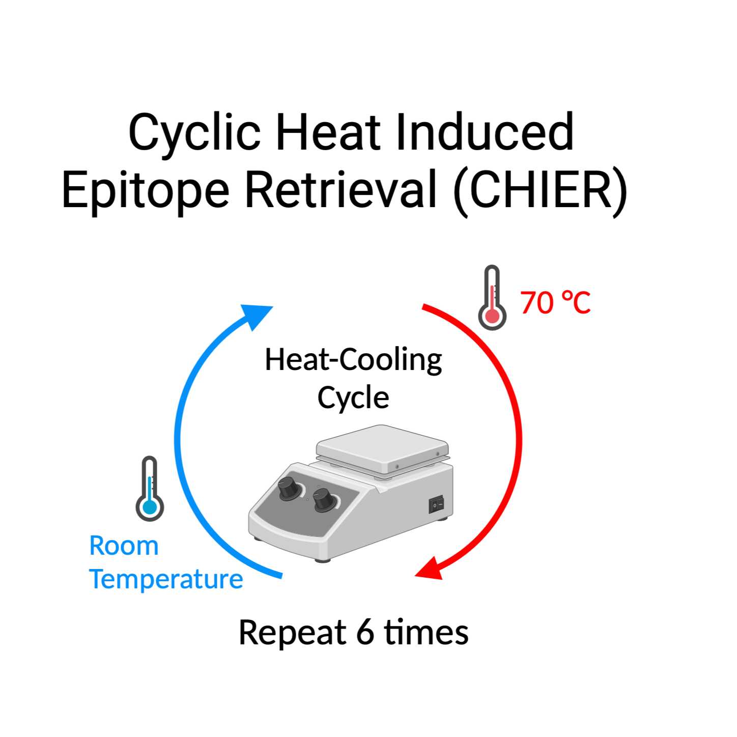
Mounted slides with Formalin-fixed paraffin-embedded tissue sections were placed tissue side up
Mounted slides with Formalin-fixed paraffin-embedded tissue sections were placed, on a 70 °C hotplate (Leica HI1220) for 00:05:00 followed by 00:05:00 at room temperature.
This heat-cooling cycle was repeated six times.
Note
Incubation time and temperature are critical.
1h
Immediately following the heat-cooling cycle, slides were placed on a 60 °C hotplate (Leica HI1220) for 00:30:00 .
Note
If using the same hotplate then make sure the temperature drops to 60 °C before placing slides.
30m
Dewaxing
1h 20m
Immediately following hotplate incubation, sections were submerged in
00:30:00 2 x 100% xylene baths to remove paraffin wax.
Subsequently, tissue sections were rehydrated by submerging slides in a series of alcohol baths
00:15:00 2 x 100% EtOH
00:05:00 95% EtOH
00:05:00 80% EtOH
00:05:00 75% EtOH
00:05:00 3 x distilled H2O (dH2O) to wash of any remaining EtOH.
1h 5m
Antigen retrieval in pressure cooker
1h 40m
The tissue on the slides is now ready for subsequent steps. Continue with antigen retrieval as normal, followed by blocking and subsequent antibody incubation.