Feb 17, 2026
Version 2
Cultivating Melanized Fungi from Biological Soil Crust and Rock Surfaces V.2
- Tania Kurbessoian1,
- Jason E. Stajich1,
- Renata Haro1
- 1University of California, Riverside
- stajichlab



Protocol Citation: Tania Kurbessoian, Jason E. Stajich, Renata Haro 2026. Cultivating Melanized Fungi from Biological Soil Crust and Rock Surfaces. protocols.io https://dx.doi.org/10.17504/protocols.io.4r3l25z2ql1y/v2Version created by Anita Broellochs
License: This is an open access protocol distributed under the terms of the Creative Commons Attribution License, which permits unrestricted use, distribution, and reproduction in any medium, provided the original author and source are credited
Protocol status: Working
We use this protocol and it's working
Created: October 13, 2020
Last Modified: February 17, 2026
Protocol Integer ID: 43125
Keywords: Melanized fungi, black yeasts, culturing, biological soil crust, rock surfaces, melanized fungi from biological soil crust, melanized fungi, isolating fungi, cultivating melanized fungi, growing fungi, fungi in question, fungi, fungi from the soil, biocrust soils onto microbiological media, strains of organism, microorganism, other microorganism, different microbiological technique, living strains of organism, utilizing different microbiological technique, starting spore, present in the biocrust, biocrust, living strain, microbiological media, plating biocrust soil, organism, life strain, strain, oligotrophic media, soil slurry, soil, antibiotic, culture independent analysis, pathological disease
Abstract
As the interest to understand melanized fungi becomes more of a focus due to pathological diseases, there needs to be a clearer method to isolate and identify the fungi from their surroundings. Culture independent analysis has helped bloom scientist understandings of the vast quantities of microorganisms around us, but culturing and identifying has always been a struggle. We must look into utilizing different microbiological techniques to help better understand, isolate, and to apply Koch’s postulates to prove they are the cause of said disease. Growing and isolating fungi has always been an issue especially if the fungi in question is a slow growing fungi, where fast growing fungi or other microorganisms can grow and surpass the field of view and compete with the slow growing fungi. A series of dilutions, antibiotics and oligotrophic media can all counteract the issues to provide you with a clear window to help isolate your fungi in question.
There is still a need to verify that living strains of organisms are actually present in the environment. One approach is to use “culture dependent” method to obtain strains of organisms present in the biocrust. This is achieved by plating biocrust soils onto microbiological media in order to further isolate life strains. Serial dilution is used to isolate the fungi from the soil. This method involves making a soil slurry by diluting the soil sample with increasing higher proportion of water or media in order to get a low starting concentration of starting spores or material growing on the Petri dishes.
Materials
MATERIALS
Asparagine
GlucoseP212121Catalog #Glucose
Potassium phosphate (dibasic)P212121
Petri dish, 100x15mmFisher ScientificCatalog #FB0875712
Falcon® Serological Pipettes, 1 mL 1000 Pipettes
STEMCELL Technologies Inc.Catalog #38001
1.5mL Microtubes
Prepare Media
1h 46m 15s
Prepare Nutrient Rich Media Malt Extract Yeast Extract Agar (MEYE)1 L
20 g Glucose
1 g Peptone
20 g Malt Extract
20 g Agar
15m
Prepare Nutrient Poor Media Glucose Asparagine Agar (GAA) - Krainsky's Media/1 L
0.500 g Dibasic Potassium Phosphate
0.500 g Asparagine
10.00 g
15.00 g Agar
Autoclave
Prepare filtered antibiotics for 1 L agar solution. Either Gentamycin, Tetracycline, Kanamycin, Streptomycin or Chloramphenicol will work.
15m
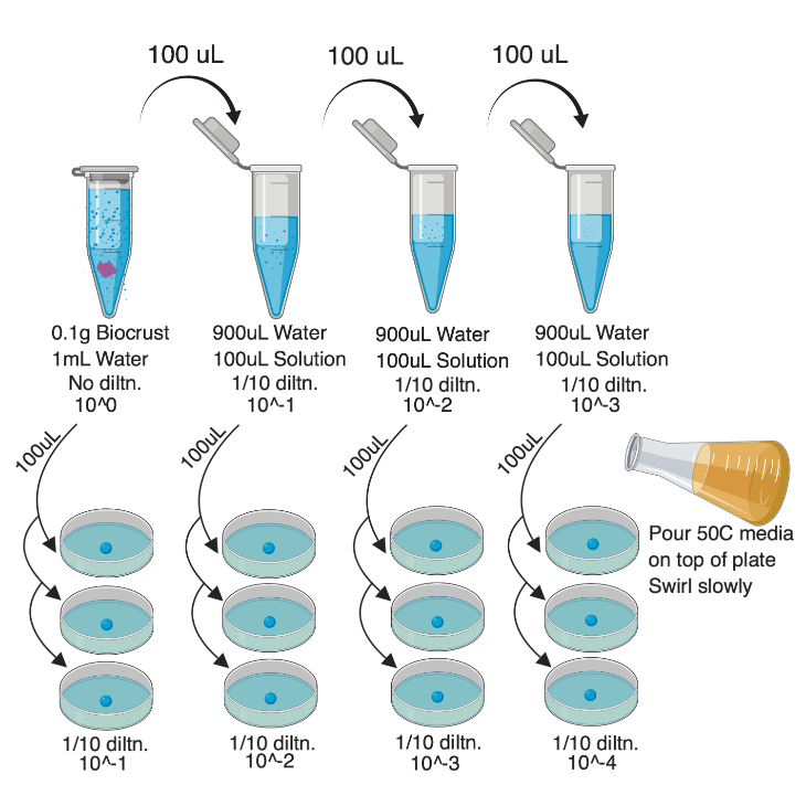
Visual protocol for clarity.
1h
0.1 g of sample (either biocrust, soil or rock surface can be used) placed in microcentrifuge tube.
1m
Add 1000 µL Sterile H2O into the same tube.
30s
Label this 10^0.
30s
Vortex (on highest setting) sample for 10 minutes until solution becomes a slurry.
Note
Rock surface samples may not become a slurry.
10m
Preparing the Serial Dilution
Prepare 3 other microcentrifuge tubes with 900 µL sterile H2O .
2m
Label them 10^-1, 10^-2, 10^-3.
45s
Take100 µL slurry solution from 10^0 and add it to 10^-1. Vortex 10^-1.
30s
Then take 100 µL 10^-1 and add it to 10^-2. Vortex 10^-2.
30s
Then take 100 µL 10^-2 and add it to 10^-3. Vortex 10^-3.
30s
Set up 12 empty Petri Plates and label them with appropriate labels. I.e. Date, Media (list antibiotic used), Crust type, Dilution, Iteration (Plate 1, Plate 2 or Plate 3).
Note
For more statistical power we must set them up in triplicate.
Pipette out 100 µL 10^0 into first three plates that were properly labeled for 10^0.
Pipette out 100 µL 10^-1 into next three plates labeled for 10^-1.
Pipette out 100 µL 10^-2 into next three plates labeled for 10^-2.
Pipette out 100 µL 10^-3 into next three plates labeled for 10^-3.
Make sure to have media cooled (to 50 °C *) and ready to go at this point.
Note
*which means you can touch it with your hands without burning yourself.
Add the media slowly to avoid bubbles. Fill until agar closes all gaps on plate. Fill up to three plates at a time.
When three plates have been filled start to slowly mix the plate and slurry solution. Do a very careful "Figure 8" motion. Make sure to mix slowly and evenly without touching the lid of the plate.
Note
If it does touch, just replace with a new lid from an unused empty plate.
Continue repeating 5.1 and 5.2 until all plates have been filled and are cooling.
Let plates air dry for 24 hours, then parafilm to allow for long-term storage.
New fungi should start popping up on undiluted and next dilution plates in the next couple of days. Most dilute plates will also have sparser filamentous fungi growing.
Melanized fungi will take about 1-2 weeks to show up on plates.
Make sure to check on the underside of plates for black dots. Those are the fungi you are looking for!
This is an example of the least diluted plate fungal growth.
This is the back of a plate. Notice the little black dots. These are the melanized fungi/yeasts to collect.