Jul 25, 2025
Version 2
Crystallization of Zika virus NS3 helicase PDB V.2
- 1University of Sao Paulo, ASAP Discovery Consortium;
- 2Diamond Light Source;
- 3Research Complex at Harwell, ASAP Discovery Consortium;
- 4Centre of Medicines Discovery, University of Oxford, ASAP Discovery Consortium
- ASAP Discovery



Protocol Citation: Andre Schutzer de Godoy, Peter Marples, Lizbé Koekemoer 2025. Crystallization of Zika virus NS3 helicase PDB. protocols.io https://dx.doi.org/10.17504/protocols.io.e6nvw152dlmk/v2Version created by Mary-Ann Xavier
License: This is an open access protocol distributed under the terms of the Creative Commons Attribution License, which permits unrestricted use, distribution, and reproduction in any medium, provided the original author and source are credited
Protocol status: Working
We use this protocol and it's working
Created: June 20, 2025
Last Modified: July 25, 2025
Protocol Integer ID: 220635
Keywords: crystallisation, XChem, ASAP, AViDD, CMD, Diamond Light Source, i04-1, Research complex at Harwell, Zika NS3, NS3 helicase, crystallization of zika virus ns3 helicase pdb, zika virus ns3 helicase pdb, zika ns3 helicase crystal, throughput crystallographic fragment screening on zikv ns3 helicase, zika ns3 crystal, zika virus, zikv ns3 helicase, other flavivirus, ns3 helicase, critical roles in viral replication, crystallographic fragment screening, viral replication, protocol for protein production, crystallization protocol, protein, targeting ns3hel, crystallization, protein production, promising drug target, ns3hel, development of specific inhibitor, heterologous protein expression, zikv
Funders Acknowledgements:
National Institutes of Health/National Institute Of Allergy and Infectious Diseases (NIH/NIAID)
Grant ID: Grant ID: U19AI171399
Disclaimer
The content is solely the responsibility of the authors and does not necessarily represent the official views of the National Institutes of Health.
Acknowledgements:
Diamond Light Source Ltd, Harwell Science and Innovation Campus, Didcot OX11 0QX, UK
Sao Carlos Institute of Physics, University of Sao Paulo, Av. Joao Dagnone, 1100 - Jardim Santa Angelina, Sao Carlos, 13563-120, Brazil
Research Complex at Harwell, Harwell Science and Innovation Campus, Didcot OX11 0FA, UK
Oxford Lab Technologies crystal shifter https://doi.org/10.1107/S2059798320014114
Abstract
The crystallization protocol and buffer conditions used to obtain Zika NS3 helicase crystals suitable for XChem fragment screening. The Zika virus (ZIKV), discovered in Africa in 1947, swiftly spread across continents, causing significant concern due to its recent association with microcephaly in newborns and Guillain-Barré syndrome in adults. Despite a decrease in prevalence, the potential for a resurgence remains, necessitating urgent therapeutic interventions. Like other flaviviruses, ZIKV presents promising drug targets within its replication machinery, notably the NS3 helicase (NS3Hel) protein, which plays critical roles in viral replication. However, a lack of structural information impedes the development of specific inhibitors targeting NS3Hel. This protocol was used to grow Zika NS3 crystals that were applied high-throughput crystallographic fragment screening on ZIKV NS3 Helicase. In this new version, we added the Addgene information for the construct used for heterologous protein expression and the protocol for protein production and purification
Materials
SwissCI 3 lens crystallization plates https://swissci.com/product/3-lens-crystallisation-plate/ Codes:
Midi: UVXPO-3LENS 3W96T-PS 3W96T-UVP
1 Molarity (M) NPS Mix (consisting of 0.3 M Sodium phosphate dibasic dihydrate, 0.3 M Ammonium sulphate, and 0.3 M Sodium nitrate from Molecular Dimensions), Catalog # MD2-250-72
1 Molarity (M) MES, Molecular Dimensions, Catalog # MD2-013-PH
Precipitant Mix 4 (11% MPD, 11% PEG 1,000, and 11% PEG 3,350 from Molecular Dimensions), Catalog # MD2-250-84
Purified Zika NS3 protein (5 mg/mL ) in 20 millimolar (mM) Bis-Tris 7 , 500 millimolar (mM) NaCl, 10% glycerol
Protocol materials
Zika virus (ZIKV) isolate PRVABC59 NS3 helicase domainaddgeneCatalog #228664
Safety warnings
Follow all handling warning for the chemicals used in the crystalllisation screen composition.
Protein expression and purification
The protein expresssion and purifaiction of the plasmid Zika virus (ZIKV) isolate PRVABC59 NS3 helicase domainaddgeneCatalog #228664 was done following the protocol below.
Equipment needed
Crystallization experiment
1d
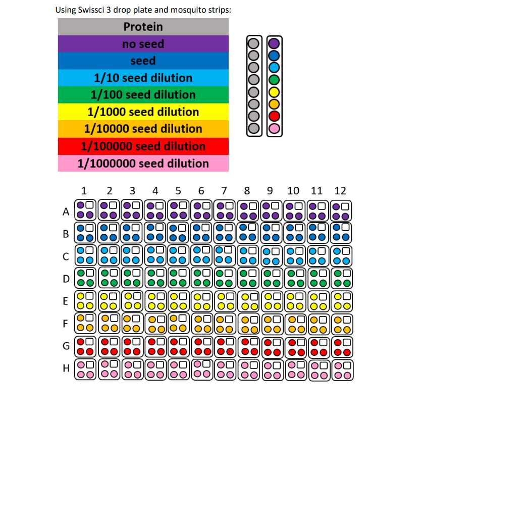
1: 100 dilution Sample seeds
Protein and buffer requirements:
43.2 µL 5 mg/mL Sample
4.8 mL Crystallization screen
4.8 mL seeds, dilution 1:100
Crystallisation screen composition:
0.12 Molarity (M) NPS Mix
0.1 Molarity (M) MES 6.5
33% Prcipitant Mix 4
Stock solutions used:
1 Molarity (M) NPS Mix (consisting of 0.3 M Sodium phosphate dibasic dihydrate, 0.3 M Ammonium sulphate, and 0.3 M Sodium nitrate from Molecular Dimensions)
1 Molarity (M) MES (Molecular Dimensions)
Precipitant Mix 4 (11% MPD, 11% PEG 1,000, and 11% PEG 3,350 from Sigma Aldrich)
Note
The crystallisation screen can be stored in a duran bottle or aliquoted into 96 deep well block for easy dispensing into SwissCI 3 lens plates.
For long term storage keep the Crystallisation screen in the fridge at 4°C.
Dispense 50 µL Crystallisation screen into SwissCI 3 lens plate reservoir wells using a 100 µl multi-channel pipette.
Dispense 150 µL 5 mg/mL Sample to each lens using the SPT mosquito.
Dispense 100 µL Crystallisation screen to each lens using the SPT mosquito.
Dispense 50 µL Seeds to each lens using the SPT mosquito.
Drop ratio: 3:2:1 ratio (150 nL Sample : 100 nL reservoir solution: 50 nL seeds)
Final drop volume: 300 nl
Incubate at 20 °C for 24:00:00 h in Formulatrix Rock Imager.
Imaging Schedule: The first images are taken after 12 h and the imaging schedule follows a Fibonacci sequence of days for further collections.
1d
Expected result
The crystals reach their maximum size after 48 h.
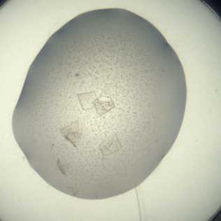
Crystals typically form either as single crystals or overlapping thin plates
Morphology: typically thin square plates
Size: ~100 μm in length and ~100 μm in width, depth of the crystals is ~10 μm
Appearance: glass shard.
Average resolution: 1.8 Å
Space group: P121
Unit cell: 53 Å, 69 Å, 57 Å
90.00°, 92.00°, 90.00°
An example of a drop containing Zika NS3 helicase crystals.
Data collection at Synchrotron
Diamond Light Source
Unattended Data Collection (UDC)
Data Collection Temperature: 100K
Detector: DECTRIS EIGER2 X 9M
Beamline: I04-1
Wavelength: 0.9212 Å
Resolution (Å): 1.62
Beam Size (μm): 60 X 50
Number of images: 3600
Oscillation: 0.10°
Exposure (s): 0.0020
Transmission (%): 100
Flux (ph/s): 3.80e+12
Solvent tolerance
Fragment Screen
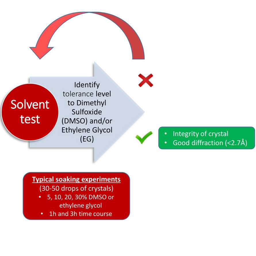
The compounds were soaked at 20% DMSO for 2hs

Protocol references
N/A