Jun 10, 2026
Crystallisation of Zika NS5 RdRp
Forked from Crystallisation of Zika NS5 RdRp
- Anu V. Chandran1,2,
- Peter Marples1,2,
- Tracy Keates1,2,
- Charlotte Chinn1,2
- 1Diamond Light Source;
- 2Research Complex at Harwell
- Anu V. Chandran: The principle crystallographer for the Zika NS5 RdRp polymerase project.;
- OpenBind Consortium


Protocol Citation: Anu V. Chandran, Peter Marples, Tracy Keates, Charlotte Chinn 2026. Crystallisation of Zika NS5 RdRp. protocols.io https://dx.doi.org/10.17504/protocols.io.4r3l2x5y4v1y/v1
License: This is an open access protocol distributed under the terms of the Creative Commons Attribution License, which permits unrestricted use, distribution, and reproduction in any medium, provided the original author and source are credited
Protocol status: Working
We use this protocol and it's working
Created: June 10, 2026
Last Modified: June 10, 2026
Protocol Integer ID: 318843
Keywords: crystallisation, XChem, CMD, Diamond Light Source, i04-1, Zika NS5 RdRp, NS5 RdRp, Research complex at Harwell, zika ns5 rdrp, bind zika ns5 rdrp, zika ns5 rdrp domain, zika ns5 rdrp the main aim, catalytic rna, rna, suitable crystals for fragment screening, dependent rna polymerase domain, fragment screening, OpenBind, Open AI, OpenBind Consortium, Open Bind, Zika, NS5, crystallisation of zika ns5 rdrp, bind zika ns5 rdrp, zika ns5 rdrp, zika ns5 rdrp domain, catalytic rna, rna, suitable crystals for fragment screening, dependent rna polymerase domain, fragment screening
Funders Acknowledgements:
National Institutes of Health/National Institute Of Allergy and Infectious Diseases (NIH/NIAID)
Grant ID: Grant ID: U19AI171399
UK Department of Science, Innovation and Technology
Grant ID: G2-SCH-2025-06-16537
Disclaimer
The content is solely the responsibility of the authors and does not necessarily represent the official views of the National Institutes of Health.
Acknowledgements:
Diamond Light Source Ltd, Harwell Science and Innovation Campus, Didcot OX11 0QX, UK
Research Complex at Harwell, Harwell Science and Innovation Campus, Didcot OX11 0FA, UK
Oxford Lab Technologies crystal shifter https://doi.org/10.1107/S2059798320014114
Abstract
The main aim of this work was to identify small molecules that bind Zika NS5 RdRp (catalytic RNA-dependent RNA polymerase domain) through X-ray fragment-based screening. The Zika NS5 RDRP domain was cloned, expressed, purified, and crystallised. Suitable crystals for fragment screening were produced and optimised allowing an extensive fragment campaign to be performed. A native high-resolution structure was determined at 1.8Å and formed the basis for the fragment campaign. This has been used as the basis for work on the target for OpenBind
Materials
SwissCI 3 lens crystallization plates https://swissci.com/product/3-lens-crystallisation-plate/ Codes:
Midi: UVXPO-3LENS 3W96T-PS 3W96T-UVP
Morpheus HT-96 single reagent 250mL Catalog # MDSR-47-250-2-10
Purified Zika NS5 polymerase protein (5 mg/mL ) in 20 millimolar (mM) HEPES 7.5 , 300 millimolar (mM) NaCl, 2.5% Glycerol, 10 micromolar (µM) ZnCl2, 2 millimolar (mM) TCEP
306-903 residues- construct 2A1 (6 Hist sumo tag)
Safety warnings
Follow all handling warning for the chemicals used in the crystalllisation screen composition.
Zika NS5 RdRp expression and purification
The protein used for crystallisation was expressed and purified using the following protocol.
Protocol
CREATED BY
korvus.wang
Note
For best results, thawed protein is exchanged into the base buffer described in the protocol above freshly before crystallisation using a 10 kDa MWCO spin column and concentrated to 13 mg/ml
Equipment needed
Formulatrix Rock Imager (or incubator of choice)
Equipment
Mosquito HV
NAME
High Volume 16-Channel Robotic Liquid Handler
TYPE
SPT LabTech
BRAND
3097-01057
SKU
LINK
P100 8 multi-channel pipette
Crystallisation experiment
1d
Protein and buffer requirements:
28.8 µL 5 mg/mL Sample
3.264 mL Crystallisation screen
Crystallisation screen composition:
Morpheous I E10 condition
0.12 Molarity (M) Ethylene glycols
0.1 Molarity (M) Buffer system 3 8.5
30 % v/v Precipitant Mix 2
Stock solutions used:
Morpheous I E10 condition
Note
The crystallisation screen can be stored in a duran bottle.
For long term storage keep the crystallisation screen in the fridge at 4°C.
Dispense 34 µL Crystallisation screen into SwissCI 3 lens plate reservoir wells using a 100 µl multi-channel pipette.
Dispense 100 µL 5 mg/mL Sample to each lens using the SPT mosquito.
Dispense 50 µL Crystallisation screen to each lens using the SPT mosquito.
Drop ratio: 2:1 ratio (100 nl Sample : 50 nl reservoir solution)
Final drop volume: 150 nl
Incubate at 20 °C for 24:00:00 h in Formulatrix Rock Imager.
Imaging Schedule: The first images are taken after 12 h and the imaging schedule follows a Fibonacci sequence of days for further collections.
1d
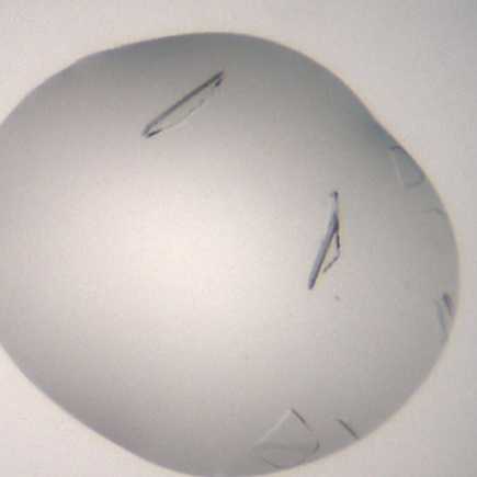
Crystal form after ~24 h.
Expected result
The crystals reach their maximum size after 96 h and the precipitant has gone.
Crystals grown inconsistently to 2 sizes,one being half the size described below, but both sizes achieve the same results
Morphology: typically plates.
Size: ~ 250 μm in length and ~60 μm in width, depth of the crystals is ~2 μm
Appearance: glass shard.
Average resolution: 2.0 Å
Space group: P43212
Unit cell: 79 Å, 79 Å, 210 Å
90.00°, 90.00°, 90.00°
An example of a drop containing Zika NS5 RdRp ploymerase crystals.
Data collection at Synchrotron
Diamond Light Source
Unattended Data Collection (UDC)
Data Collection Temperature: 100K
Detector: DECTRIS EIGER2 X 9M
Beamline: I04-1
Wavelength: 0.9212 Å
Resolution (Å): 1.78
Beam Size (μm): 60 X 50
Number of images: 3600
Oscillation: 0.10°
Exposure (s): 0.0020
Transmission (%): 100
Flux (ph/s): 9.50e+11
Protocol references
N/A