Jun 05, 2025
Version 2
Crystallisation of West Nile virus NS2B-NS3 protease in P64 (PDB ID: 8CO8) V.2
Version 1 is forked from Crystallisation of SARS-CoV-2 Mpro
- 1Diamond Light Source;
- 2Research Complex at Harwell, ASAP Discovery Consortium;
- 3Centre of Medicines Discovery, University of Oxford, ASAP Discovery Consortium;
- 4Diamond Light Source, Research Complex at Harwell, ASAP Discovery Consortium
- ASAP Discovery


External link: https://asapdiscovery.org/outputs/target-enabling-packages/
Protocol Citation: Blake Balcomb, Peter Marples, Lizbé Koekemoer, Daren Fearon, Michael Fairhead 2025. Crystallisation of West Nile virus NS2B-NS3 protease in P64 (PDB ID: 8CO8). protocols.io https://dx.doi.org/10.17504/protocols.io.x54v9293zl3e/v2Version created by Mary-Ann Xavier
License: This is an open access protocol distributed under the terms of the Creative Commons Attribution License, which permits unrestricted use, distribution, and reproduction in any medium, provided the original author and source are credited
Protocol status: Working
We use this protocol and it's working
Created: June 05, 2025
Last Modified: June 05, 2025
Protocol Integer ID: 219617
Keywords: crystallisation, XChem, ASAP, AViDD, CMD, Diamond Light Source, i04-1, West Nile Virus, protease, 8CO8, NS2B-NS3, crystallisation of west nile virus ns2b, west nile virus ns2b, ns3 protease in p64, ns3 protease, ns3 innactive fusion protease, protein expresssion, crystallisation, addgene id of the plasmid, innactive fusion protease, protein, shaped crystal, protease, crystal, pdb id 8co8, plasmid, purification
Funders Acknowledgements:
National Institutes of Health/National Institute Of Allergy and Infectious Diseases (NIH/NIAID)
Grant ID: Grant ID: U19AI171399
Disclaimer
The content is solely the responsibility of the authors and does not necessarily represent the official views of the National Institutes of Health.
Acknowledgements:
Diamond Light Source Ltd, Harwell Science and Innovation Campus, Didcot OX11 0QX, UK
Research Complex at Harwell, Harwell Science and Innovation Campus, Didcot OX11 0FA, UK
Oxford Lab Technologies crystal shifter https://doi.org/10.1107/S2059798320014114
Abstract
West Nile virus NS2B-NS3 innactive fusion protease was crystallized using vapor diffusion in Morpheus screen conditions at pH 8.5. Hexagonal rod-shaped crystals grew to ~50 μm in length after 14 days at 20°C. The crystals belonged to space group P64 and diffracted to 1.91 Å resolution at Diamond Light Source beamline I04. The structure has been deposited as PDB ID 8CO8. In this version, we added the Addgene id of the plasmid used for the protein expresssion and purification.
Materials
Morpheus screen single reagent F12 2-24, Catalog # MDSR-46-250-
SwissCI 3 lens crystallization plates https://swissci.com/product/3-lens-crystallisation-plate/ Codes:
Midi: UVXPO-3LENS 3W96T-PS 3W96T-UVP
Protocol materials
WNV NS2B-NS3 protease (catalytically active, self cleave); West Nile NS2B-NS3 fusionaddgeneCatalog #204795
Safety warnings
Follow all handling warning for the chemicals used in the crystalllisation screen composition.
Protein expression and purification
3d
The protein was purified and expressed using the following protocol using an inactive construct of NS2B-NS3. Details in the protocol below. WNV NS2B-NS3 protease (catalytically active, self cleave); West Nile NS2B-NS3 fusionaddgeneCatalog #204795
Protocol
CREATED BY
Michael Fairhead
3d
Equipment needed
Formulatrix Rock Imager (or incubator of choice)
Liquidator 96 5-200 µl pipette (or pipette of choice)
Crystallisation experiment
1d
Protein and buffer requirements:
30 µL 94 mg/mL Sample
30 µL Morpheus screen well F12 or tube 2-24
Crystallisation screen composition:
0.12 Molarity (M) Monosaccharides
0.1 Molarity (M) buffer system 3 8.5
37.5 % v/v precipitant mix 4
Drop ratio: 1:2 (50 nL Sample : 100 nL reservoir solution)
Final drop volume: 150 nL
Dispense 30 µL Crystallisation screen into SwissCI 3 lens plate reservoir wells using the Liquidator.
Dispense 50 µL 94 mg/mL Sample to each lens using the SPT mosquito.
Dispense 100 µL Crystallisation screen to each lens using the SPT mosquito.
10m
Incubate at 20 °C for 336:00:00 hours (14 days) in Formulatrix Rock Imager.
Imaging Schedule: The first images are taken after 12 h and the imaging schedule follows a Fibonacci sequence of days for further collections.
2w
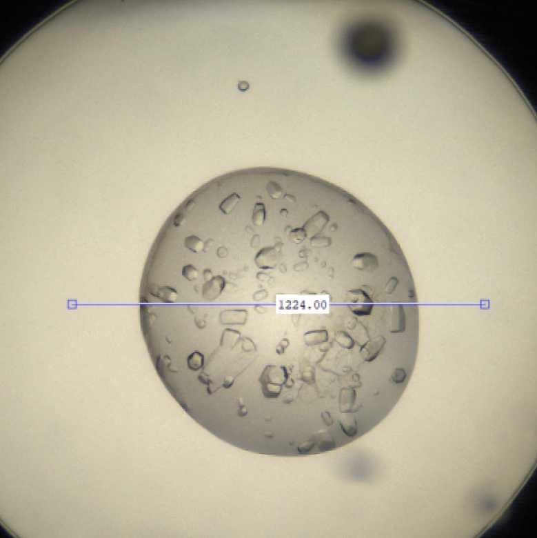
Crystal first observed and harvested on day 14.
Expected result
Crystals typically form hexagonal rods approximately 50 μm in size.
Morphology: Hexagonal rods.
Size: ~50 μm in length, 20 μm in width, 20 μm in depth
Appearance: varied sized of hexagonal rods
Average resolution: 1.9 Å
Space group: P64
Unit cell: 56 Å, 56 Å, 103 Å
90.00°, 90.00°, 120.00°
Exemplar picture of West Nile NS2B-NS3 protease crystals.
Data collection at Synchrotron
Diamond Light Source
Unattended Data Collection (UDC)
Data Collection Temperature: 100K
Detector: DECTRIS EIGER2 X 9M
Beamline: I04
Wavelength: 0.9537 Å
Resolution (Å): 1.91
Beam Size (μm): 30 X 20
Number of images: 3600
Oscillation: 0.10°
Exposure (s): 0.0053
Transmission (%): 100
Flux (ph/s): 9.50e+11
Protocol references
N/A