Apr 25, 2025
Version 2
Crystallisation of Enterovirus D68 3C protease apo structure and fragment screen in space group P212121 (PDB code 8CNX) V.2
- Ryan Lithgo1,2,3,
- Peter Marples1,2,3,
- Lizbé Koekemoer4,5,3,
- Daren Fearon1,3
- 1XChem, Diamond Light Source;
- 2Research complex at Harwell;
- 3ASAP Discovery Consortium;
- 4Centre of Medicines Discovery;
- 5University of Oxford
- Ryan Lithgo: The principle crystallographer on the Enterovirus 3C protease project.;
- ASAP Discovery


Protocol Citation: Ryan Lithgo, Peter Marples, Lizbé Koekemoer, Daren Fearon 2025. Crystallisation of Enterovirus D68 3C protease apo structure and fragment screen in space group P212121 (PDB code 8CNX). protocols.io https://dx.doi.org/10.17504/protocols.io.n92ldm6jnl5b/v2Version created by Mary-Ann Xavier
License: This is an open access protocol distributed under the terms of the Creative Commons Attribution License, which permits unrestricted use, distribution, and reproduction in any medium, provided the original author and source are credited
Protocol status: Working
We use this protocol and it's working
Created: February 08, 2025
Last Modified: April 25, 2025
Protocol Integer ID: 119833
Keywords: crystallisation, 3C protease, 3CNY, XChem, ASAP, AViDD, Diamond Light Source, Enterovirus D68, crystallisation of enterovirus d68 3c protease, enterovirus d68 3c protease crystal, enterovirus d68 3c protease, enterovirus d68, crystallization protocol, fragment screen in space group p212121, space group p212121, crystal structure, rcsb pdb with code 8cnx, crystal form, pdb code 8cnx, crystal form suitable for fragment screen, crystallisation, rcsb pdb, protease, protein expression, purification protocol, protein
Funders Acknowledgements:
National Institutes of Health/National Institute Of Allergy and Infectious Diseases (NIH/NIAID)
Grant ID: Grant ID: U19AI171399
Abstract
The crystallization protocol and buffer conditions used to obtain Enterovirus D68 3C protease crystals. The crystal structure was depostited to RCSB PDB with code 8CNX.
In this new version we added the Addgene ID and the protein expression and purification protocol. This construct gave us a crystal form suitable for fragment screen and initial compound soaking.
Materials
EV-D68 3C protease; aliases: Enterovirus A71 3C protease, non cleavable C-term 6xHis tagaddgeneCatalog #(Plasmid #204817)
SwissCI 3 lens crystallization plates https://swissci.com/product/3-lens-crystallisation-plate/
1 M Tris-HCl pH 8.14
1 M Ammonium acetate
50% w/v PEG 3350
Protocol materials
EV-D68 3C protease; aliases: Enterovirus A71 3C protease, non cleavable C-term 6xHis tagaddgeneCatalog #(Plasmid #204817)
Protein expression and purification
2d
Small scale protein purification and expression protocols used for this assayEV-D68 3C protease; aliases: Enterovirus A71 3C protease, non cleavable C-term 6xHis tagaddgeneCatalog #(Plasmid #204817)
Protocol
CREATED BY
korvus.wang
Equipment needed
Crystallisation experiment
1d
Protein and buffer requirements:
32 µL 1 millimolar (mM) Sample
2.88 mL Crystallization screen
16 µL Sample seeds, dilution 1:100 000
Crystallisation screen composition:
0.1 M Tris-HCl (pH 8.14)
0.2 M Ammonium acetate
25% w/v PEG 3350
Stock solutions used:
1 M Tris-HCl adjusted to pH 8.14 with HCl
1 M Ammonium acetate
50% w/v PEG 3350
Note
The crystallisation screen can be stored in a duran bottle or aliquoted into 96 deep well block for easy dispensing into SwissCI 3 lens plates.
For long term storage keep the Crystallisation screen in the fridge at 4°C.
1: 100 000 dilution Sample seeds
Dispense 30 µL Crystallisation screen into SwissCI 3 lens plate reservoir wells using a 100 µl multi-channel pipette.
Dispense 100 µL 1 millimolar (mM) Sample to each lens using the SPT mosquito.
Dispense 200 µL Crystallisation screen to each lens using the SPT mosquito.
Dispense 50 µL Seeds to each lens using the SPT mosquito.
Drop ratio: 1:2:05 ratio (100 nl Sample : 200 nl reservoir solution: 50 nl seeds)
Final drop volume: 350 nl
Incubate at 20 °C for 24:00:00 h in Formulatrix Rock Imager.
Imaging Schedule: The first images are taken after 12 h and the imaging schedule follows a Fibonacci sequence of days for further collections.
1d
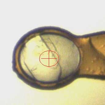
Crystal form after ~12 h.
Expected result
The crystals reach their maximum size after 24 h and most of the precipitant has gone.
Crystals typically form either as single crystals or in small clusters containng 4-6 crystals in a flower-like arrangement. Upon mounting a gentle nudge dissociates the cluster.
Morphology: typically thin rectangles with pointed ends.
Size: ~100 μm in length and ~50 μm in width, depth of the crystals is ~10 μm
Appearance: glass shard.
Average resolution: 1.5 Å
Space group: P212121
Unit cell: 42.82 Å, 62.53 Å, 147.36 Å
90.00°, 90.00°, 90.00°
An example of a drop containing EV D68 3C protease crystals.
Soaking tolerance
2h 30m
Final condition idenitified: 20% DMSO, 2hr 30mins using the protocol bellow.
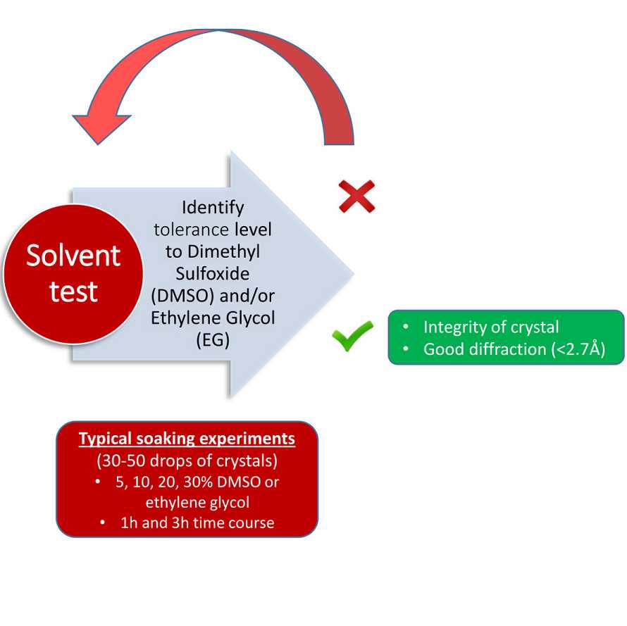
2h 30m
Fragment and compound soaking
2h 30m
20% fragment/compound, 2hr 30mins using the protocol bellow.

2h 30m
Protocol references
N/A