Apr 30, 2026
Crystallisation of coxsackievirus A16 2A protease in space group C2 used for apo, fragment screen and follow-up compounds
- Milo Cooper1,2,3,
- Ryan Lithgo1,2,4,
- Peter Marples1,2,3,
- Lizbé Koekemoer5,3,
- Korvus Wang5,3,
- Eda Capkin1,2,3,
- Mathew Golding1,2,3,
- Cedric Vallee1,2,3,
- Tracy Keates1,2,3,
- Charlotte Chinn1,2,3,
- Anu V. Chandran1,2,3,
- Ali Ebrahim1,2,3,
- Eleanor Williams5,3,
- Mary-Ann Xavier1,2,3,
- Daren Fearon1,2,4,
- Warren Thompson1,2,3,
- Jasmin Aschenbrenner1,2,3,
- Frank von Delft5,1,2,6,3,4
- 1Diamond Light Source;
- 2Research Complex at Harwell;
- 3OpenBind Consortium;
- 4ASAP Discovery Consortium;
- 5Centre of Medicines Discovery, University of Oxford;
- 6University of Johannesburg
- OpenBind Consortium


Protocol Citation: Milo Cooper, Ryan Lithgo, Peter Marples, Lizbé Koekemoer, Korvus Wang, Eda Capkin, Mathew Golding, Cedric Vallee, Tracy Keates, Charlotte Chinn, Anu V. Chandran, Ali Ebrahim, Eleanor Williams, Mary-Ann Xavier, Daren Fearon, Warren Thompson, Jasmin Aschenbrenner, Frank von Delft 2026. Crystallisation of coxsackievirus A16 2A protease in space group C2 used for apo, fragment screen and follow-up compounds . protocols.io https://dx.doi.org/10.17504/protocols.io.rm7vz4r1rlx1/v1
License: This is an open access protocol distributed under the terms of the Creative Commons Attribution License, which permits unrestricted use, distribution, and reproduction in any medium, provided the original author and source are credited
Protocol status: Working
We use this protocol and it's working
Created: April 10, 2026
Last Modified: April 30, 2026
Protocol Integer ID: 314826
Keywords: crystallisation, XChem, ASAP, AViDD, CMD, Diamond Light Source, i04-1, Coxsackievirus, A16, crystallisation of enterovirus coxsackievirus a16, coxsackievirus a16 crystal, 2a protease of the virus, enterovirus coxsackievirus a16, picornaviridae coxsackievirus a16, assembly of capsid protein, final stages of viral replication, cleavage from the poly protein, capsid protein, antiviral activity, virus, crystallographic fragment screening, poly protein, throughput crystallographic fragment screening, viral replication, protein production protocol, pdb 8poa, 2a protease in space group c2, 2a protease, formation of mature virion, protein production, protease, crystallisation, target for pandemic preparedness, solvent, CVA16, EV-A71, Enterovirus A71, EV-A71-2A, CVA16-2A, crystallisation of coxsackievirus a16, coxsackievirus a16, surrogate for enterovirus a71, enterovirus a71, 2a protease crystal, cleavage from the polyprotein, protein, picornaviridae, crystallographic screening, polyprotein, host cell, cva16
Funders Acknowledgements:
UK Department of Science, Innovation and Technology
Grant ID: G2-SCH-2025-06-16537
Abstract
This protocol was used to grow coxsackievirus A16 (CVA16) 2A protease crystals that were used as a surrogate for enterovirus A71 (EV-A71) 2A protease in high-throughput crystallographic fragment screening and in the crystallographic screening of follow-up compounds against the target. (PDB ID of apo-structure solved using sulfur phasing: pdb_000029jc)
Picornaviridae, primarily CVA16 and EV-A71, are the causative agents of paediatric hand-foot-and-mouth disease. These viruses are a target for pandemic preparedness due to the risk of higher-order complications in a large-scale outbreak. The 2A protease of the viruses is responsible for self-cleavage from the polyprotein, allowing for correct folding and assembly of capsid proteins in the final stages of viral replication. Inhibition deranges capsid folding and assembly, preventing formation of mature virions in host cells and making the protease a valuable target for antiviral activity.
Materials
Midi: UVXPO-3LENS 3W96T-PS 3W96T-UVP
1 Molarity (M) MES 6.7 , Molecular Dimensions, Catalog # MD2-013-PH 6.7
50% w/v PEG 20000, Molecular Dimensions, Catalog # MD2-250-16
Purified Coxsackievirus A16 protein (20 mg/mL ) in 10 millimolar (mM) HEPES, 7.5 , 0.5 Molarity (M) NaCl, 5% glycerol, 0.5 millimolar (mM) TCEP
Protein construct https://www.addgene.org/204809/
Hampton Research Crystal Crusher
Protocol materials
Human Coxsackievirus A16 strain G10 2A proteaseaddgeneCatalog #228632
Safety warnings
Follow all handling warning for the chemicals used in the crystalllisation screen composition.
Wear proper PPE at all times, including when handling cryogenic liquid nitrogen.
Protein expression and purification
We used the following protein expression and purification protocol for the plasmid:
Human Coxsackievirus A16 strain G10 2A proteaseaddgeneCatalog #228632
Protocol
CREATED BY
korvus.wang
Equipment needed
Seed stock preparation
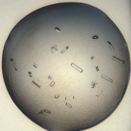
Select 5 well-crystallised drops of CVA16 2A protease (example image below). Prepare a Crystal Crusher and a small foam dewar of liquid nitrogen. It is important to complete all steps of the seed stock preparation as quickly as possible, as post-crushing the seeds may degrade or grow, and so ideally the stock should be frozen as fast as possible.
Use the Crystal Crusher to crush the crystals until a fine shimmer remains and no large pieces are visible.
Pipette the crushed solution into a microcentrifuge tube and use the crystal screen reservoir solution (~1 µL) to wash the well. Pipette the wash solution into the same tube. Repeat this step for all 5 wells.
Once all drops are crushed and pooled, make the seed stock up to 50 µL total volume.
Complete a serial dilution of the seed stock down to a 1:1000 dilution. This should produce sufficient seed stock for a large number of crystal plates.
Split the serially diluted seed stock into aliquots to avoid freeze-thawing the seed stock.
Note
30 µL is a good aliquot size, providing sufficient stock for two 288-well crystallisation plates.
Snap freeze each aliquot by immersing the PCR tube into cryogenic liquid nitrogen with tweezers. The aliquots can be left floating in the LN2 while you continue pipetting.
Store all aliquots at -80 °C .
Crystallization experiment
1d
Protein and buffer requirements:
21.6 µL 20 mg/mL Sample
2.40 mL Crystallization screen
7.2 µL Sample seeds, dilution 1:1000
Crystallisation screen composition:
13-19% (w/v) PEG 20000
0.1 Molarity (M) MES 6.7
Stock solutions used:
1 Molarity (M) MES 6.7
50% (w/v) PEG 20000
Note
The crystallisation screen can be stored in a Duran bottle or aliquoted into a 96 deep-well block for easy dispensing into SWISSCI 3 lens plates.
For long-term storage, keep the crystallisation screen in the fridge at 4°C.
Dispense 30 µL Crystallisation screen into SWISSCI 3 lens plate reservoir wells using a 100 µL multi-channel pipette.
Dispense 75 nL 20 mg/mL Sample to each lens using the SPT mosquito.
Dispense 75 nL Crystallisation screen to each lens using the SPT mosquito.
Dispense 25 nL Seeds to each lens using the SPT mosquito.
Drop ratio: 3:3:1 ratio (75 nL Sample : 75 nL reservoir solution: 25 nL seeds)
Final drop volume: 350 nL
Incubate at 20 °C for 24:00:00 h in Formulatrix Rock Imager.
Imaging Schedule: The first images are taken after 12 h and the imaging schedule follows a Fibonacci sequence of days for further collections.
1d
Crystals typically form after ~24 h.
Expected result
Crystals typically reach their maximum size after ~24 h.
Morphology: typically rectangles.
Size: ~75 μm in length and ~10 μm in width, depth of the crystals is ~10 μm, giving a
rectangular appearance
Average resolution: 1.6 Å
Space group: C2
Unit cell: 86 Å, 57 Å, 32 Å
90°, 95°, 90°
An example of a drop containing coxsackievirus A16 2A crystals.
Data collection at Synchrotron
Diamond Light Source
Unattended Data Collection (UDC)
Data Collection Temperature: 100K
Detector: DECTRIS EIGER2 X 9M
Beamline: I04-1
Wavelength: 0.9212 Å
Resolution (Å): 1.21
Beam Size (μm): 60 X 50
Number of images: 3600
Oscillation: 0.10°
Exposure (s): 0.0020
Transmission (%): 100
Flux (ph/s): 9.50e+11
Solvent tolerance
Best conditions were: 10% (v/v) DMSO, for 2 h.
Best cryoprotectant was: 25% (v/v) Glycerol.
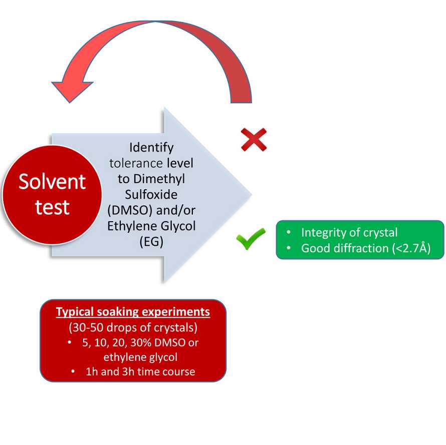
Fragment and compound soaking
Conditions used 5-10% (v/v) DMSO, for 2 h

Acknowledgements
Diamond Light Source Ltd, Harwell Science and Innovation Campus, Didcot OX11 0QX, UK
Research Complex at Harwell, Harwell Science and Innovation Campus, Didcot OX11 0FA, UK
Oxford Lab Technologies Crystal Shifter https://doi.org/10.1107/S2059798320014114
OpenBind received funding from the UK Department of Science, Innovation and Technology under grant number G2-SCH-2025-06-16537.