Protocol Citation: Hanna Edler 2025. Clearing for Lightsheet Microscopy. protocols.io https://dx.doi.org/
Manuscript citation:
This protocol is part of the following dissertation and publicly available under a Creative Commons license (CC BY-SA 4.0):
http://dx.doi.org/10.25673/114391
License: This is an open access protocol distributed under the terms of the Creative Commons Attribution License, which permits unrestricted use, distribution, and reproduction in any medium, provided the original author and source are credited
Protocol status: Working
We used this protocol and it's working
Created: April 28, 2025
Last Modified: May 05, 2025
Protocol Integer ID: 184756
Keywords: fluorescence lightsheet microscopy, lightsheet microscopy this protocol, lightsheet microscopy, organic clearing of mouse brain, whole mount nanobody, mouse brain, organic clearing
Disclaimer
DISCLAIMER – FOR INFORMATIONAL PURPOSES ONLY; USE AT YOUR OWN RISK
The protocol content here is for informational purposes only and does not constitute legal, medical, clinical, or safety advice, or otherwise; content added to protocols.io is not peer reviewed and may not have undergone a formal approval of any kind. Information presented in this protocol should not substitute for independent professional judgment, advice, diagnosis, or treatment. Any action you take or refrain from taking using or relying upon the information presented here is strictly at your own risk. You agree that neither the Company nor any of the authors, contributors, administrators, or anyone else associated with protocols.io, can be held responsible for your use of the information contained in or linked to this protocol or any of our Sites/Apps and Services.
Abstract
This protocol was developed for whole mount nanobody staining and organic clearing of mouse brains for fluorescence lightsheet microscopy.
Materials
Please read the safety data sheets before use and handle reagents with care.
Handle toxic reagents (e.g. paraformaldehyde, sodium hydroxide, diclormethane) with care! Read the safety data sheets before use and follow the recommendations for safe handling and storage.
Ethics statement
For conducting animal experiments as in this protocol, prior approval by the users' Institutional Animal
Care and Use Committee (IACUC) or equivalent ethics committee is needed.
Before start
Prepare the needed buffers and solutions in advance.
fill up to 1 L with any distilled water and stirr until dissolved.
For 1xPBS, 10xPBS is diluted 1:10 with any distilled water. 7.2 -7.4
4% Paraformaldehyde solution (4% PFA solution)
40 gparaformaldehydeAppliChemCatalog #A3813.1000
100 mL 10xPBS (prepared in step 1.1)
Heat up (<70°C!) while stirring under the hood for ~ 04:00:00 -05:00:00 ; let it cool down; add ~ 250 µL10M sodium hydroxide solutionMerck MilliporeSigma (Sigma-Aldrich)Catalog #72068 (adjust to 8 to preserve endogenous fluorescent proteins). Store in aliquots at -20 °C and thaw slowly just before use (overnight in fridge and directly use).
when permeabilization buffer is prepared in aliquots for several days in advance: add goat serum freshly just before use!
Start
1h
In case of intravenous vessel labeling:
Anesthetize mouse and inject vessel dye i.v. or a dye combination in accordance with legal requirements for animal experiments.
Euthanize mouse in accordance with legal requirements for animal experiments until it shows no pedal reflexes anymore, open the thorax, expose the heart.
1h
Perfusion
4h 30m
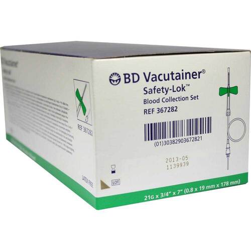
Prick the BD Vacutainer Blood Collection Set into the left ventricle (light red) and snip with small scissors in the right atrium (the bigger the snip the better it runs and clears).
25 mL heparinized 1xPBS (10 U/mLHeparin sodium salt from porcine intestinal mucosaMerck MilliporeSigma (Sigma-Aldrich)Catalog #H3149 ) at Room temperature
Expected result
liver becomes pale, yellowish and/or light brown
150 mL 4% PFA solution (cf. step 2), ice-cold (fresh or freshly thawed aliquot only)
Dissect the organ of interest immediately, postfix it in 4% PFA solution for 2 h (for bones: 4 h) at 4 °C
Note
check individual fixation times here:
Citation
Klingberg A, Hasenberg A, Ludwig-Portugall I, Medyukhina A, Männ L, Brenzel A, Engel DR, Figge MT, Kurts C, Gunzer M (1970). Fully Automated Evaluation of Total Glomerular Number and Capillary Tuft Size in Nephritic Kidneys Using Lightsheet Microscopy.
Wash 3x with 1xPBS at 40 rpm, Room temperature , 00:30:00 orbital shaker
1h 30m
Store in 1xPBS + 0.05 % NaN3 at 4 °C for up to 6 months OR start immediately.
Decolorization
2d 1h 30m
Incubate the dissected organ in 30 % volume decolorization solution (cf. step 3) for 48:00:00 at Room temperature while gentle shaking
2d
Note
clears hemoglobin, if necessary, should not be combined with tomato lectin i.v. staining
Wash afterwards 3x 00:30:00 at Room temperature
1h 30m
Decalcification
2d 1h 30m
Incubate the whole skull (cleared from jaws and muscles) in PBS/EDTA (22.5 mL0.5M EDTAInvitrogenCatalog #AM9260G + 27.5 mL 1xPBS) at Room temperature for 48:00:00, gentle shaking
2d
Note
for bony tissues only; adjust time if needed
Wash afterwards 3x 00:30:00 at Room temperature
1h 30m
Permeabilization and staining
1w 4d
Incubate in permeabilization buffer containing normal goat serum at 37 °COvernight , mild shaking/rotating improves the result
1d
Nanobody staining (repeat the following steps depending on sample size):
Note
I repeated this once for a total of 2 staining steps for whole mouse brain.
1 µLTO-PRO™-1 Iodide (515/531) - 1 mM Solution in DMSOThermo FisherCatalog #T3602
at37 °C for 120:00:00 , mild shaking/rotating
Note
Recommendations for nanobodies:
http://discotechnologies.org/vDISCO/
Please note:
Antibodies labeled with fluorophores emitting at longer wavelength decrease background signals.
No fluorescein/cyanine-based fluorophores (organic dyes) – there are publications using FITC (?)
Antibody penetration time and efficiency strongly depend on tissue composition.
Antibody concentration strongly depends on availability/expression of antigen.
Small antibodies like nanobodies (Chromotek/NanoTag) strongly improve the staining outcome.
Washing
2d
Wash 1
3x wash for 02:00:00 at Room temperature with 1xPBS + 0.1 % volumeTriton® X 100Carl RothCatalog # 3051 and mild shaking/rotating
repeat Overnight
1d
Wash 2
3x wash for 02:00:00 at Room temperature with 1xPBS and mild shaking/rotating
repeat Overnight
1d
Dehydration and Clearing
5d 4h
Dehydration
Recommended for bone
50% EtOH/dH2O for 12:00:00
70% EtOH for 12:00:00
100%EtOH for 12:00:00
100% EtOH for 12:00:00
Recommended for brain
In 50% EtOH/dH2O Overnight , rotation at Room temperature
1d
In 70% EtOH/dH2O Overnight , rotation at Room temperature
1d
In 100% EtOH Overnight , rotation at Room temperature
1d
In 100% EtOH Overnight , rotation at Room temperature
1d
Note
Please note:
at least butanol, isopropanol, methanol or tetrahydrofurane can also be used or even combined (one by one) but the effects on the tissue (e.g. shrinkage) can differ.
Delipidation
(optional, recommended for lipid-rich organs as brains)
incubate for 04:00:00 in dichloromethane Dichlormethane
4h
Clearing
Incubate in DibutyletherMerck MilliporeSigma (Sigma-Aldrich)Catalog #5.89583 or Ethyl cinnamateMerck MilliporeSigma (Sigma-Aldrich)Catalog #112372 for at least 24:00:00
Note
replace the solvent at least once after 24 h to improve clearing result
1d
Note
Samples can be stored quite long in the organic solvent (keep water-free!) without losing quality @ RT in the dark.
Samples are imaged in the same solution or solutions with equal refractive index and characteristics.
Note
In general:
Use only hand-made PFA solutions (without stabilizing reagents).
Dehydrating reagents can be changed/combined.
Organic solvents can be changed/combined.
Dehydrating and clearing are reversible.
Organize your panels as following:
Use fluorophores emitting at longer wavelength (red or far-red).
Small structures: as “red” as possible, bigger structures can eventually be stained with “green” fluorophores.
Avoid lipid-based/organic dyes (=fluorescein and cyanine-based dyes; better to use AF dyes, DyLight or new synthetic dyes (Atto/Abberior Star)).
Never ever bring the cleared organs in contact with water!
Keep fluorescently labeled samples in the dark.
organic solvents are not compatible with PE or PS plastic ware! Solely use PP or glass ware! Always test solvent stability first. Always handle reagents with care.
This protocol is a so-called „organic-based clearing“ and parts may be exchangeable with comparable protocols like iDisco, uDisco, FluoClearBABB, PEGASOS, THF/DBE,…
Miltenyi offers kits and special antibodies for this procedure as well.
Protocol references
Citation
Cai R, Pan C, Ghasemigharagoz A, Todorov MI, Förstera B, Zhao S, Bhatia HS, Parra-Damas A, Mrowka L, Theodorou D, Rempfler M, Xavier ALR, Kress BT, Benakis C, Steinke H, Liebscher S, Bechmann I, Liesz A, Menze B, Kerschensteiner M, Nedergaard M, Ertürk A (2019). Panoptic imaging of transparent mice reveals whole-body neuronal projections and skull-meninges connections.
Klingberg A, Hasenberg A, Ludwig-Portugall I, Medyukhina A, Männ L, Brenzel A, Engel DR, Figge MT, Kurts C, Gunzer M (2017). Fully Automated Evaluation of Total Glomerular Number and Capillary Tuft Size in Nephritic Kidneys Using Lightsheet Microscopy.
Klingberg A, Hasenberg A, Ludwig-Portugall I, Medyukhina A, Männ L, Brenzel A, Engel DR, Figge MT, Kurts C, Gunzer M. Fully Automated Evaluation of Total Glomerular Number and Capillary Tuft Size in Nephritic Kidneys Using Lightsheet Microscopy.
Cai R, Pan C, Ghasemigharagoz A, Todorov MI, Förstera B, Zhao S, Bhatia HS, Parra-Damas A, Mrowka L, Theodorou D, Rempfler M, Xavier ALR, Kress BT, Benakis C, Steinke H, Liebscher S, Bechmann I, Liesz A, Menze B, Kerschensteiner M, Nedergaard M, Ertürk A. Panoptic imaging of transparent mice reveals whole-body neuronal projections and skull-meninges connections.
Klingberg A, Hasenberg A, Ludwig-Portugall I, Medyukhina A, Männ L, Brenzel A, Engel DR, Figge MT, Kurts C, Gunzer M. Fully Automated Evaluation of Total Glomerular Number and Capillary Tuft Size in Nephritic Kidneys Using Lightsheet Microscopy.