License: This is an open access collection distributed under the terms of the Creative Commons Attribution License, which permits unrestricted use, distribution, and reproduction in any medium, provided the original author and source are credited
Protocol status: Working
We use this collection and it's working
Created: February 02, 2023
Last Modified: February 02, 2023
Collection Integer ID: 76314
Keywords: electrophysiology, Neuropixels, silicon probe, hippocampus, entorhinal cortex, spatial navigation, freely moving recording, electrode, chronic recoverable neuropixels in mice, chronic recoverable neuropixel, neuropixels 2021 course lecture, other chronic recoverable design, probes for future use, neuropixel, probe during surgery, mice, beta probe, same probe, implantation into any brain region, probe, probe adjustable before gluing,
Funders Acknowledgements:
Simons Collaboration on the Global Brain
Grant ID: 542987SPI
Stanford School of Medicine Dean's Postdoctoral Fellowship
A.P. Giannini Foundation Postdoctoral Fellowship
Abstract
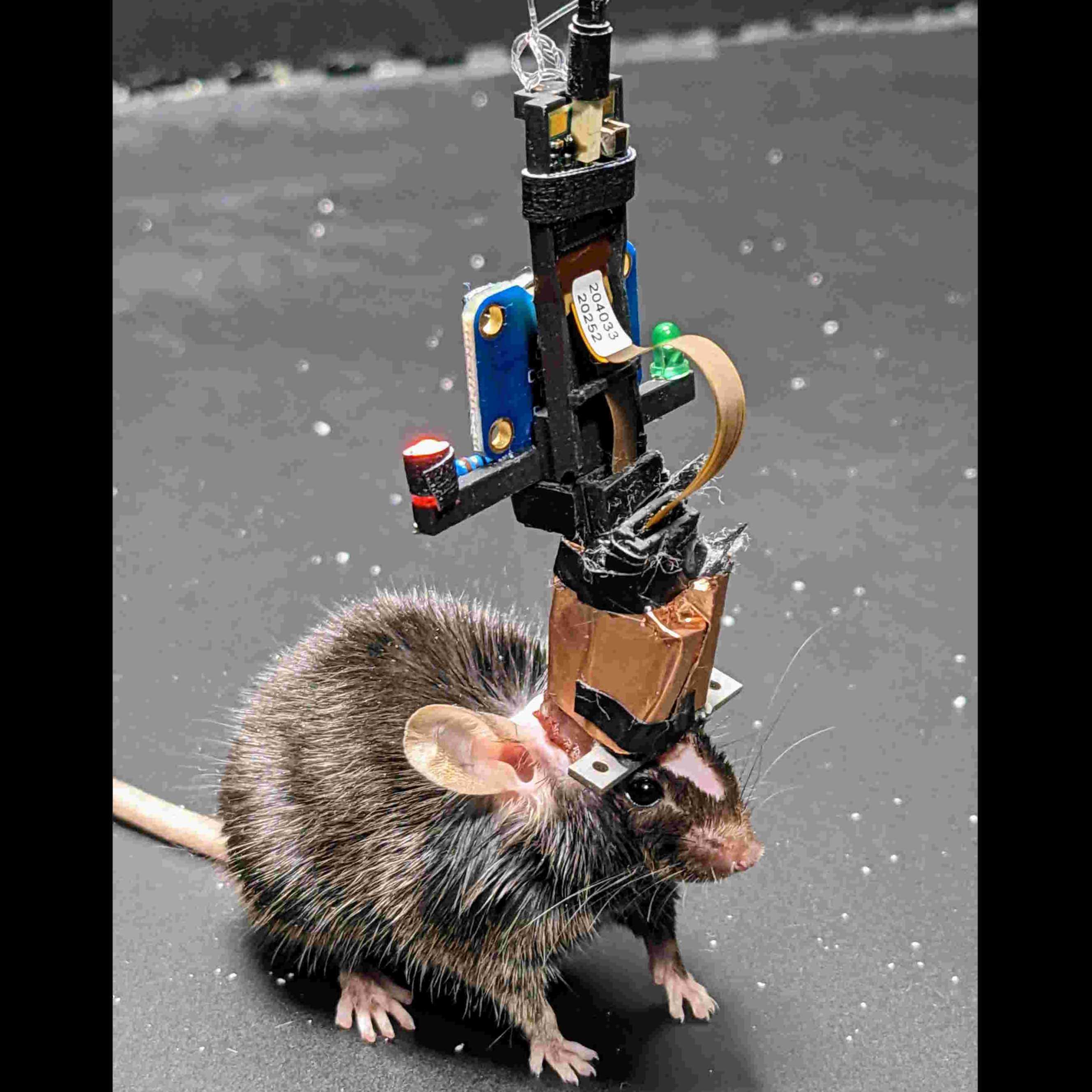
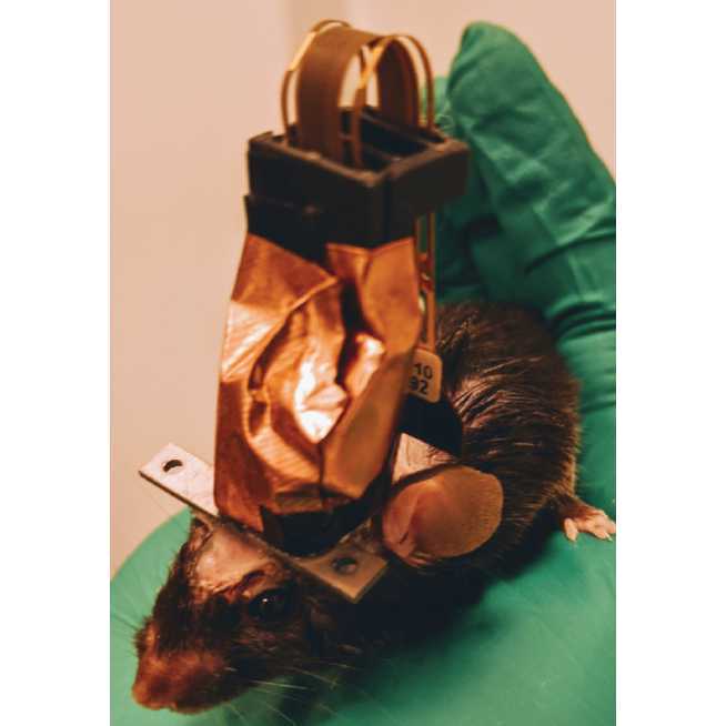
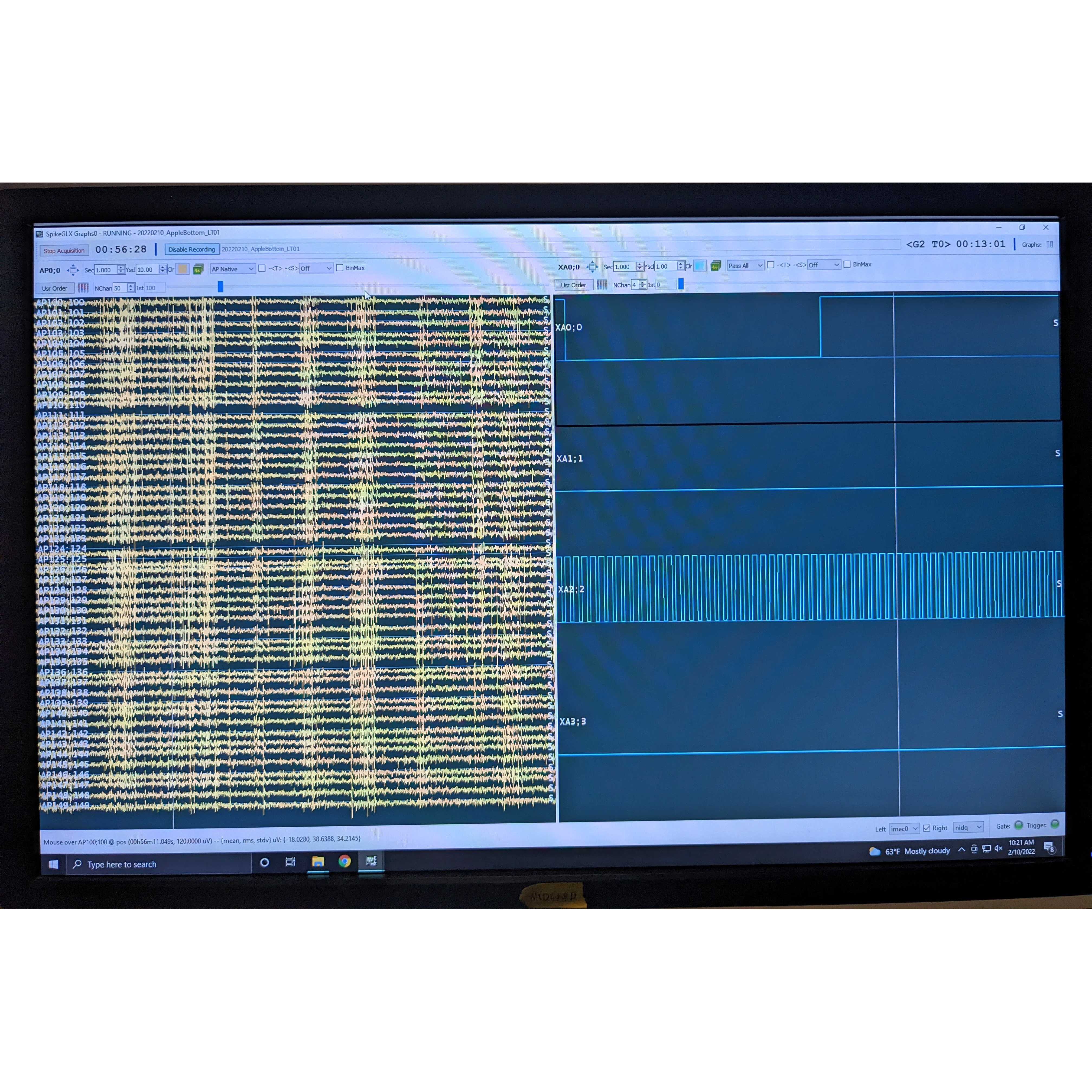
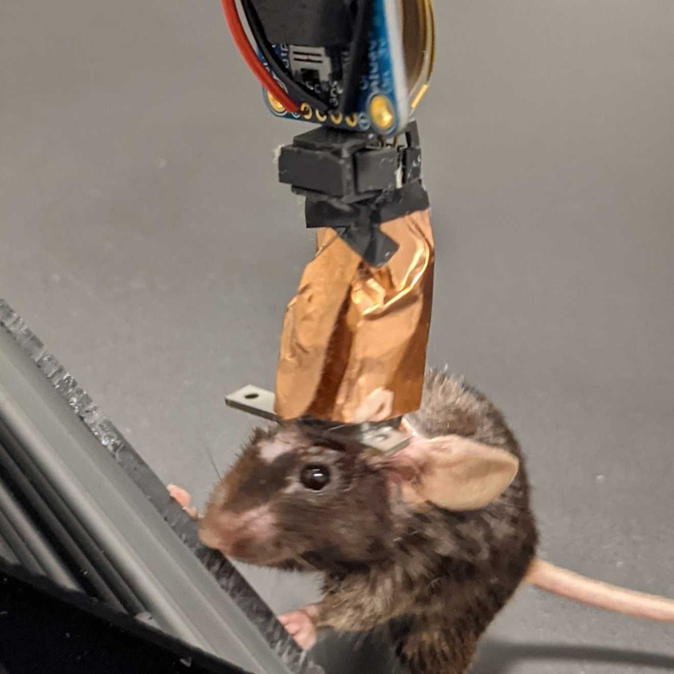
This protocol collection explains how to build a low-cost, lightweight system to implant 1 Neuropixels 1.0 probe or 2 Neuropixels 2.0 probes into mice, record during freely moving behavior, then recover the probes for future use.
Lightweight (entire assembly, including headbar, dental cement, and tape, weighs <3g for single 1.0 probe or <4g for dual 2.0 probes)
Allows headfixed or freely moving recordings
Quickly attaches to a headstage holder, which provides LEDs for tracking and a counterweight to encourage running
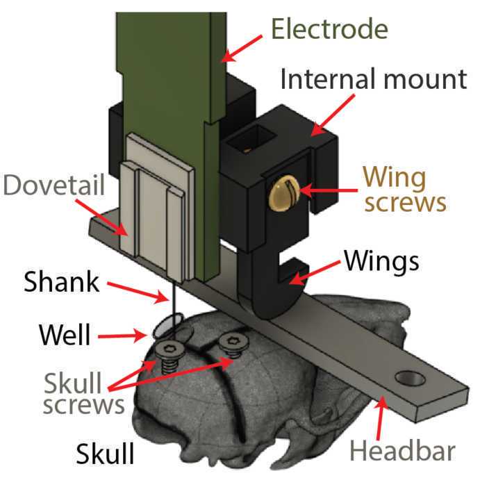
Position and angle of internal mount on the probe adjustable before gluing, allowing implantation into any brain region, but custom adjustment is not required, allowing the same probe to be re-inserted into a variety of sites
Unlike completely enclosed designs, the shank is uncovered during insertion for better visualization, yet doesn't require delicately surrounding the shank with a glue column
Dual 2.0 probes: probes are independently insertable, so can set custom positions, depths, and angles for each
Disadvantages of this design:
Not suitable for larger animals (lightweight design likely can't withstand larger forces, flex cable remains exposed, moisture known to wick up shank into PCB in rats)
External components are assembled around the probe during surgery rather than during assembly, so surgeries take slightly longer
Less elegant than completely enclosed designs and requires a larger skull surface area for gluing
Dual 2.0 probes: how close together the probes can get is limited by some of the components