Dec 01, 2021
Chlamydia pneumoniae-Induced Neuroinflammation Cell Model Using Lyophilized Cell-Free Supernatant
- Elif Kaya Tilki1
- 1Anadolu University


External link: https://doi.org/10.1371/journal.pone.0260633
Protocol Citation: Elif Kaya Tilki 2021. Chlamydia pneumoniae-Induced Neuroinflammation Cell Model Using Lyophilized Cell-Free Supernatant . protocols.io https://dx.doi.org/10.17504/protocols.io.bzw2p7ge
Manuscript citation:
Kaya-Tilki E, Dikmen M (2021) Neuroprotective effects of some epigenetic modifying drugs’ on Chlamydia pneumoniae-induced neuroinflammation: A novel model. PLoS ONE 16(11): e0260633. doi: 10.1371/journal.pone.0260633
License: This is an open access protocol distributed under the terms of the Creative Commons Attribution License, which permits unrestricted use, distribution, and reproduction in any medium, provided the original author and source are credited
Protocol status: Working
Created: November 09, 2021
Last Modified: December 01, 2021
Protocol Integer ID: 54970
Keywords: chlamydia pneumoniae, induced neuroinflammation cell model, bacterial products into the host tissue, infected cell culture, role in alzheimer, cell free supernatant, negative intracellular pathogen, using lyophilized cell, lyophilized cell, introducing bacterial product, alzheimer, epigenetic regulatory system, pathogenesi, pulmonary disease, variety of pulmonary disease
Abstract
Chlamydia pneumoniae (Cpn) is a gram-negative intracellular pathogen that causes a variety of pulmonary diseases, and there is growing evidence that it may play a role in Alzheimer's disease (AD) pathogenesis. Cpn can interact functionally with host histones, altering the host's epigenetic regulatory system by introducing bacterial products into the host tissue and inducing a persistent inflammatory response. Because Cpn is difficult to propagate, isolate, and detect, a modified LPS-like neuroinflammation model was established using lyophilized cell free supernatant (CFS) obtained from infected cell cultures, and the effects of CFS were compared to LPS.
Materials
HEp-2 human epithelial carcinoma cell line (ATCC CCL-23)
Chlamydia pneumoniae (ATCC 53592)
Pathfinder Chlamydia Culture Confirmation System (Cat. No. 30701, Bio-Rad, Germany)
Troubleshooting
HEp-2 cells were used as a host to inoculate Chlamydia pneumoniae. A 6-well plate of 1X10⁶ HEp-2 cells was seeded 48 hours prior to inoculation with Chlamydia pneumoniae (ATCC 53592).
The suspension of elementary bodies diluted in infection medium was added directly to wells.
The mixture was centrifuged at 1500 × g for 1 h.
The plate was incubated for 1 h at 37 °C in the presence of 5% CO2.
Current medium was discarded.
Cells were washed with 300 μL Hanks Balanced Salt Solution.
500 μL fresh medium was added to the wells.
The plate was incubated for 72 hours.
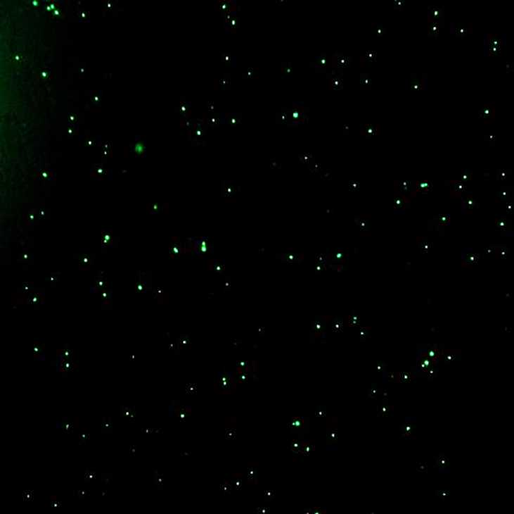
The inclusion bodies were confirmed using the Pathfinder Chlamydia Culture Confirmation System (Cat. No. 30701, Bio-Rad, Germany) according to the kit manual.
The cells were imaged using the Cytation 3 Cell Imaging Multi-Mode Reader (BioTek, USA).
The number of inclusion-forming units per milliliter (IFU/ml) in HEp-2 cells was used to determine the infectivity titers of chlamydial stocks and 1x10⁶ HEp-2 monolayers in 6-well plate were contaminated with Chlamydia pneumoniae suspended in inoculating media at 1 multiplicity of infection (MOI) ratio.
Cell-free supernatant was collected from the wells, lyophilized and stored in aliquots at -80 °C.
The lyophilized cell-free supernatant was weighed to prepare a main stock in the desired ratio and used to trigger the inflammatory response.