Aug 05, 2024
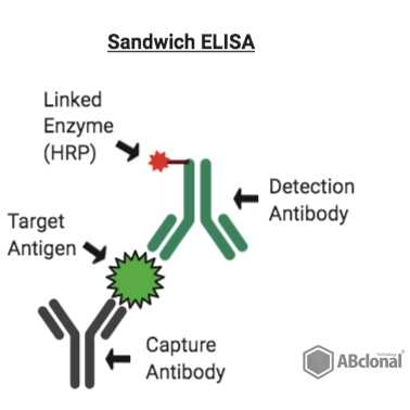
Chemiluminescence-enhanced ELISA measurements for α-Synuclein fibrils
- 1Duke University
- West lab protocols


External link: https://doi.org/10.1126/sciadv.adq3539
Protocol Citation: Arpine Sokratian 2024. Chemiluminescence-enhanced ELISA measurements for α-Synuclein fibrils. protocols.io https://dx.doi.org/10.17504/protocols.io.5qpvob8odl4o/v1
Manuscript citation:
Sokratian A, Zhou Y, Tatli M, Burbidge KJ, Xu E, Viverette E, Donzelli S, Duda AM, Yuan Y, Li H, Strader S, Patel N, Shiell L, Malankhanova T, Chen O, Mazzulli JR, Perera L, Stahlberg H, Borgnia M, Bartesaghi A, Lashuel HA, West AB Mouse α-synuclein fibrils are structurally and functionally distinct from human fibrils associated with Lewy body diseases. Science Advances 10(44). doi: 10.1126/sciadv.adq3539
License: This is an open access protocol distributed under the terms of the Creative Commons Attribution License, which permits unrestricted use, distribution, and reproduction in any medium, provided the original author and source are credited
Protocol status: Working
We use this protocol and it's working
Created: May 23, 2022
Last Modified: August 05, 2024
Protocol Integer ID: 63016
Keywords: ASAPCRN, synuclein aggregate elisa kit, synuclein aggregate antibody, syn fibril levels in neuronal lysate, syn fibril level, synuclein, syn fibril, enhanced elisa measurement, fibril, days of fibril, monomeric protein
Funders Acknowledgements:
ASAP
Grant ID: Grant ID: 020527
Abstract
This protocol is optimized The BioLegend LEGEND MAX α-Synuclein Aggregate ELISA Kit in a 96-well strip plate precoated with rat monoclonal anti-α-synuclein aggregate antibody. We use in-house a-syn fibrils and monomeric protein as a control. We use ELISA to detect a-syn fibril levels in neuronal lysates after 14 days of fibril-treatment.
Guidelines
Before running the ELISA kit, it is crucial to define the correct dilutions for the specific samples. This can be achieved by conducting a pilot ELISA with a control standard curve alongside different dilutions of the pooled sample. This preliminary step ensures the accuracy and reliability of the results obtained from the ELISA kit. Please follow this guideline to ensure optimal results.
Protocol materials
LEGEND MAX™ α-Synuclein Aggregate ELISA KitBioLegendCatalog #449407
Safety warnings
Hazard Identification and Risk of Exposure to the
Hazards:
Inhalation or spread through food or drink that contain fibrils aerosols or fibrils.
Protective gloves, safety glasses and lab coat must always be used when handling anything that possibly could contain α-synuclein fibrils. Food or drink is strictly prohibited in any environment where α-syn fibrils are used.
Routes of Transmission: Prior to assigning containment requirements, it is imperative to
understand the routes of transmission.
Some issues to address:
- What are the exposure routes/risks of most concern:
Inhalation or spread through food or drink that contain fibril aerosols or fibirls accordingly. Fibrils possibly might reach the brain regions through the olfactory epithelium; Risk of accidental needlestick/droplet splash while handling fibrils for in vitro or in vivo work.
- What are the consequences of exposure (potential illness, etc)
Fibrils may be considered as infectious material. Minimum to no hazard is expected from α-syn protein. There is no evidence that transmission of fibrils can lead to development Parkinson’s disease. However, taking into account prion-like properties of α-syn fibrils should therefore be handled cautiously and wisely. Strictly recommended using disposable materials and Personal Protective Equipment (PPE) such as gloves, face mask, etc.
PRECAUTIONS:
Laboratory work where high concentration of fibrils (more than 300 uM) is needed must comply with biosafety level 2 (BSL2) containment as described in the current edition of the CDC/NIH’s
Biosafety in the Microbiological and Biomedical Laboratories: http://www.cdc.gov/od/ohs/biosfty/bmbl5/bmbl5toc.htm
Sharps safety precautions:
The use of sharps (glass pipettes, glass slides and cover slips, scalpels and
lancets) should be eliminated, when possible. Appropriate precautions should be
taken to avoid percutaneous injuries. These items should be disposed of
immediately in a puncture-resistant sharps container. Bending, recapping or
clipping of needles is prohibited. As described in CDC’s sharps safety website:
https://www.cdc.gov/sharpssafety/index.html
Procedural Methods
and Materials:
- Laboratory work where high concentration of fibrils (more than 300 uM) is needed must comply with biosafety level 2 (BSL2) containment. This means all aerosol generating procedures must be performed within the biosafety cabinet.
- All the fibrils work involves using PPE, aerosol-tight centrifuges, water bath sonicator in a closed cabinet, homogenization of frozen brain samples using probe-tip sonicator under the hood (collection of protein fractions in BSL2 cabinets), chromatography equipment in a closed-door fridge, sealed plates, safe lock microfuge tubes (or tubes wrapped/sealed with parafilm), and use of filtered tips for pipettes. All personnel must strictly adhere to these procedures.
- Use of proper PPE as stated in the section below. Use of available N95 respirators is voluntary (same for the use of available sleeve protectors). Follow safety precautions for sharps (for e.g., to avoid accidental needle sticks) while working with PFFs in the lab and for doing in vivo work.
Personal Protective
Equipment (PPE): Appropriate PPE includes gloves, lab coat and safety glasses,
face mask (voluntary N95 respirator use and sleeve protectors), face / bench
top splash shield for specific procedures as stated above.
Methods to minimize personal exposure: Strictly adhere to sharps safety precautions using needles or any material that can potentially cause wounds. Use disposable supplies where possible. Use the minimal amounts of α-fibrils needed for an experiment. Keep fibrils in closed tubes. 10% of SDS solution in water must be used for decontaminating work areas. Do not use NaOH or Sodium Hypochlorite or ethanol. Do not leave samples containing fibrils unattended at the bench.
Methods to prevent the release of fibrils/protect workers from aerosols,
splashes, splatters: protective gloves and clothing always be always be worn when handling frozen vials. High concentration of fibrils(>1mg/mL) always be handled under Biosafety cabinet and containment caps will be used while centrifugation. Centrifuge cups will be opened inside a biosafety cabinet. Face shield or benchtop splash shield will be used when working at the open bench.
Specimen transport
and removal of material(s) from the laboratory: Transported in secondary
container (plastic/Styrofoam) in a closed box. The closed box is carried in a
bag.
Standard
microbiological methods: hand washing after removal of gloves and before
leaving the work area, no mouth pipetting, strictly no food or drink in
refrigerators where material is stored, no eating in work area.
Cleaning &
Disinfection: Work area must be
cleaned with 10% SDS in water. Wipes used must be immediately disposed into
biohazard waste container. Any piece of equipment or supplies that possibly
have been exposed to fibrils must be wiped with 10% of SDS.
Waste Generation and Disposal Methods: The solutions that contain α-syn fibrils must be
decontaminated with 10% of SDS in water for 30 minutes and be thrown as a
biohazard waste in a sealed container/bag (use a minimal volume of fibrils needed
for an experiment, do not generate large volumes of fibril-containing liquids).
Use small biohazard bags to collect tips and consumables of experiment
performed, appropriately tie neck of bag in single knot and place in into
secondary biohazard waste container.
Spill and Accident Response Procedure: Describe all emergency procedures including spill clean-up. Describedisinfectants and
environmental decontamination. (ex., Outside of a BSC: If spill is a
respiratory hazard, evacuate 30 minutes to allow aerosols to settle. Place absorbent towels over the spill, apply freshly
prepared 10% SDS solution to entire area of spill starting on the outer edges
and working inward, pick up sharp items with mechanical device (not hands),
place all clean-up materials in a biohazard bag)
LEGEND MAX™ α-Synuclein Aggregate ELISA KitBioLegendCatalog #449407
Preparation of buffers
Preparation of 1X Reagent Diluent
1. Label an appropriate sized bottle as “1X Reagent Diluent”.
2. Dilute 2X Reagent Diluent to 1X by adding 30mL of 2X Reagent Diluent to 30mL of lab grade water in the bottle labeled “1XReagentDiluent”.
3. Mix well by vortex.
Preparation of 1X Wash Buffer
1. Label a 1L bottle as “1X Wash Buffer”.
2. Dilute 5X Wash Buffer 1:5 using lab grade water* and mix well.
Preparation of standard intermediates
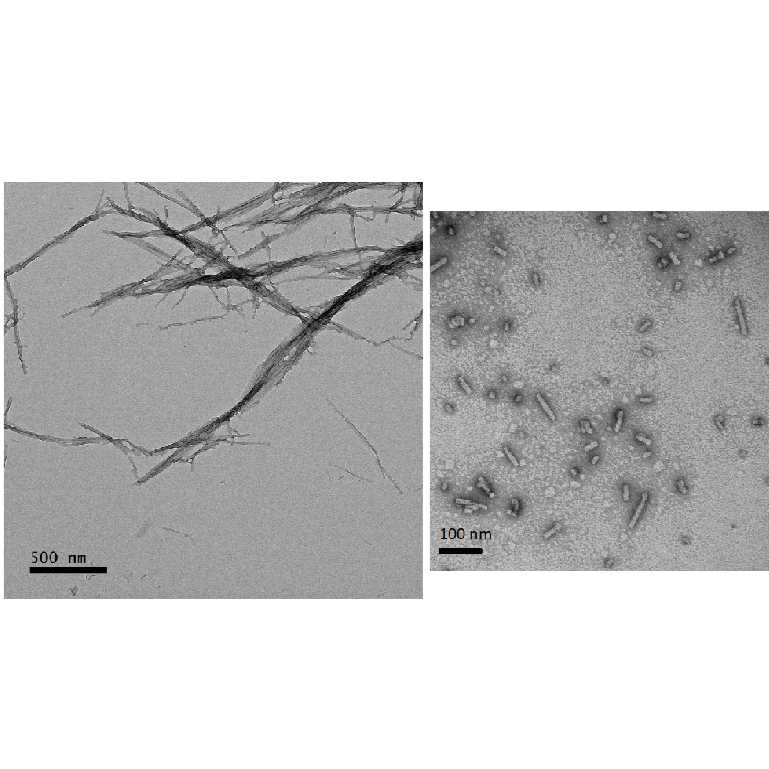
Thaw down On ice monomer/sonicated fibril preparation aliquots stored at -80 °C
Measure monomer concentration via Nanodrop
Add Amount of 5x, 10x or 20x diluted aliquots in PBS onto nanodrop piedestal;
Parameters: other proteins; coefficient extinction:
5.98; MW: 14.4 kDA for human a-synuclein
7.45; MW: 14.4 kDA for mouse a-synuclein
Perform two measurements for each dilution and confirm <10% standard error between two measurements
For fibril preparations, dilute in 6M guanidine HCL and follow the aforementioned instructions derscribed for monomeric protein
Equipment
NanoDrop™ One/OneC Microvolume UV-Vis Spectrophotometer
NAME
UV-Vis Spectrophotometer
TYPE
Thermo Scientific
BRAND
ND-ONE-W
SKU
Use 1X Reagent Diluent to prepare the standard intermediates as follows:
Assay reaction 0 : dilute monomer stock to a concentration 50ng/mL
Assay reaction // Volume of standard (uL) // Volume of 1X reagent diluent (uL) // Final concentration (pg/mL)
1 40 uL of #0 1280 1500
2 550 uL of #1 825 600
3 550 uL of #2 825 240
4 550 uL of #3 825 96.0
5 550 uL of #4 825 38.4
6 550 uL of #5 825 15.4
7 550 uL of #6 825 6.1
8 0 825 0
Sample preparation
Dilute samples in 1X Reagent Diluent; if correct dilution is unknown - prepare a standard dilutions as follows: 10X; 20X, 40x;100X; 500X;
Once evaluated - use correct dilution factor to run an experiment;
Minimum recommended dilution is 1:10;
we recommend dilution for neuronal lysates to be set at 1:100
ELISA procedure
1d
Remove the plate from the foil pouch.
Add 300 µL / well of 1X Wash Buffer (use multichannel pipette and ELISA racks to easily access the wash buffer).
Dump out wash buffer and pat dry.
Repeat 3 more times for a total of 4 washes.
Add 50 μL of 1X Reagent Diluent to each well that will contain either standard dilutions or samples.
Add 50 µL of each standard to the plate in duplicate or triplicate. Follow the plate layout below.
Note: The volumes in Table 2 are sufficient for running the standard curve in triplicate.
Add 50 µL of each sample dilution to the plate.
Cover the plate with the plate sealer provided and incubate for 2 hours at Room temperature , with shaking
EXAMPLE LAYOUT:
Add 100 µL μL of α-Synuclein Aggregate Detection Antibody to each well, seal the plate, and incubate at room temperature for 1 hour with shaking.
Dump the content of the plate and repeat
Dump the content and add Avidin-HRP 100 µL / well to each used well, cover and incubate for 00:30:00 at Room temperature
30m
Remove plate from incubation and dump components; repeat
Discard the contents of the plate into a sink, then wash the plate 5 times with 1X Wash Buffer as in step 4. For this final wash, soak wells in 1X Wash Buffer for 30 seconds to 1 minute for each wash. This will help minimize background.
Add 100 µL / well of Substrate Solution F to each well and incubate at room temperature for 15 minutes in the dark.* Wells containing α-synuclein aggregates should turn blue in color with an intensity proportional to its concentration. It is not necessary to seal the plate during this step.
Stop the reaction by adding 100 μL of Stop Solution to each well. The
solution color should change from blue to yellow.
Read plate immediately using ClarioStar plate reader
Equipment
Microplate Reader
NAME
BMG Labtech CLARIOstar
BRAND
None
SKU
LINK
Basic settings
Measurement type: Luminescence
Microplate name: SBS STANDARD 96
Endpoint settings
Measurement interval time [s]:0.35
Scan mode: orbital averaging
Scan diameter [mm]:4
Optic settings
Emission: No filter
Gain:2000
Well(s) used for gain adjustment:A1
Focal height [mm]:12
General settings
Top optic used
To allow comparison of measurements with different measurement interval times values are normalized to 0.5 sec.
Aperture spoon:-
Injection needle holder type:-
Reading direction: bidirectional, horizontal left to right, top to bottom
Target temperature [°C]:set off
Target concentration O2 [%]:set off
Target concentration CO2 [%]:set off
Example signal AUs:
Extrapolate standard curve values to recreate a linear regression curve and extract an equation to use for defining concentration of the experimental samples
Example standard curve:
Expected result