May 25, 2026
Cell Killing Assay Protocol for Recombinant DT3C Protein (CSB-EP360556CQR1) Complexed with Anti-CCR8 Antibody
- Rosie Liu1
- 1CUSABIO
- CUSABIO TECHNOLOGY LLC


Protocol Citation: Rosie Liu 2026. Cell Killing Assay Protocol for Recombinant DT3C Protein (CSB-EP360556CQR1) Complexed with Anti-CCR8 Antibody. protocols.io https://dx.doi.org/10.17504/protocols.io.kxygxjrdkl8j/v1
License: This is an open access protocol distributed under the terms of the Creative Commons Attribution License, which permits unrestricted use, distribution, and reproduction in any medium, provided the original author and source are credited
Protocol status: Working
We use this protocol and it's working
Created: May 25, 2026
Last Modified: May 25, 2026
Protocol Integer ID: 317856
Keywords: DT3C protein; antibody internalization; CCR8 Recombinant Monoclonal Antibody; CT26/Human CCR8 Stable Cell Line; antibody-drug conjugate (ADC); cytotoxicity, cell killing assay protocol for recombinant dt3c protein, recombinant dt3c protein, cell killing assay protocol, dt3c protein, human ccr8 cell, internalizing antibody, cell death, cytotoxic effect, induced cell death, dt3c
Abstract
This protocol aims to evaluate the cytotoxic effect of the CUSABIO Recombinant DT3C protein(CSB-EP360556CQR1) when complexed with anti-CCR8 recombinant monoclonal antibody (CSB-RA004847MA3HU) against CT26/Human CCR8 cells (CSB-SC004847HU3). This assay determines the half-maximal effective dose (ED50) of the anti-CCR8 antibody required to mediate DT3C-induced cell death, validating the DT3C protein as a robust tool for screening internalizing antibodies and evaluating the internalization efficiency of anti-CCR8 antibodies.
Image Attribution
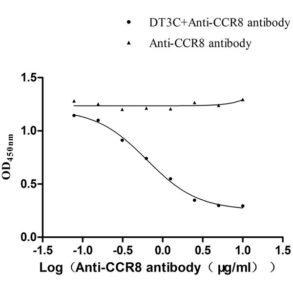
Figure 1. Representative dose-response curve of DT3C-mediated cytotoxicity. Black circles: CT26/CCR8 cells treated with DT3C (10 µg/ml) + varying concentrations of anti-CCR8 antibody. Black triangles: Cells treated with anti-CCR8 antibody only (no DT3C).
Guidelines
Principle of the Assay
DT3C consists of the catalytic and translocation domains of diphtheria toxin (DT) fused to the 3C domain of Streptococcus protein G (SPG), which binds with high affinity to the Fc region of IgG antibodies.
- Complex Formation: When mixed with anti-CCR8 antibody, DT3C binds to the antibody's Fc region, forming a stable DT3C-antibody complex.
- Target Binding: The complex specifically binds to CCR8 receptors expressed on the surface of CT26/CCR8 cells via the antibody's Fab region.
- Internalization: The receptor-bound complex is internalized via clathrin-mediated endocytosis into endosomes.
- Cytotoxicity: The acidic environment of endosomes induces a conformational change in the diphtheria toxin translocation (DT T) domain, which facilitates the translocation of the diphtheria toxin catalytic (DT C) domain into the cytoplasm. The DT C domain possesses ADP-ribosyltransferase activity, which catalyzes the ADP-ribosylation of elongation factor 2 (EF-2), thereby inhibiting protein synthesis and inducing cell death.
- Detection: The degree of cell viability reduction is positively correlated with the internalization efficiency of the antibody. Cell viability is measured using the CCK-8 assay, which detects the activity of mitochondrial dehydrogenases in viable cells. The amount of orange formazan dye produced is directly proportional to the number of living cells.
Experimental Precautions
- All cell culture operations must be performed in a Class II biological safety cabinet under strict aseptic conditions.
- Use only cells in the logarithmic growth phase with �>95% viability as determined by trypan blue exclusion.
- Do not blot dry the wells during washing steps. Always leave approximately 20µL of medium in each well to prevent cell detachment and death.
- DT3C-antibody complexes should be prepared immediately before use and not stored for long periods.
- Strictly control incubation time and temperature to ensure experimental reproducibility.
- Ensure uniform cell seeding density to avoid edge effects. Do not use the outermost wells of the 96-well plate for experimental samples; fill them with sterile PBS instead.
- Perform all experiments in triplicate to ensure statistical significance and reproducibility.
- Include appropriate positive and negative controls in each assay run to validate the results.
- Record all experimental details, including reagent lot numbers, cell passage numbers, and incubation times, for traceability.
Materials
Biological Reagents:
· Cell Line: CT26/Human CCR8 Stable Cell Line (CSB-SC004847HU3)
· Recombinant Protein: Recombinant DT3C Protein (6xHis Tag, Low Endotoxin, CSB-EP360556CQR1)
· Antibody: Anti-CCR8 Recombinant Antibody (CSB-RA004847MA3HU)
· Detection Kit: Cell Counting Kit-8 (CCK-8)
· Culture Reagents:
RPMI-1640 Medium
Fetal Bovine Serum (FBS)
Puromycin Dihydrochloride (10 mg/ml Stock Solution)
0.25% Trypsin-EDTA Solution
Phosphate-Buffered Saline (PBS)
Sterile Deionized Water
Dimethyl Sulfoxide (DMSO)mycin
Consumables:
· 96-well cell culture plates (flat-bottom, tissue culture-treated)
· 15 mL and 50 mL sterile centrifuge tubes
· 1.5 mL sterile microcentrifuge tubes
· Serological pipettes (1 mL, 5 mL, 10 mL, 25 mL)
· Pipette tips (10 μL, 20 μL, 200 μL, 1000 μL), sterile
· 0.22 μm syringe filters
· Parafilm
· Disposable gloves, lab coats, safety goggles
Equipment:
· Humidified CO₂ incubator (37°C, 5% CO₂)
· Inverted microscope for cell culture observation
· Microplate reader capable of measuring absorbance at 450 nm
· Centrifuge with swinging-bucket rotor
· Biological safety cabinet (Class II)
· Water bath (37°C)
· Pipettors (single-channel and multi-channel)
· Vortex mixer
Troubleshooting
Problem
No significant cytotoxicity in DT3C+antibody group (Possible Causes: 1. DT3C protein is inactive due to improper storage or reconstitution 2. Anti-CCR8 antibody is inactive or degraded 3. CCR8 expression on cells is too low 4. Incubation time is too short 5. Complex formation step was omitted or performed incorrectly)
Solution
1. Use a new aliquot of DT3C protein; ensure proper reconstitution and storage 2. Use a new aliquot of anti-CCR8 antibody; verify antibody activity by flow cytometry 3. Use cells with lower passage numbers (<15); verify CCR8 expression by flow cytometry before the assay 4. Ensure the 48-hour post-treatment incubation time is strictly followed 5. Confirm that the protein and antibody are pre-incubated together for 30 minutes before adding to cells.
Problem
DT3C alone control shows significant cytotoxicity (>20% cell death) (Possible Causes: 1. DT3C concentration is too high 2. Cells are non-specifically sensitive to DT3C 3. DT3C protein is contaminated)
Solution
1. Test lower DT3C concentrations (2.5, 5, 10μg/mL) 2. Verify that untransfected CT26 cells do not show cytotoxicity with DT3C alone 3. Use a new batch of DT3C protein.
Problem
High variation between replicate wells (Possible Causes: 1. Inaccurate pipetting 2. Uneven cell seeding 3. Bubbles in the wells during CCK-8 detection 4. Edge effects in the 96-well plate)
Solution
1. Use calibrated pipettors; practice precise pipetting technique 2. Mix the cell suspension thoroughly before seeding; use a multi-channel pipette for seeding 3. Avoid introducing bubbles when adding CCK-8 reagent; centrifuge the plate briefly to remove bubbles 4. Add sterile water to the peripheral wells of the plate to minimize evaporation and edge effects.
Problem
Dose-response curve is not sigmoidal (Possible Causes: 1. Antibody concentration range is too narrow 2. DT3C concentration is too low or too high 3. Data fitting is incorrect)
Solution
1. Expand the antibody concentration range to include higher and lower concentrations 2. Optimize the DT3C concentration (test 5-20 μg/mL) to achieve maximum dynamic range 3. Use a four-parameter logistic (4PL) regression model for curve fitting; ensure sufficient data points are included.
Problem
ED₅₀ value is higher than expected (Possible Causes: 1. DT3C protein has reduced activity 2. Anti-CCR8 antibody has reduced binding affinity 3. CCR8 expression on cells is decreased 4. Incubation time is too short)
Solution
1. Use a fresh aliquot of DT3C protein 2. Verify antibody binding affinity by flow cytometry or ELISA 3. Use cells with lower passage numbers; confirm CCR8 expression level 4. Extend the post-treatment incubation time to 72 hours.
Problem
High background cell death in antibody-only control group (bank wells) (Possible Causes: 1. Anti-CCR8 antibody is contaminated with endotoxin or other toxins 2. Cells are unhealthy or over-confluent before seeding 3. Washing steps are too harsh, causing cell detachment 4. Contamination with bacteria or fungi)
Solution
1. Test a different batch of anti-CCR8 antibody 2. Use cells in the logarithmic growth phase with >95% viability; seed cells at the correct density 3. Perform washing steps more gently; do not aspirate all medium from the wells 4. Check for contamination under the microscope; discard contaminated cultures and start fresh.
Safety warnings
Biological Hazard Warning: DT3C is a recombinant toxin protein derived from diphtheria toxin. Although it lacks the native receptor-binding domain, it can still cause cytotoxicity if internalized into cells. Handle DT3C with extreme caution:
- Wear appropriate personal protective equipment (PPE), including nitrile gloves, a lab coat, and safety glasses at all times.
- Avoid contact with skin, eyes, and mucous membranes.
- In case of accidental contact, flush the affected area with copious amounts of water for at least 15 minutes and seek medical attention immediately.
Cell Culture Biosafety: CT26 cells are a murine colon carcinoma cell line. Handle all cell culture materials as potentially biohazardous:
- Perform all cell culture manipulations in a Class II biological safety cabinet.
- Disinfect all work surfaces with 70% ethanol before and after use.
Chemical Hazards: DMSO is a skin irritant and can penetrate the skin, carrying other chemicals into the body. Avoid direct contact with DMSO.
Reagent Storage Warning: Store all reagents according to the manufacturer's instructions. Do not use reagents beyond their expiration dates. Avoid repeated freeze-thaw cycles of protein and antibody reagents, as this can lead to denaturation and loss of biological activity.
CCK-8 Reagent Warning: CCK-8 reagent is light-sensitive. Protect from direct light during storage and use. Do not ingest or inhale CCK-8 reagent.
Ethics statement
This protocol involves the use of animal blood samples. Users must obtain prior approval from their Institutional Animal Care and Use Committee (IACUC) or equivalent ethics committee before performing this protocol. All procedures must comply with applicable institutional and governmental regulations regarding the ethical use of animals.
Before start
Maintain strict aseptic technique throughout all cell culture steps to prevent microbial contamination. Ensure that all reagents are pre-warmed to 37°C before use to avoid thermal shock to the cells.
Pre-experiment Sample Processing
Cell Line Preparation:
- Thaw the CT26/Human CCR8 Stable Cell Line (CSB-SC004847HU3) rapidly in a 37°C water bath with gentle agitation.
- Transfer the cell suspension to a 15 mL centrifuge tube containing 9 mL of pre-warmed complete culture medium.
- Centrifuge at 1000 rpm for 5 minutes at room temperature.
- Discard the supernatant and resuspend the cell pellet in 10 mL of complete culture medium.
- Seed the cells into a T75 culture flask and incubate at 37°C in a humidified 5% CO₂ incubator.
- Passage the cells every 2-3 days when they reach 80-90% confluence using 0.25% trypsin-EDTA solution.
- Use only cells in the logarithmic growth phase (passage number < 15) for the assay to ensure consistent CCR8 expression levels.
Protein and Antibody Preparation:
Recombinant DT3C Protein (CSB-EP360556CQR1) Reconstitution and Storage:
- Centrifuge the vial briefly at 12000 rpm for 30 seconds to collect all the lyophilized powder at the bottom.
- Reconstitute the protein with 20μL sterile deionized water to a final concentration of 1 mg/mL.
- Add 20μL sterile glycerol to a final concentration of 50% for long-term storage.
- Aliquot into 5μL portions and store at -20°C to -80°C; avoid repeated freeze-thaw cycles.
Anti-CCR8 Recombinant Antibody (CSB-RA004847MA3HU) Thawing and Storage:
- Centrifuge the vial briefly at 12000 rpm for 30 seconds to collect all liquid at the bottom of the tube.
- Aliquot into single-use volumes and store at -20°C to -80°C; avoid repeated freeze-thaw cycles.
- Allow the antibody to equilibrate to room temperature for 15 minutes before use and mix gently by pipetting.
Reagent Preparation:
Complete Culture Medium:
- RPMI 1640 medium supplemented with 10% fetal bovine serum (FBS) and 10 µg/mL puromycin.
- Filter-sterilize through a 0.22 µm filter and store at 4°C for up to 1 month. Pre-warm to 37°C before use.
Serum-Free Medium:
- Use plain RPMI 1640 medium without FBS or Puromycin.
- Pre-warm to 37°C before use.
DT3C Protein Working Solution:
- Dilute the reconstituted DT3C protein in pre-warmed serum-free medium to a final concentration of 20 µg/mL.
Note: This is a 2× working solution, which will be diluted 1:1 with antibody dilutions to achieve a final DT3C concentration of 10 µg/mL.
- Prepare fresh immediately before use.
Anti-CCR8 Recombinant Antibody Dilution Series:
- Prepare a 20 µg/mL stock solution of anti-CCR8 antibody in serum-free medium.
- Perform 8-point 2-fold serial dilutions to obtain concentrations of 20, 10, 5, 2.5, 1.25, 0.625, 0.3125, and 0.156 µg/mL.
Note: These dilutions will result in final antibody concentrations of 0.078-10 µg/mL after mixing with an equal volume of DT3C solution.
CCK-8 Detection Solution:
- Thaw CCK-8 reagent at room temperature and protect from light.
- Prepare sufficient CCK-8 solution (10 µl per well) immediately before use.
Assay Procedures
Cell Seeding (Day 1):
- Aspirate the old medium from the T75 flask containing logarithmic phase CT26/Human CCR8 cells. Wash the cells twice with 5mL sterile PBS.
- Add 2mL 0.25% trypsin-EDTA solution and incubate at 37°C for 2-3 minutes until cells detach. Add 8mL complete culture medium to neutralize the trypsin.
- Transfer the cell suspension to a 15mL centrifuge tube and centrifuge at 1000rpm for 5 minutes. Aspirate the supernatant and resuspend the cell pellet in complete culture medium.
- Count the cells using a hemocytometer and adjust the cell concentration to 5×10⁴ cells/mL.
- Seed 100 µL of the cell suspension into each well of a 96-well cell culture plate (5×10⁴ cells/mL × 0.1mL/well = 5×10³ cells/well).
- Incubate the plate overnight at 37°C in a humidified 5% CO₂ incubator to allow cell attachment.
Washing Step (Day 2):
- Carefully aspirate the old medium from each well without disturbing the adherent cells.
- Wash each well twice with 200 μL of pre-warmed serum-free RPMI 1640 medium.
Critical Note: Do not blot the wells completely dry. Leave approximately 20 μL of medium in each well after each wash to prevent cells from drying and detaching.
Protein-Antibody Complex Formation (Day 2):
- In a separate 96-well plate, mix equal volumes (50 μL each) of the 2× DT3C working solution and each serial dilution of the CCR8 antibody (20, 10, 5, 2.5, 1.25, 0.625, 0.3125, and 0.156 μg/mL).
- Set up the following control groups (3 replicates per group):
· Blank control: 100μL serum-free RPMI 1640 medium (no cells)
· Cell control: 100μL serum-free RPMI 1640 medium (cells only)
· DT3C alone control: 100μL 10μg/mL DT3C in serum-free medium
· Antibody alone control: 100μL 10μg/mL CCR8 antibody in serum-free medium
- Gently mix and incubate the plate for 30 minutes at 37°C in a humidified 5% CO₂ incubator to allow antibody-DT3C complex formation.
Addition of Complexes to Cells (Day 2):
- Add 100 μL of the DT3C-antibody complexes or control solutions to the corresponding wells of the cell culture plate.
- Set up 3 replicate wells for each concentration point.
- Incubate the plate for 1 hour at 37°C in a humidified 5% CO₂ incubator to allow antibody binding and complex internalization.
Post-Incubation Washing and Culture (Day 2):
- Aspirate the medium containing unbound complexes from each well.
- Wash each well twice with 200 μL of pre-warmed serum-free RPMI 1640 medium, again leaving ~20 μl of medium per well.
- Add 200 μL of pre-warmed complete culture medium to each well.
- Incubate the plate for 48 hours at 37°C in a humidified 5% CO₂ incubator to allow DT3C-mediated cytotoxicity to occur.
Cell Viability Detection (Day 4):
- Carefully aspirate 100μL of supernatant from each well, leaving 100μL of medium in the wells.
- Add 10 μL of CCK-8 solution to each well.
- Gently tap the plate to mix the reagent evenly and avoid air bubbles.
- Incubate the plate for 4 hours at 37°C in a humidified 5% CO₂ incubator (in the dark).
Absorbance Measurement (Day 4):
- Immediately place the plate in a microplate reader.
- Measure the absorbance at 450 nm (use 630 nm as a reference wavelength if available).
- Record all absorbance values for data analysis.
Result Analysis
Data Normalization:
- Calculate the average absorbance value for each set of triplicate wells.
- Calculate cell viability using the following formula:
Cell Viability (%) = [(OD450 of experimental group - OD450 of blank control) / (OD450 of cell control - OD450 of blank control)] × 100%
- Calculate the average cell viability and standard deviation for each group.
Dose-Response Curve Generation:
- Plot the logarithm of the anti-CCR8 antibody concentration (µg/mL) on the x-axis and the corresponding cell viability (%) on the y-axis.
- Use a four-parameter logistic (4PL) regression model to fit the dose-response curve.
- The curve should show a sigmoidal shape with decreasing cell viability as antibody concentration increases for the DT3C+antibody group.
ED50 Calculation:
- Determine the ED50 value from the fitted curve, which represents the anti-CCR8 antibody concentration required to kill 50% of the cells in the presence of 10 µg/ml DT3C.
- The expected ED50 range for this assay is 0.4661-0.8614 µg/mL as validated by CUSABIO.
Result Interpretation:
Figure. Representative dose-response curve of DT3C-mediated cytotoxicity. Black circles: CT26/CCR8 cells treated with DT3C (10 μg/ml) + varying concentrations of anti-CCR8 antibody. Black triangles: Cells treated with anti-CCR8 antibody only (no DT3C).
- A significant dose-dependent decrease in cell viability in the DT3C+antibody group confirms specific antibody-mediated internalization and DT3C cytotoxicity.
- No significant cytotoxicity (cell viability >90% at all concentrations) in the antibody-only control group indicates that the anti-CCR8 antibody itself has no intrinsic cytotoxic activity.
- Confirm that the DT3C-only control group shows minimal cytotoxicity (cell viability ≥90%).
- If the ED₅₀ value falls within the expected range, the assay is considered valid, and the DT3C protein is confirmed to be biologically active.
Protocol references
This assay is based on an antibody-dependent cellular cytotoxicity (ADCC)-like mechanism using a recombinant DT3C fusion protein.
○ Products:
Recombinant DT3C (Diphtheria toxin & spg 3C domain) for Antibody Internalization Assay (CSB- EP360556CQR1): purity > 90% as determined by SDS-PAGE and SEC-HPLC; endotoxin < 1.0 EU/ug as determined by LAL method; biological active
CCR8 Recombinant Monoclonal Antibody (CSB-RA004847MA3HU): ELISA, FC validated
CT26/Human CCR8 Stable Cell Line (CSB-SC004847HU3): high expression of CCR8 protein