Apr 21, 2026
CD274 Recombinant Monoclonal Antibody (CSB-RA977797A0HU) Western Blot Validation Protocol
- Rosie Liu1
- 1CUSABIO
- CUSABIO TECHNOLOGY LLC


Protocol Citation: Rosie Liu 2026. CD274 Recombinant Monoclonal Antibody (CSB-RA977797A0HU) Western Blot Validation Protocol. protocols.io https://dx.doi.org/10.17504/protocols.io.36wgqxzyolk5/v1
License: This is an open access protocol distributed under the terms of the Creative Commons Attribution License, which permits unrestricted use, distribution, and reproduction in any medium, provided the original author and source are credited
Protocol status: Working
We use this protocol and it's working
Created: April 20, 2026
Last Modified: April 21, 2026
Protocol Integer ID: 315418
Keywords: CD274 Recombinant Monoclonal Antibody; PD-L1; Western Blot Validation; WB, cd274 recombinant monoclonal antibody, endogenous cd274 protein expression, human cd274 protein, analytical performance of cusabio cd274, recombinant monoclonal antibody, cusabio cd274, routine wb detection, reliable experimental conditions for routine wb detection, western blot validation protocol, derived cell line
Abstract
This standardized Western Blot (WB) protocol is specifically designed to validate the specificity, reactivity, and analytical performance of CUSABIO CD274 (PD-L1) Recombinant Monoclonal Antibody (Cat. No. CSB-RA977797A0HU). It aims to detect endogenous CD274 protein expression in four human-derived cell lines (HepG2, K562, A549, HeLa) and establish reliable experimental conditions for routine WB detection of human CD274 protein using a semi-dry transfer system and chemiluminescent detection.
Guidelines
Principle: Western blotting separates proteins by molecular weight via SDS-PAGE, transfers them onto a PVDF membrane, and uses specific enzyme-labeled antibodies to detect the target protein.
For CD274 antibody validation
1. SDS-PAGE Separation: Proteins are denatured by SDS and separated based on their molecular weight through polyacrylamide gel electrophoresis. CD274 (PD-L1) is a transmembrane glycoprotein with a core molecular weight of ~33 kDa. Its predominant glycosylated form migrates as a diffuse band at 45-55 kDa.
2. Semi-dry Transfer: Separated proteins are electrophoretically transferred from the gel to a 0.45 μm PVDF membrane, which provides a stable solid support for subsequent antibody binding. Methanol activation of PVDF increases its hydrophobicity and protein binding capacity.
3. Blocking: Non-specific protein binding sites on the membrane are blocked with 5% non-fat milk to reduce background noise.
4. Immunodetection: The membrane is incubated with the primary antibody (CSB-RA977797A0HU) that specifically recognizes CD274, followed by an HRP-conjugated secondary antibody that binds to the primary antibody.
5. Chemiluminescence Detection: HRP catalyzes the oxidation of luminol in the presence of H2O2, producing light that is detected by a chemiluminescence imaging system. The intensity of the light signal is proportional to the amount of target protein present.
Key Notes and Precautions
1. Reagent Preparation: All reagents should be prepared with ultrapure deionized water and stored at appropriate temperatures. Prepare working solutions fresh immediately before use unless otherwise specified.
2. Temperature Control: All sample handling and storage steps must be performed at 4ºC or on ice to prevent protein degradation. Incubations at room temperature should be strictly timed.
3. Gel Running: Start at 80 V until the dye front enters the resolving gel, then increase to 120 V. Monitor bromophenol blue migration to avoid over-running.
4. Buffer Consistency: Use the same batch of buffers throughout the experiment to ensure reproducibility. Check the pH of all buffers before use.
5. Membrane Handling: Always handle PVDF membranes with clean tweezers only at the edges. Avoid touching the protein-binding surface to prevent contamination and fingerprints.
6. Membrane Activation: Pre-wet PVDF membrane in methanol (or alcohol solution) for 1 min, then equilibrate in transfer buffer. This step is critical for protein binding.
7. Air Bubble Elimination: Ensure no air bubbles are trapped between the gel and membrane during transfer, as this will result in blank areas on the blot.
8. Blocking: Incubate with 5% milk in PBS at 25 ºC for 1 h with gentle shaking. Do not exceed 1 h to avoid masking epitopes.
9. Complete Solution Removal: Aspirate all solutions completely after each incubation and washing step to prevent dilution of subsequent reagents.
10. Gentle Shaking: Perform all incubations with gentle orbital shaking (50-70 rpm) to ensure uniform reagent distribution and prevent membrane drying.
11. Antibody Storage: Aliquot antibodies into small volumes and store at -20ºC. Avoid repeated freeze-thaw cycles, which significantly reduce antibody activity.
12. Negative Control: Always include a negative control lane where the primary antibody is omitted to assess non-specific binding of the secondary antibody.
13. Housekeeping Protein: Load equal amounts of total protein and include a housekeeping protein (β-actin, GAPDH) for normalization of loading and transfer variations.
14. Chemiluminescence: Apply substrate evenly and image immediately for optimal signal-to-noise ratio.
15. Membrane Handling: Handle membranes with clean forceps, and avoid touching the membrane surface. Use an orbital shaker at 50–100 rpm for all incubation steps.
Materials
Biological Samples
- HepG2 human liver cancer cell lysate
- K562 human chronic myeloid leukemia cell lysate
- A549 human lung adenocarcinoma cell lysate
- HeLa human cervical cancer cell lysate
- Pre-stained protein ladder
Antibodies and Reagents
- CD274 Recombinant Monoclonal Antibody (CUSABIO, Cat. No. CSB-RA977797A0HU)
- Peroxidase AffiniPure® Goat Anti-Rabbit IgG (H+L)
- Non-fat milk powder
- Tris, glycine, SDS, Methanol (HPLC grade)
- Cell lysis buffer (RIPA)
- Laemmli sample buffer, β-mercaptoethanol
- Protease inhibitor cocktail
- Phosphatase inhibitor cocktail
- 5× SDS-PAGE loading buffer
- Luminol-based HRP chemiluminescent substrate
Instruments and Consumables
- SDS-PAGE electrophoresis system
- Semi-dry transfer apparatus
- Chemiluminescence imaging system
- Incubator shaker (4ºC and RT)
- Refrigerated microcentrifuge
- Vortex mixer
- Heating block (95-100ºC)
- Plastic incubation boxes or trays
- Cell scraper
- 1.5 mL microcentrifuge tubes
- Pipettes and filtered pipette tips
- SDS-PAGE gel (8–12% acrylamide, 1.0 mm thickness)
- PVDF membrane (0.45 μm)
- Transfer filter paper (8.5 cm × 5.2 cm)
Troubleshooting
Problem
No visible bands (Cause: Insufficient protein loading;Incorrect antibody dilution;Transfer failure;Target protein not expressed in tested cells; Expired chemiluminescent substrate)
Solution
Increase loading to 40-60 μg per well; Optimize primary antibody dilution (1:500-1:2000); Confirm membrane activation, check transfer stack orientation, and extend transfer time to 60-90 min; Include a CD274-positive control cell line; Prepare fresh substrate working solution.
Problem
Faint or smeared bands (Cause: Over- or under-cooked samples; Gel percentage inappropriate;Uneven transfer;Excessive washing)
Solution
Denature samples at 95 °C for exactly 5 min and avoid boiling; Use a 10–12% gel for 33–40 kDa proteins and optimize the acrylamide concentration; Remove all bubbles from the transfer stack; ensure the filter papers are saturated; Reduce the number of washes to 3 × 10 min.
Problem
High background (Cause: Inadequate blocking; Too high antibody concentration; Insufficient washing; Non-specific secondary antibody binding)
Solution
Block for 2 h at RT or overnight at 4°C. Reduce primary/secondary antibody concentration. Increase washing time to 15 min per wash. Use a human serum-adsorbed secondary antibody.
Problem
Non-specific bands (Cause: Excess primary antibody; Non-fat milk cross-reactivity; Protein degradation; Over-transfer)
Solution
Dilute primary antibody to 1:2000-1:5000; Use 5% BSA in PBS as blocking buffer; Add fresh protease inhibitors to lysis buffer and avoid repeated freeze-thaw cycles; Reduce transfer time to 30 min and use lower current (40 mA per gel).
Problem
Uneven signal (Cause: Uneven substrate application; Membrane contamination; Inconsistent shaking during incubation)
Solution
Pipette substrate evenly over the entire membrane; avoid air bubbles. Wear gloves, use clean forceps, and avoid touching the membrane surface. Use an orbital shaker at 50–100 rpm for all incubation steps.
Safety warnings
1. Chemical Safety: Methanol is flammable and toxic. Handle only in a fume hood wearing nitrile gloves, safety goggles and lab coat. Avoid inhalation, skin contact and ingestion. SDS powder is an irritant—wear a mask and gloves when weighing.
2. Biological Safety: All cell and lysate handling must be performed in a biosafety cabinet. Dispose of biological waste according to institutional regulations.
3. Antibody Stability: Aliquot antibodies and store at -20ºC. Avoid repeated freeze-thaw cycles, which will significantly reduce antibody activity.
4. Light Sensitivity: Chemiluminescent substrate is light-sensitive. Prepare and use in the dark. Do not expose to light for prolonged periods.
5. Membrane Handling: Always handle PVDF membrane with clean tweezers only at the edges to avoid protein contamination and fingerprints.
6. Electrical Safety: Ensure proper grounding of electrophoresis and transfer apparatus. Do not touch electrodes while power is on.
Before start
All sample preparation steps should be performed on ice to prevent protein degradation.
Pre-experiment Sample Processing
Cell Culture
Culture HepG2 (human liver cancer), K562 (human chronic myeloid leukemia), A549 (human lung adenocarcinoma), and HeLa (human cervical cancer) cells in their respective complete culture media at 37ºC with 5% CO₂ in a humidified incubator. Harvest cells when they reach 80-90% confluence (adherent cells) or logarithmic growth phase (suspension cells).
Cell Harvest
Adherent cells: Aspirate culture medium, wash twice with pre-chilled 1× PBS. Add pre-chilled RIPA lysis buffer supplemented with protease inhibitors (1:100) and phosphatase inhibitors (1:100). Scrape cells gently and transfer the suspension to pre-chilled 1.5 mL microcentrifuge tubes.
Suspension cells: Collect by centrifugation at 1000 × g for 5 min at 4ºC. Discard supernatant and resuspend the cell pellet in the same lysis buffer as above.
Cell Lysis
Lyse cells using RIPA buffer supplemented with protease and phosphatase inhibitors. Incubate samples on ice for 30 min, gently vortex every 10 min to ensure complete lysis, then centrifuge at 12,000 × g for 15 min at 4 °C to collect the supernatant.
Protein Quantification
Centrifuge lysates at 12000 × g for 15 min at 4ºC to remove cell debris. Transfer supernatants to new pre-chilled tubes. Determine protein concentration of each lysate using a Bradford, BCA, or Lowry assay.
Sample Preparation
Dilute lysates to equal protein concentrations (e.g., 20–40 µg per lane) in Laemmli sample buffer (containing SDS and β-mercaptoethanol). Mix with 5× SDS-PAGE loading buffer at a 4:1 volume ratio. Heat at 95-100ºC for 5 min to denature proteins, cool immediately on ice, and centrifuge briefly before loading.
Loading
Load equal amounts of each lysate into separate wells of an SDS-PAGE gel alongside a pre-stained protein ladder.
Reagent Preparation
10× PBS (pH 7.4): Dissolve 80 g NaCl, 2 g KCl, 14.4 g Na₂HPO₄ and 2.4 g KH₂PO₄ in 800 mL water. Adjust pH to 7.4 with HCl/NaOH, bring volume to 1 L. Autoclave and store at room temperature (RT). Dilute to 1× before use.
10× PBST (0.5% Tween-20 in 10× PBS): Add 5 mL Tween-20 to 1 L 10× PBS, mix thoroughly. Store at RT. Dilute to working concentrations:
- 0.05% PBST: 1:20 dilution with water
- 0.1% PBST: 1:10 dilution with water
10× SDS-PAGE Electrophoresis Buffer (Running Buffer): Dissolve 30.3 g Tris, 144 g glycine, and 10 g SDS in 800 mL water. Bring volume to 1 L. Store at RT. Dilute to 1× before use.
10× Semi-dry Transfer Buffer: Dissolve 30.3 g Tris and 144 g glycine in 800 mL water. Add 200 mL of methanol, bring volume to 1 L. Store at 4°C. Dilute to 1× before use.
Blocking Buffer: 5% (w/v) non-fat milk powder in 1× PBS (pH 7.4).
Antibody Dilution Buffer: 2.5% (w/v) non-fat milk powder in 0.05% PBST (PBS + 0.05% Tween‑20, for both primary and secondary antibodies).
Wash Buffer: 0.1% PBST (PBS + 0.1% Tween‑20).
Primary Antibody Working Solution: Dilute CD274 Recombinant Monoclonal Antibody (CSB-RA977797A0HU) at 1:1000 in antibody dilution buffer. Prepare 10 mL per incubation box.
Secondary Antibody Working Solution: Dilute Peroxidase AffiniPure® Goat Anti-Rabbit IgG (H+L) at 1:50000 in antibody dilution buffer.
HRP Chemiluminescent Substrate: Prepare Luminol/H₂O₂ working solution according to the manufacturer's instructions immediately before detection.
Assay Procedures
Sample preparation and running the Gel
Sequentially, add the HepG2 cell lysate, K562 cell lysate, A549 cell lysate, and HeLa cell lysate to the wells. Run the gel. Add the electrophoresis buffer after the samples are added. The running conditions are as follows: at a low voltage of 80V for approximately 30 minutes, then adjust the voltage to 120V for electrophoresis for about 90 minutes. The actual time should be based on when the bromophenol blue indicator reaches the bottom of the gel.
Transfer
Cut the transfer filter paper (8.5cm x 5.2cm) and the 0.45 μm PVDF membrane in advance. Activate the PVDF membrane with an alcohol solution and then immerse it in the transfer buffer. Remove the backing, place the sandwich, with 3 sheets of filter paper on each side, and sandwich the gel and the membrane in between. Eliminate air bubbles for each layer. After preparing, the transfer conditions are as follows: semi-dry transfer, 60mA current for each piece of gel, constant current for 40-60 minutes.
Blocking
Place the incubation box containing the PVDF membrane in a blocking buffer (5% non-fat milk powder in PBS) and let it incubate at 25ºC for 1 hour. After blocking, aspirate the solution in the box.
Adding antibody
Add 10ml CD274 Recombinant Monoclonal Antibody (CSB-RA977797A0HU) by 2.5% non-fat milk powder in 0.05% PBST at 1:1000 to each box, and seal the plate with sealing film. Then incubate at 4ºC for overnight.
Washing
Remove the protein solution by decanting or aspirating. Make sure that the solution in the box is completely removed. Then wash the PVDF membrane with 10ml/box 0.1%PBST for 4 times for 10 mins and aspirate the solution.
Adding detection antibody
Dilute the Peroxidase AffiniPure® Goat Anti-Rabbit IgG (H+L) by 2.5% non-fat milk powder in 0.05% PBST at 1:50000, and add it to the incubation box. Shake and incubate at 25ºC for 2 hours.
Washing
Repeat step 5.
Chemiluminescence
Luminol, which is one of the most classical HRP chemiluminescent substrates, can generate enzyme catalysis reaction with horseradish peroxidase in the presence of H_2O_2. It has high sensitivity and good imaging characteristic, and can be directly imaged and developed using a chemiluminescence device.
Result Analysis
Observed Band Characteristics
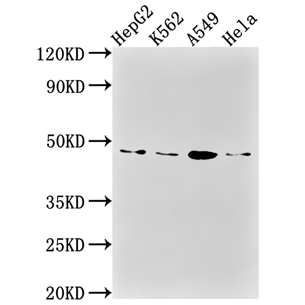
Consistent Specific Band: A clear, distinct immunoreactive band at 45 kDa is consistently detected in all tested cell lines, including HepG2, K562, A549, and HeLa.
Glycosylated Isoform Confirmation: The observed band size of 45 kDa aligns with the reported molecular weight of the glycosylated form of CD274/PD-L1 (40-55 kDa is a common position for its main glycosylated form), consistent with the predicted core sequence (21-21 kDa) undergoing post-translational modification.
Uniform Expression Across Panel: The intensity of the 45 kDa band is relatively consistent across HepG2, K562, A549, and HeLa cell lysates, indicating broad expression of CD274 in these cell lines.
Validation of Antibody Performance
Specificity: The absence of non-specific bands or background smearing in any lane confirms the high specificity of the CD274 Recombinant Monoclonal Antibody (CSB-RA977797A0HU) for its target antigen. No cross-reactivity with unrelated proteins was observed.
Sensitivity: Successful detection at a primary antibody dilution of 1:1000 demonstrates excellent analytical sensitivity of the antibody, enabling reliable identification of endogenous CD274 even in standard cell lysate preparations.
Assay Reproducibility: The uniformity of band intensity across replicate lanes and different cell lines highlights the high reproducibility of the developed Western Blot protocol.
Result Interpretation
CD274 (PD-L1) Recombinant Monoclonal Antibody (Cat. No. CSB-RA977797A0HU) exhibits exceptional performance in Western Blot applications, characterized by high specificity, strong sensitivity, and consistent signal intensity. The antibody reliably detects the endogenous glycosylated form of CD274 at the expected molecular weight (45 kDa) in multiple human cell lines, thereby fulfilling the objective of validating the antibody for routine CD274 detection.
Positive WB detected in: HepG2 whole cell lysate, K562 whole cell lysate, A549 whole cell lysate, HeLa whole cell lysate
All lanes: CD274 Antibody at 1:1000
Secondary Goat polyclonal to rabbit IgG at 1/50000 dilution
Predicted band size: 34, 21, 21 kDa
Observed band size: 45 kDa
Protocol references
○ Product Name: CD274 Recombinant Monoclonal Antibody
○ Catalog Number: CSB-RA977797A0HU
○ Target Species: Homo sapiens (Human)
○ Target Analyte: CD274 (PD-L1)