Mar 21, 2024
Cassiopea xamachana Cellular Dissociation
Forked from a private protocol
- 1University of Miami;
- 2Pennsylvania State University;
- 3Texas A&M University
- Cassiopea protocolsTech. support phone: +1 (814) 321-5684 email: [email protected]


Protocol Citation: Anthony Bonacolta, Victoria Sharp, Marta Mammone 2024. Cassiopea xamachana Cellular Dissociation. protocols.io https://dx.doi.org/10.17504/protocols.io.3byl4qnnovo5/v1
License: This is an open access protocol distributed under the terms of the Creative Commons Attribution License, which permits unrestricted use, distribution, and reproduction in any medium, provided the original author and source are credited
Protocol status: Working
We use this protocol and it's working
Created: February 09, 2024
Last Modified: March 21, 2024
Protocol Integer ID: 94965
Keywords: dissociation, cassiopea, cytometry, calcein, cnidaria, cnidarian, enzyme, fluorescence, cassiopea xamachana cellular dissociation this protocol, cassiopea xamachana cellular dissociation, cassiopea xamachana cells for cell, cassiopea xamachana cell, viable cell, cassiopea xamachana, cell, lyse cell, scrna
Abstract
This protocol is to optimized to dissociate and fix Cassiopea xamachana cells for cell sorting and scRNA-seq.
The dissociation by itself results in 53-55% of viable cells.
Cells cannot be sorted without fixative, unless your machine can sort a seawater solution. Any other solution will lyse cells.
Guidelines
Make sure to work in an RNAse free-environment when able to. Use RNAse-ZAP or work in a UV sterilized hood if possible.
The tissue should be less than 1 cm long
Materials
- Sterile razors
- Sterile forceps
- Ice
- Wide-bore pipette tips, or cut 1000 uL pipette tips
- 15 mL tube for digestion buffer
- 2 petri dishes to wash and incubate tissue in Ca2+ and Mg 2+ free seawater
- 2 70-um filters
- 2 mL microcentrifuge tubes
- pipettes
Equipment:
microcentrifuge
rocking plate
Reagents:
NaCl
KCl
NaSO4
NaHCO3
Dispase II
Liberase
L-cysteine
PBS
BSA
DNase/RNase-free distilled water
methanol
glacial acetic acid
glycerol
RNAse Inhibitor
Before start
Treat reagents and materials with UV-light for ~15 mins before beginning protocol.
Set 15 mL and microcentrifuge to 4° C.
Prepare Reagents:
Ca2+ Mg2+ free seawater (Roger et al. 2021)
To 1 L Distilled Water add:
- 23 g NaCl
- 0.763 g KCl
- 3 g NaSO4
- 0.25 g NaHCO3
- Dissociation Mix:
To Ca2+ Mg2+ free ASW add:
- 3.6 mg/mL Dispase II
- 0.25 mg/mL of Liberase
- 4% L-cysteine
- 1x PBS 0.5% bovine serum albumin (BSA)
- Add 0.25 g to 50 mL 1x PBS.
- Fresh ACME Solution
- 13:3:3:2 ratio of DNase/RNase-free distilled water, methanol, glacial acetic acid, and glycerol
-Prep about 15 mL, FRESH, per sample each time
- RNAse Inhibitor
Dissociation
1h
Cut the jellyfish tissue with a sterile razor to encourage permeability of reagents.
2m
Gently wash the jellyfish tissue in 10 mL Ca-Mg-Free SW for 00:01:00 then transfer to fresh 10 mL Ca-Mg-Free SW and let incubate at Room temperature for 00:02:30 .
3m 30s
Using sterilized forceps, place the jelly tissue into a clean 15 mL tube then add 1 mL dissociation mix on top of the jelly, or enough to submerge the tissue.
30s
Incubate the tissue on a rocker for 00:30:00 at room temperature.
30m
Pipette up and down using a wide-bore tip 10 times.
5m
Repeat steps 4 and 5.
35m
Add 80 µL fetal bovine serum to the cell suspension to create a 8% FBS solution to halt enzyme digestion.
2m
After the incubation period, filter the sample through a 70 µm filter Keep sample On ice moving forward.
2m
Resuspend in 1000 µL Ca-Mg-free SW . Gently pipette up and down 10 times with a wide-bore tip to dissociate clumps.
2m
Staining and Fixation
Add 1 micromolar (µM) Calcein Violet 450 AM to cells. Incubate in the dark for 00:30:00 .
30m
After the incubation period, filter the sample through a 70 µm filter
5m
Resuspend in 500 µL ACME . Incubate for 00:30:00 .
35m
Centrifuge 350 x g, 4°C, 00:07:00 then carefully discard the supernatant.
7m
Re-suspend the pellet in 800 µL 1x PBS w/ 1% BSA using gentle pipetting with a wide-bore pipette then add 1 µL RNA Inhibitor .
1m
FACS & Cryopreservation
Pre-chill chambers of FACS Machine to 4 °C
5m
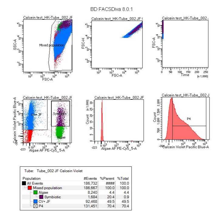
Sort at the slowest rate (High-purity) with less than 50 PSI at 4 °C . Gate for Calcein Violet (450 nm) and chlorophyll autofluorescence (650-700 nm) for viable jelly cells and symbiont cells.
2h
After sorting, cryopreserve by adding 10 % volume DMSO and 1 µL RNA Inhbitor and immediately putting the sample at -80 °C .
1m
Thaw and Sample Submission
38m
Thaw the sample On ice
30m
Centrifuge 350 x g, 4°C, 00:07:00 then carefully remove the supernatant.
7m
Re-suspend the cells in 1 mL 3.3x PBS w/ 1% BSA then add 1 µL RNA Inhbitor .
1m
Submit to sequencing center On ice .
Protocol references
Roger LM, Reich HG, Lawrence E, Li S, Vizgaudis W, Brenner N, et al. (2021) Applying model approaches in non-model systems: A review and case study on coral cell culture. PLoS ONE 16(4): e0248953. https://doi.org/10.1371/journal.pone.0248953