Mar 21, 2020
Brooks Lab Western Blotting Protocol
- Brooks Lab University of California1,
- Eva Robinson1,
- Alison Tang1
- 1University of California, Santa Cruz
- BrooksLabUCSC


External link: https://doi.org/10.1186/s13059-024-03301-y
Protocol Citation: Brooks Lab University of California, Eva Robinson, Alison Tang 2020. Brooks Lab Western Blotting Protocol. protocols.io https://dx.doi.org/10.17504/protocols.io.bcsmiwc6
Manuscript citation:
Tang AD, Felton C, Hrabeta-Robinson E, Volden R, Vollmers C, Brooks AN (2024) Detecting haplotype-specific transcript variation in long reads with FLAIR2. Genome Biology 25(). doi: 10.1186/s13059-024-03301-y
License: This is an open access protocol distributed under the terms of the Creative Commons Attribution License, which permits unrestricted use, distribution, and reproduction in any medium, provided the original author and source are credited
Protocol status: Working
We use this protocol and it's working
Created: February 21, 2020
Last Modified: March 21, 2020
Protocol Integer ID: 33325
Keywords: western blot, western blotting, protein expression, proteins, brooks lab western blotting protocol, western blotting protocol for brooks lab, western blotting protocol, brooks lab, department of biomolecular engineering, protocol, biomolecular engineering,
Abstract
This is a Western Blotting Protocol for Brooks Lab, Department of Biomolecular Engineering, University of California, Santa Cruz.
Attachments

Materials
MATERIALS
Pierce BCA Protein Assay KitThermo Fisher ScientificCatalog #23225
4–15% Mini-PROTEAN® TGX™ Precast Protein Gels, 10-well, 30 µl Bio-Rad LaboratoriesCatalog #4561083
Pierce RIPA Lysis and Extraction BufferThermo Fisher ScientificCatalog #P189900
cOmplete™ Mini Protease Inhibitor Cocktail (Roche)Merck MilliporeSigma (Sigma-Aldrich)Catalog #04693124001
4x Laemmli Sample Buffer Bio-Rad LaboratoriesCatalog #1610747
2-Mercaptoethanol Bio-Rad LaboratoriesCatalog #1610710
Precision Plus Protein™ Dual Color Standards 500 µl Bio-Rad LaboratoriesCatalog #1610374
Precision Plus Protein™ Dual Color Standards 500 µl Bio-Rad LaboratoriesCatalog #1610374
Western Blot Box (black size 8.9 cm × 6.5 cm × 2.5 cm)Merck MilliporeSigma (Sigma-Aldrich)Catalog #Z742099-5EA
β-Actin Antibody (C4)Santa Cruz BiotechnologyCatalog #sc-47778
SuperSignal™ West Pico PLUS Chemiluminescent SubstrateThermo Fisher ScientificCatalog #34577
Note: You can use Pierce BCA Protein Assay Kit #23225 or #23227
Additional Reagents required:
- 100 mM Tris-Cl, pH 8.0
- 300 mM NaCl
- 10% NP-40 in ddH2O
- 10% Na-deoxycholate in ddH2O (light sensitive!)
- 10% SDS
- 10x PBS
- Tween-20
- 190 proof ethanol
- 10x TGX running buffer
- Safeway brand non-fat milk pouch
- PBST
- BioRad Transblot kit
- LoBind microcentrifuge tubes Protein, 1.5 ml
Additional Equipment
- Sonicator or Bioruptor
- Microplate reader
- 200 μl pipettor
- Varioskan
- BioRad Gel Doc EZ Gel Documentation System
- BioRad ChemiDoc XRS+
Troubleshooting
Safety warnings
Please refer to the Safety Data Sheets (SDS) for safety and environmental hazards.
RIPA Lysis Buffer
Make 100 mL RIPA w/o protease inhibitor , cover, and freeze in 10 ml aliquots at -20 °C .
OR
Use pre-made Pierce RIPA buffer, 100 ml, which is aliquoted in 10 mL aliquots and stored in -20 °C
Before use, thaw and add 1 tablet of protease inhibitor (PI) per 10 ml aliquot. (Roche cOmplete™, Mini Protease Inhibitor Cocktail).
Harvesting Cells and Preparing Lysate
Note
Always keep everything on ice, unless otherwise indicated.
Wash confluent 10 cm plate of cells 2X in ice cold PBS.
Add 1 mL cold RIPA with PI to cells, and scrape the cells to remove them from the dish. Transfer to a pre-chilled 2 ml tube On ice .
Incubate On ice 00:00:20 , with periodic vortexing.
Pellet the insoluble material by spinning at max speed in refrigerated (4 °C ) microcentrifuge 00:00:10 .
Transfer supernatant to a clean LoBind Protein 1.5 ml tube as lysate. To avoid multiple freeze-thaw cycles, make aliquots (generally 200 µL each).
Note
Additional Sonicator or BioEruptor sonication step often needed here for more complete nucleic acid removal. See below.
Store lysates aliquots at -80 °C .
Sonication of Lysate to break up nucleic acids
Place samples in a 1.5 ml LoBind Protein microfuge tube and prepare a volume balance tube.
Bring a timer, ear cuffs, On ice samples, and balance to second floor to Kamakaka Lab to get the plastic tube adaptor (in back of second drawer in 2nd bay from the back of the lab.)
Sonicator is in cold room down the hall. Put the outflow tube up on the shelf and fill glass reservoir with water to top of white line.
Sonicator settings: timer on hold, use max setting (10), constant, then flip on.
Hold tubes in the adaptor in the sonicating water bath for 00:00:30 .
Place tubes On ice to cool for 00:01:00 .
Repeat the 30’ sonication (= total of two rounds on max at 00:00:30 .)
BCA Assay for Protein Concentration Determination
Pierce BCA Protein Assay Kit #23225 or #23227, Thermo Sci with 2 mg/ml BSA standard.
Use instructions from kit.
Equilibrate reagents, samples, and standards to Room temperature . Use the Microplate reader instructions.
Prepare a dilution series of BSA in working range of 20 μg/ml – 2000 μg/ml from 1 glass vial of stock 2 mg/ml BSA in the kit for the standard curve:
| Vial | RIPA buffer, μl | 2 mg/ml BSA or standard, μl | Final con μg/ml | |
| A | 0 | 300 of stock | 2000 | |
| B | 125 | 375 of stock | 1500 | |
| C | 325 | 325 of stock | 1000 | |
| D | 175 | 175 of B | 750 | |
| E | 325 | 325 of C | 500 | |
| F | 325 | 325 of E | 250 | |
| G | 325 | 325 of F | 125 | |
| H | 400 | 100 of G | 25 | |
| I | 400 | 0 | 0 |
Prepare fresh working reagent 50:1 of A:B, enough for all standards, unknowns, and replicates of the unknowns (should have n=3).
Add 25 µL of unknown or standard to 96-well plate well followed by 200 µL working reagent using 200 μl pipettor and gently mix avoiding spillover.
Cover plate with foil and incubate 37 °C for 00:30:00 in shaking incubator, gently shaking.
Cool to Room temperature .
Measure Absorbance on the VarioSkan at 562 nm.
Determine protein concentration of your unknown from the standard curve (the Skanit software will plot the curve and the unknowns on it if you edit the standards in the plate layout to include the concentrations above in the table.)
Export report as an excel doc.
SDS PAGE
Prepare the following solutions for SDS-PAGE, Transfer, and Antibody Incubations:
| Running Buffer (Tris-Glycine) | ||
| 100 ml | 10x TGX running buffer | |
| 900 ml | dd water |
| Transfer Buffer | ||
| 200ml | 5x Biorad Transfer Buffer | |
| 200ml | 190 proof ethanol | |
| 600ml | dd water |
| PBST | ||
| 50 ml | 10x PBS | |
| 0.5 ml | Tween-20 | |
| bring vol to 500 ml with dd water | ||
| 5 % Milk Block (prepare immediately before use!) | ||
| 5 g | Safeway brand non-fat milk pouch | |
| 100 ml | PBST |
SDS PAGE: Denaturing samples in Laemmli reducing buffer
In a fume hood, Add 100 µL 2-mercaptoethanol to 900 µL 4x Laemmli sample buffer to make MLB.
Dilute samples with MLB at a ratio of 3 parts sample to 1 part MLB. So for 30 µL final volume, add 7.5 µL MLB to 22.5 µL sample in RIPA in 1.5 ml LoBind safety lock tube.
Denature at 95 °C for 00:05:00 .
Transfer to ice.
SDS PAGE: Loading and Running Gel
Remove gel from package, take off green strip at the bottom, and rinse the wells three times with about 1 mL running buffer (use 1 ml pipettor).
Place gel, tall plate facing out in outer side of a holder and the reservoir block at the other to make a running buffer reservoir. Put the other gel holder in place to take up space.
Fill the chamber to the 2-gel mark. Make sure there are no leaks before testing the circuit. Test the circuit by checking for bubbles after turning on power to 70 volts.
Using a 20 ul conte-tipped pipettor, load 30 µL of the samples in MLB onto gel, as well as 10 µL of Biorad Precision Plus protein ladder Dual Color.
Run at 100 – 120V, depending on desired resolution, until adequate separation of ladder lanes in sizes regions of interest.
Photograph the gel after the run with your phone to help keep track of orientation.
Semi-dry Transfer to PVDF Membrane
Pre-soak blotting stacks in 1X transfer buffer made according to directions on the bottle.
Pre-wet PVDF membrane from Biorad Transblot kit in 190 proof ethanol, then in transfer buffer.
Layer into Transblot drawer (stack, membrane, gel, second stack) before rolling out bubbles gently with conical tube
Run the transfer on Biorad setting, standard Mininigel TGX, 25mA, 25V, 00:03:00 .
While running, pour 5% milk block in 1X PBST directly into cleaned small black Western blot box.
Incubate transferred blot for 01:00:00 at Room temperature in 5% milk block in 1X PBST.
Photograph the blot after the transfer to help keep track of orientation.
Antibody Incubations and ECL visualization of target protein bands: Primary and secondary antibody binding
Primary and secondary antibody binding
- β-Actin (C4): sc-47778
- ECL SuperSignal West Pico Chemiluminescent Substrate
Use 1:500 Dilutions for primary antibodies, in 5% milk/PBST
Note
Note: Primary antibody (unconjugated with HRP) can be re-used 3x if kept frozen.
Drain milk block from blots and add primary Ab (in milk block).
Incubate in cold room Overnight covered and shaking on orbital shaker, covered with plastic.
The next day, wash blot 3x/00:15:00 in PBST.
Freshly prepare secondary antibody at 1:10,000 dilution (2 µL to 20 mL 5% milk/PBST ).
Incubate in secondary antibody for 01:00:00 , Room temperature .
Discard secondary and wash blot 3x/00:05:00 in 1xPBST.
Discard secondary and in the same box or moving the blot to a new box, add ECL working solution (1 mL of both reagents mixed in a 15 mL conical and applied to blot.)
Incubate in ECL for 00:05:00 , shaking occasionally by hand, covered.
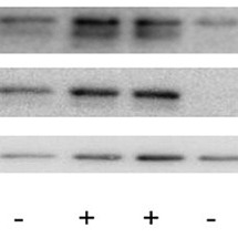
Drain blot and place between plastic sheets for imaging using Biorad GelDoc, as below. After imaging, the blot can be probed for actin as a protein loading control without stripping the blot, using anti-actin-HRP as described below.
Antibody Incubations and ECL visualization of target protein bands: Actin staining with anti-actin-HRP primary
Actin staining with anti-actin-HRP primary
Wash blot 3x with 1X PBST < 00:10:00 .
Incubate 00:20:00 , Room temperature in the primary actin antibody 1:500 freshly made dilution in 5% Milk block.
Wash 2 – 3x in 1x PBST 00:05:00 each.
Incubate in ECL as above (step 51) and image.
Antibody Incubations and ECL visualization of target protein bands: Imaging Blot on BioRad ChemiDoc XRS+ (Vollmers lab)
Imaging Blot on BioRad ChemiDoc XRS+ (Vollmers lab)
Place blot in between sheet protector plastic and transport to imager in cassette to keep dark.
Place blot on white screen.
Place screen into chamber drawer.
Select new protocol, then blot, then chem hi sensitivity and position blot under live focus setting.
Under Applications, select chemidoc hi sensitivity to photograph target bands. Note the image size, the gain (2X), and bin (2x2) to make sure the image size is the same when the protein standards are photographed in order to be able to merge the images.
Under Live Acquire, select acquisition settings: 1, 600, for range and total =100 acquisitions (the 100 can be lowered for longer exposure), starting at 0.25 start time for high expression target.
Freeze and save the image of your choice as it comes up.
Without moving the blot, take a picture to visualize the protein standards by selecting Custom under Applications, then create an Epi illumination protocol with the same gain and binning as used for photographing the bands (double check the image size prediction under the options…)
Merge the target bands and the protein standards pictures in Image Lab software using Image tools.
Note
Note: Imaged blots can be stored at 4ºC in PBST for stripping and re-probing. Some folks also store them frozen in -20ºC flat in plastic bag.