Feb 26, 2026
BIDMC TMC / STU - Methodological compendium for Singular Genomics G4X spatial multiomics platform
- Shuoshuo Wang1,2,3,
- Antonella Arruda de Amaral1,3,
- Athanasios Ploumakis1,2,
- Sheethal Umesh Nagalakshmi1,3,
- Ioannis Vlachos1,2,3
- 1Beth Israel Deaconess Medical Center;
- 2Harvard Medical School;
- 3Broad Institute of MIT and Harvard
- Human BioMolecular Atlas Program (HuBMAP) Method Development CommunityTech. support email: [email protected]


Protocol Citation: Shuoshuo Wang, Antonella Arruda de Amaral, Athanasios Ploumakis, Sheethal Umesh Nagalakshmi, Ioannis Vlachos 2026. BIDMC TMC / STU - Methodological compendium for Singular Genomics G4X spatial multiomics platform. protocols.io https://dx.doi.org/10.17504/protocols.io.81wgbokeylpk/v1
License: This is an open access protocol distributed under the terms of the Creative Commons Attribution License, which permits unrestricted use, distribution, and reproduction in any medium, provided the original author and source are credited
Protocol status: Working
We use this protocol and it's working
Created: February 26, 2026
Last Modified: February 26, 2026
Protocol Integer ID: 244109
Keywords: Tissue mapping, Spatial, Spatial transcriptomics, smFISH, single molecule FISH, Imaging, Tissue preparation, Sample preparation, Spatial Transcriptomics, In Situ Hybridization, Lymphatic Vasculature, Tissue Mapping, Human Reference Atlas, HuBMAP, reproducible workflow for in situ spatia..., situ spatial transcriptomic, spatial gene expression data into atlas, hubmap tissue mapping center, spatial transcriptomic, spatial gene expression data, scale tissue mapping initiative, tissue architecture, subcellular transcript detection in both..., subcellular transcript detection, resolution molecular map, reconstruction of tissue architecture, localization of transcriptional program, human cervix, lymphatic network, interoperable spatial biology, Spatial Multiomics, In situ Sequencing, Singular Genomics G4X, G4X, Sequencing-by-Synthesis, SBS, Multiplexed Spatial Proteomics, Fluorescent Hematoxylin and Eosin, fH&E, TissuStamp, TissuGrip, FFPE, Formalin-Fixed Paraffin-Embedded, Tissue Transfer Workflow, High-Throughput Histology, Rolling Circle Amplification, RCA,
Funders Acknowledgements:
National Heart Lung and Blood Institute
Grant ID: U54HL165440
National Institutes of Health National Institute of Allergy and Infectious Diseases
Grant ID: P01AI179405
National Cancer Institute
Grant ID: P30CA006516
Disclaimer
DISCLAIMER – FOR INFORMATIONAL PURPOSES ONLY; USE AT YOUR OWN RISK
The protocol content here is for informational purposes only and does not constitute legal, medical, clinical, or safety advice, or otherwise; content added to protocols.io is not peer reviewed and may not have undergone a formal approval of any kind. Information presented in this protocol should not substitute for independent professional judgment, advice, diagnosis, or treatment. Any action you take or refrain from taking using or relying upon the information presented here is strictly at your own risk. You agree that neither the Company nor any of the authors, contributors, administrators, or anyone else associated with protocols.io, can be held responsible for your use of the information contained in or linked to this protocol or any of our Sites/Apps and Services.
Abstract
This protocol describes the application of the Singular Genomics G4X Spatial Sequencer for high-resolution, in situ spatial multiomics on formalin-fixed, paraffin-embedded (FFPE) tissue sections within the HuBMAP consortium. Tissue sections are transferred using a hydrogel-pad based mechanism that separates sectioning from slide assembly, enabling profiling of up to 128 millimeter-scale regions of interest per run across approximately 40 cm² of imageable area. The platform integrates spatial transcriptomics (up to 500-plex), multiplexed proteomics (up to 18-plex), and automated fluorescent Hematoxylin and Eosin (fH&E) imaging, with molecular and histological data registered at subcellular resolution. This workflow allows tissue cutting, storage, and transfer to occur independently, with gel-mounted sections storable at 4 °C and transportable between locations. This modular approach supports multi-institutional studies in which tissue collection centers and analytical sequencing sites are geographically separate, facilitating the generation of high-density spatial datasets across distributed research networks.

Materials
| A | B | C | |
| Reagent or Resource | Source | Identifier | |
| Hardware and Instrumentation | |||
| G4X Spatial Sequencer | Singular Genomics | RRID: SCR_026683 | |
| Singular Control System (SCS) | Singular Genomics | N/A | |
| G4X Primary and Secondary Compute Nodes | Singular Genomics | N/A | |
| HistoCore Autocut Microtome | Leica Biosystems | RRID: SCR_023603; Cat# 149AUTO00C1 | |
| Tissue Flotation Water Bath | Boekel Scientific | Cat# 15-464-81 | |
| High-Pressure Cooker (Duo 60 v5) | Instant Pot | N/A | |
| G4X Flow Cell Press | Singular Genomics | Cat# 800110 | |
| G4X Pipette Aid | Singular Genomics | Cat# 800111 | |
| TissuStamp Flow Cell Assembly Tool | Singular Genomics | Cat# 800109 | |
| TissuStamp Gel Pad Drying Rack | Singular Genomics | Cat# 800104 | |
| Refrigerated Microcentrifuge | Thermo Fisher Scientific | Cat# 75002447 | |
| Kits and Consumables | |||
| X4 Spatial Multiomic Kit | Singular Genomics | Cat# 800035 | |
| X2 Spatial Multiomic Kit | Singular Genomics | Cat# 800034 | |
| X4 Spatial Transcriptomic Kit | Singular Genomics | Cat# 800038 | |
| TissuGrip Sample Slide | Singular Genomics | Cat# 500061; 800031 | |
| TissuStamp Gel Pads | Singular Genomics | Cat# 500047; 800028 | |
| TissuStamp Small Blades (4.5×4.5 mm2) | Singular Genomics | Cat# 500056; 800030 | |
| TissuStamp Large Blades (10×10 mm2) | Singular Genomics | Cat# 500057; 800029 | |
| Coplin Staining Jar | Epredia | Cat# 194 / 19-4 | |
| Low-profile Microtome Blades | Leica Biosystems | Cat# NC9948287 | |
| Desiccant Packets | McMaster Carr | Cat# 3492T34 | |
| Chemicals, Reagents, and Macromolecules | |||
| Nuclease-free DEPC-treated water | Thermo Fisher Scientific | Cat# AM9906 | |
| Xylene, Molecular Biology Grade | Thermo Fisher Scientific | Cat# X5-1 | |
| HistoClear II (Alternative to Xylene) | Thermo Fisher Scientific | Cat# 50-899-90150 | |
| Ethanol (100%), Molecular Biology Grade | Thermo Fisher Scientific | Cat# BP2818-4 | |
| Sodium Hypochlorite (5%) | Sigma-Aldrich | Cat# 239305 | |
| Software and Algorithms | |||
| Sequencing Operating System (SeqOS) | Singular Genomics | N/A | |
| Cellpose (Single-cell Segmentation) | Stringer et al., Nature Methods | RRID: SCR_022744 | |
| DeepCell (Single-cell Segmentation) | Bannon et al. | N/A | |
| G4X Analysis Viewer | Singular Genomics | N/A | |
| Sample Sheet Generator | Singular Genomics | N/A | |
| Target Panels (Examples) | |||
| Breast IMX Transcriptomic Panel | Singular Genomics | Cat# 800018 | |
| Colon IMX Transcriptomic Panel | Singular Genomics | Cat# 800015 | |
| Kidney IMX Transcriptomic Panel | Singular Genomics | Cat# 800017 | |
| Lung IMX Transcriptomic Panel | Singular Genomics | Cat# 800016 | |
| IMX Proteomic Panel | Singular Genomics | Cat# 400149 |
Safety warnings
Hazardous Waste Management: The SBS chemistry utilizes formamide for DNA denaturation between sequencing rounds. Formamide is a regulated developmental toxin requiring dedicated disposal protocols within the reagent cartridge.
Introduction
Human BioMolecular Atlas Program (HuBMAP) requires technological platforms that provide high molecular sensitivity, subcellular resolution, and the throughput necessary for cohort-scale studies. The Singular Genomics G4X Spatial Sequencer is an in situ sequencing system that integrates spatial transcriptomics, multiplexed spatial proteomics, and automated fluorescent Hematoxylin and Eosin (fH&E) imaging on formalin-fixed, paraffin-embedded (FFPE) tissue sections. A critical component of this workflow is the TissuStamp system, a gel-pad-based tissue transfer mechanism that decouples histological sectioning from final slide assembly, allowing for the arraying of up to 128 samples per run across 40 cm2 of imageable area.
The G4X platform utilizes Sequencing-by-Synthesis (SBS) to quantify and localize molecular analytes directly within their native histological context. The technology employs circularizable padlock probes (PLPs) that hybridize to target mRNA or oligonucleotide-conjugated antibodies. These probes are enzymatically ligated and then amplified via Rolling Circle Amplification (RCA) to create dense DNA nanoballs (amplicons). These amplicons remain anchored within the tissue matrix and are interrogated through iterative cycles of fluorescent nucleotide incorporation. Following molecular detection, the system performs automated fH&E staining, which is mathematically registered to the molecular coordinates, facilitating histological validation and cell segmentation.
The execution of this analysis demands a rigorous, multi-day preparatory workflow. The protocol is conceptually divided into critical sequential phases:
- microtome sectioning and tissue transfer,
- biochemical target reconstitution,
- microfluidic flow cell assembly,
- pre-sequencing macromolecular assays (involving padlock probe hybridization, enzymatic ligation, and rolling circle amplification),
- tissue conditioning,
- and automated high-throughput imaging and sequencing-by-synthesis (SBS).
The G4X's primary distinction is its high sample density and the use of SBS chemistry for decoding.
| A | B | C | D | E | |
| Feature | G4x | Xenium | MERSCOPE | CosMx | |
| Detection Method | Sequencing-by-Synthesis (SBS) | Cyclic smFISH Hybridization. Barcode imaging after probe + RCA | MERFISH (Combinatorial FISH) | Cyclic FISH / Branching Hyb | |
| Signal Amplification | Rolling Circle Amplification (RCA) | Rolling Circle Amplification (RCA) | Transcript Tiling (No RCA) | Branch Chain Hybridization | |
| Plexity (RNA) | Up to 500-plex | 300 to 5,000-plex | Up to 1,000-plex | Up to 18,000-plex | |
| Max Samples/Run | 128 | 2 (Standard) | 1-2 | 4 | |
| Histology | Automated fH&E | Post-assay H&E | Integrated segmentation | Morphology markers (IHC) |
Technical distinction across comparable platforms.
| Platform | Samples/Run | Imaging Area / ROI | Typical Run Time | |
| G4X (Singular Genomics) | Up to ~128 samples | ~40 cm² total per run | ~5 days sample‑to‑answer (scalable) | |
| 10x Xenium Analyzer | 2 slides | ~472 mm² (~4.7 cm²) | <3–6 days depending on gene panel (10x Genomics) | |
| Vizgen MERSCOPE / Ultra | ~1 slide per run | ~1–3 cm² per slide; ~9 cm²/week | ~1 day imaging per slide (plus prep) (Vizgen) | |
| Bruker CosMx SMI | Up to ~4 slides | FOV‑driven (tens–hundreds mm²) | Days to >10 days depending on FOVs and panel (Bruker Spatial Biology) |
Throughput across comparable platforms
General Information
Materials Availability
The protocol uses commercially available reagents, spatial transcriptomic and proteomic panels, and hardware manufactured by Singular Genomics. Core spatial reagent kits (e.g., X4 and X2 Spatial Multiomic Kits) and specialized tooling (e.g., TissuStamp Gel Pads, TissuGrip Sample Slides) are procured through the vendor.
Custom transcriptomic panels (e.g., 500-gene panels or 150-gene expansions) and oligonucleotide-conjugated proteomic add-ons are designed for specific experimental objectives and are provided under standard material transfer agreements (MTAs) or commercial procurement terms.
Data and Code Availability
The G4X platform generates standardized, structured outputs, including multidimensional image files (.ome.tiff), decoded spatial transcript tables (.parquet), and single-cell feature matrices (.csv.gz). These outputs are compatible with open-source analysis tools. Any specialized computational scripts for downstream spatial processing, neighborhood clustering, or comparative statistics should be deposited in public repositories (e.g., GitHub, Zenodo) with persistent identifiers (DOIs) to support reproducibility and open science standards.
Subject Details
The protocol is optimized for formalin-fixed, paraffin-embedded (FFPE) tissue specimens, which are widely used in archival clinical pathology and allow access to retrospective tissue collections. The protocol is robust to variable cellularity, necrosis, and dense extracellular matrices. All use of primary human tissue must comply with Institutional Review Board (IRB) approval, applicable national and international ethical guidelines, and documented informed consent.
Pre-Analytical Tissue Processing and Environmental Controls
The preservation of RNA integrity and native spatial architecture begins immediately at the microtome stage. To eliminate the introduction of exogenous ribonucleases, the protocol mandates that all histology equipment, including the microtome stage, specimen holder clamps, flywheel handles, and forceps, must be meticulously decontaminated using 70% Molecular Biology Grade ethanol prior to sectioning.
Furthermore, the tissue flotation bath must be filled exclusively with nuclease-free, MilliQ-filtered water.
The thermal stabilization of the flotation bath requires precise calibration between 37 °C and 42 °C . A slightly reduced operational temperature 37 °C to 40 °C is strongly recommended to safely accommodate the extended flotation times required when manipulating and aligning delicate tissue sections onto transfer gel pads without inducing thermal artifacts, structural separation, or excessive paraffin expansion.
FFPE tissue blocks must undergo a controlled hydration phase. Blocks are placed on a HistoCool block or an ice bath filled with RNase-free water for exactly 10 to 20 minutes before active sectioning begins. Prolonged exposure to the hydration source (exceeding 20 minutes) risks severe overhydration, which induces tissue swelling, microscopic wrinkling, and catastrophic delamination during downstream molecular processing.
Note
Overhydration Artifacts: During sample preparation, FFPE blocks must be hydrated for no more than 10 to 20 minutes. Excessive hydration risks microscopic wrinkling and tissue delamination during the heated precedures.
The microtome section thickness is explicitly dictated by the tissue density and cellularity:
Lymphoid tissues (e.g., tonsil, lymph node, spleen, thymus) are sectioned at 3 µm to mitigate the optical crowding of densely packed nuclei, whereas non-lymphoid, solid-organ tissues are sectioned at a standard 5 µm thickness.
Utilizing a low-profile microtome blade secured within the stage clamp, the operator continuously rotates the flywheel to advance the block, generating a uniform, contiguous tissue ribbon. High-profile blades are reserved exclusively for bony, highly calcified, or otherwise brittle tissue matrices. The generated ribbon is carefully transferred to the heated flotation bath using chilled forceps to prevent paraffin adherence to the instrumentation. The section floats on the thermal gradient until the paraffin matrix relaxes and all microscopic wrinkles dissipate. This flotation process is strictly limited to a maximum duration of two minutes; exceeding this temporal threshold triggers overhydration and compromises the structural integrity of the sample.
Expected result
Intermediate Tissue Mounting via TissuStamp Gel Pads
1h 45m
The transition of the delicate tissue section into the microfluidic flow cell environment necessitates a specialized intermediate transfer mechanism utilizing proprietary TissuStamp Gel Pads. The gel pad, stored at 4 °C and subsequently equilibrated to room temperature for a minimum of 00:30:00 , is gently submerged into the flotation bath at a shallow angle to prevent turbulence. The floating tissue section is maneuvered directly over the submerged pad. The protocol strictly dictates that the operator handle only the surrounding paraffin border using forceps, fundamentally avoiding any mechanical tear or compression to the biological specimen itself. Once aligned, the gel pad is slowly extracted from the bath, and excess water is permitted to drain.
Drying the mounted tissue requires environmental precision to prevent desiccation artifacts. The gel pad is placed vertically into the specific slots of a TissuStamp Gel Pad Drying Rack. This vertical orientation is maintained for exactly 00:15:00 to facilitate the active capillary drainage of underlying water trapped between the tissue and the gel matrix. Complete evacuation of moisture from this interface is critical, as residual water initiates premature dissolution of the hydrogel matrix or inhibits covalent-like adherence to the glass slide in subsequent steps. Following the drainage phase, the gel pad is transferred to a flat surface where it undergoes a secondary desiccation phase for 01:00:00 at room temperature.
The pad must achieve a specific morphological state: a well-dried gel extends smoothly to the edges of the tray casing, and the tissue lacks any visible condensation. Over-drying causes the gel to shrink and physically detach from its casing, while under-drying leaves visible moisture droplets that severely compromise the fidelity of the subsequent tissue transfer.
1h 45m
Precision Region of Interest (ROI) Excision and Slide Adherence
35m
Targeted spatial multiomic profiling relies on isolating highly specific biological context while maximizing the area. The TissuStamp workflow enables tissue cutting, storage, and transfer to occur at different times and locations. Gel-mounted sections can be stored at 4 °C and shipped between facilities without the risk of degradation typical of pre-cut slides. This modularity supports multi-institutional studies where tissue collection centers (TMCs) and analytical sequencing sites are geographically separated.
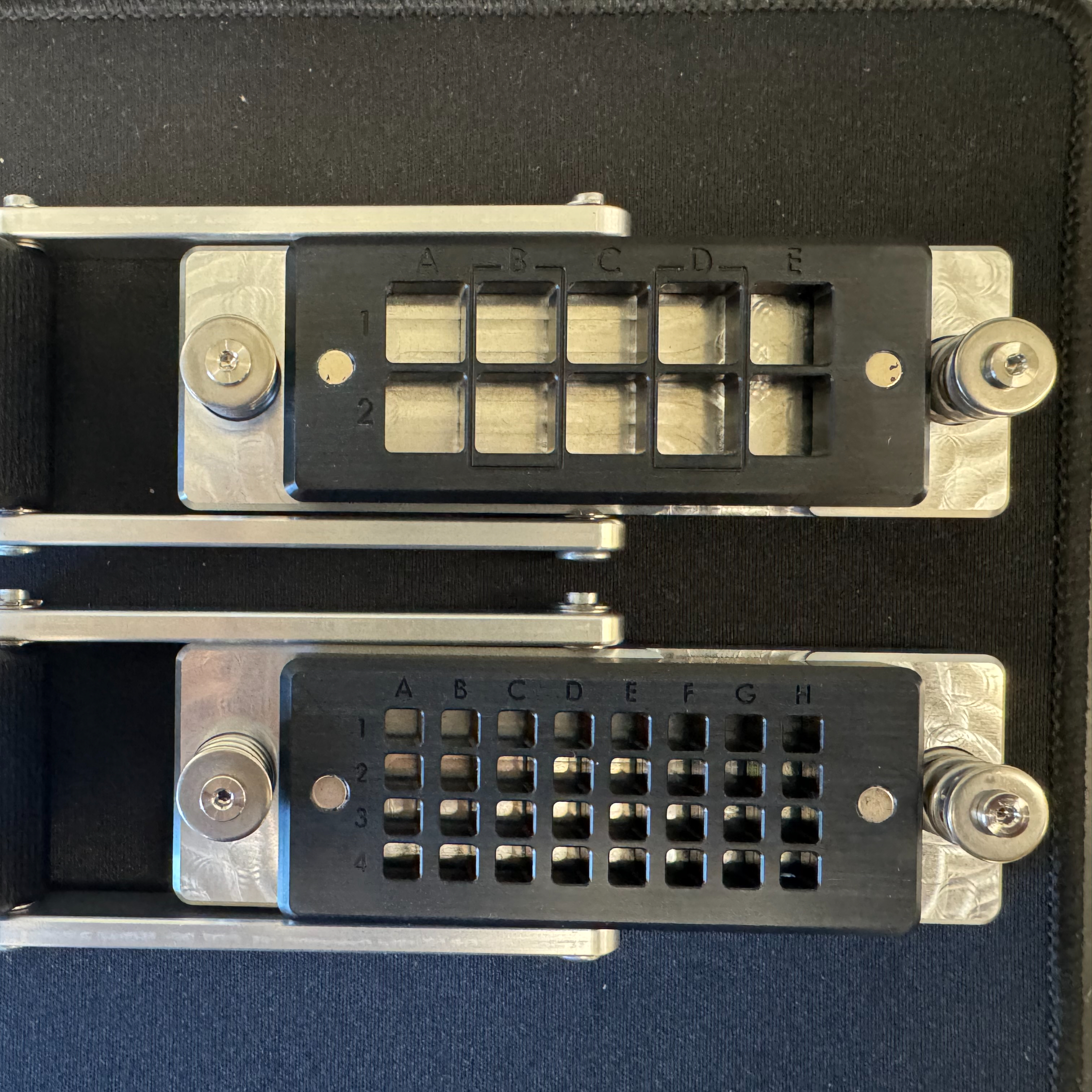
Using the TissuStamp Punch Tool equipped with stainless steel TissuStamp Blades, specific Regions of Interest (ROIs) are physically excised directly from the desiccated gel pad. The protocol provides two distinct blade geometries dictated by the target flow cell architecture: a small 4.5 x 4.5 mm format (capable of highly multiplexed analyses up to 32 sections per X4 flow cell lane, or 128 samples per run) and a large 10 x 10 mm format (accommodating up to 10 larger sections per X2 flow cell).
The operator hovers the blade precisely over the target tissue and depresses it with a firm, vertical force. Lateral movement or rotational torsion during this punch motion is strictly prohibited, as it induces microscopic shearing of the tissue architecture.
The excised tissue plug, securely retained within the hollow cavity of the blade, is then aligned onto the functionalized surface of a TissuGrip Sample Slide using a template-guided transfer base. The TissuGrip slide features a proprietary chemical coating on one side, which is identified by an etched, readable slide ID and a specific chamfer orientation (located at the bottom right when oriented correctly). The blade is inverted and positioned into the transfer template with the tissue facing downward against the glass. A weighted pusher block mechanism is then carefully lowered over the blades. The entire transfer assembly is subsequently incubated on a solid metal heat block at exactly 37 °C for 00:30:00 . This precisely controlled thermal exposure induces the complete chemical adherence of the tissue section to the TissuGrip surface, while the gel matrix serves as a protective backing preventing desiccation. Following the thermal incubation, the assembly is cooled on a laboratory benchtop for 00:05:00 , the lever arm is released, and the pusher block is retracted. Residual gel plugs and debris are meticulously extracted using forceps with a sideways tilting motion, leaving only the naked, firmly affixed tissue ROI on the glass slide. The slide is thoroughly inspected for moisture; if any water remains, a brief re-incubation at 37 °C is performed until complete desiccation is achieved.
Expected result
Note
35m
Deparaffinization
3h 17m
Xylene (or the aliphatic hydrocarbon alternative, HistoClear II) – 00:05:00 (10 minutes if substituting HistoClear).
5m
Xylene (or HistoClear II) – 00:05:00 (10 minutes if substituting HistoClear).
5m
100% Ethanol (Molecular Biology Grade) – 00:03:00 .
3m
100% Ethanol (Molecular Biology Grade) – 00:03:00 .
3m
95% Ethanol (formulated from 237.5 mL 100% ethanol plus 12.5 mL DEPC-treated water) – 00:03:00 .
3m
70% Ethanol (formulated from 02:55:00 100% ethanol plus 75 mL DEPC-treated water) – 00:03:00 .
2h 58m
Microfluidic Flow Cell Assembly and Pipette Aid Integration
The TissuStamp Flow Cell Assembly Tool and G4X Flow Cell Press convert a dried histological slide into a sealed sequencing chamber. The TissuGrip sample slide is placed in one half of the assembly tool, and a laser-drilled glass slide with fluidic inlet and outlet ports is placed in the other half. Alignment pins ensure parallel positioning of the slides, separated by a patterned adhesive microfluidic spacer.
The paired slides are then passed through the G4X Flow Cell Press. Rotation of the actuating knob moves the slides under precision rollers over 2–3 seconds, applying uniform pressure to fuse the glass layers and form a hermetic seal, preventing fluidic leakage during the sequencing run.
Note
Improper seating or debris can lead to fluidic leakage or vacuum seal failure, affecting data acquisition.
Expected result
The assembled flow cell is mounted into the G4X Pipette Aid, which enables manual reagent exchange while minimizing shear stress and bubble formation over the tissue. The Pipette Aid base secures the flow cell, and inlet and outlet adapters, fixed with steel clamp bars, establish a fluid-tight connection to the glass ports. An integrated vacuum pump connected to the outlet ports aspirates excess fluid, ensuring complete and uniform lane clearance between incubation steps. The pipette tip must be inserted vertically into the inlet adapter to maintain proper fluidic contact.
Proteomic Targeting: Tissue Passivation and Oligonucleotide-Conjugated Antibody Binding
2h 35m
For multiomic investigations, the spatial mapping of the proteome precedes transcriptomic probing. Background tissue autofluorescence and non-specific antibody adsorption are mitigated by infusing a Tissue Passivation Working Mix into the microfluidic lanes. This working mix consists of Tissue Passivation Solution (TPS) supplemented with RNase Inhibitor 2 (RNI 2).
Prior to formulating the working mix, the raw TPS and Antibody Binding Solution (ABS) undergo a critical centrifugation step at 4 °C for 00:10:00 at 10.000 x g . This high-speed spin ensures that any micro-precipitates or aggregated proteins in the heavily concentrated buffers are pelleted, preventing steric hindrance or catastrophic microfluidic occlusion during the delicate infusion phase. The supernatant is carefully transferred to a new Protein low-bind tube, avoiding the bottom of the tube to leave any invisible pellets undisturbed. The Tissue Passivation Working Mix is then synthesized on ice by combining 1247.0 µL of centrifuged TPS with 12.5 µL of RNI 2.
Mixing is performed by slow pipetting; vortexing is avoided to maintain enzyme integrity. After infusion into the flow cell, the passivation buffer incubates for 30 minutes at room temperature before being removed.
Simultaneously, the Antibody Binding Working Mix is prepared. This solution incorporates highly specific antibodies pre-conjugated to unique oligonucleotide barcodes mapping to a targeted epitope library (e.g., an 18-plex spatial proteomics panel). Table 2 details the exact volumetric formulation of this binding mixture.
| A | B | |
| Centrifuged Antibody Binding Solution | 330.0 µL | |
| RNase Inhibitor 2 | 3.3 µL | |
| Total Volume | 333.3 µL |
Table 2: Standard formulation for the Antibody Binding Working Mix.
If the experimental design requires the integration of custom protein targets, custom antibody add-ons (up to an additional 2-plex) may be computationally integrated. The baseline recommended concentration for these proprietary add-ons is 5 µL .
The finalized Antibody Binding Working Mix is infused into the flow cell, and the entire Pipette Aid assembly is incubated on a flat, calibrated thermal block at 37 °C for precisely 02:00:00 . Following targeted binding, a secondary Tissue Passivation wash is performed for 00:15:00 at 37 °C , after which the heat block is ramped to 45 °C , and the lanes are flushed extensively with Wash Buffer. An Antibody Treatment Solution (ATS) is subsequently applied for 00:10:00 at the elevated 45 °C temperature. This critical treatment phase biochemically locks the oligonucleotide tags, reducing off-target dissociation and preparing the molecular environment for downstream DNA ligation mechanisms.
2h 35m
Transcriptomic Targeting: Padlock Probe Hybridization and Ligation
1h 16m
Transcript detection on the G4X platform relies on single-molecule padlock probes (PLPs) combined with in situ rolling circle amplification (RCA). Padlock probes are linear DNA oligonucleotides with target-specific complementary arms at the 5′ and 3′ ends and a backbone containing a spatial detection sequence.
The Transcript PLP Working Mix is synthesized On ice . The PLP Reaction Buffer must be vigorously vortexed for a full 00:01:00 prior to combining to ensure solute homogenization. Table 3 details the precise volumetric combination required for formulating the hybridization mix.
| A | B | C | D | |
| Component | Volume per 4-Lane FC (per lane) | Volume per 2-Lane FC (per lane) | Volume per Flow Cell (Total) | |
| PLP Reaction Buffer | 82.9 μL | 165.8 μL | 331.6 μL | |
| Transcript Panel | 15.8 μL | 31.5 μL | 63.0 μL | |
| RNase Inhibitor 1 | 7.9 μL | 15.8 μL | 31.5 μL | |
| DEPC-treated water | 51.0 μL | 102.0 μL | 203.9 μL | |
| Total Volume | 157.5 μL | 315.0 μL | 630.0 μL |
Table 3: Volumetric formulation of the Transcript Padlock Probe Working Mix.
1m
For full custom panels, 63.0 µL of the custom formulation directly replaces the standard panel. If utilizing an add-on panel, 63.0 µL of the base panel and 63.0 µL of the custom add-on are integrated, and the DEPC-treated water volume is consequentially reduced to 140.9 µL to maintain the 630.0 µL total volume constraint and preserve critical molarity.
The resultant mixture is dispensed into the flow cell lanes, and the fluidic inlet/outlet ports are hermetically sealed using single-use Pipette Aid Plugs. This physical capping is critical to prevent evaporative loss and internal pressure gradients. The flow cell is then subjected to a prolonged, highly stringent Overnight incubation spanning 14 to 20 hours at a continuous 45 °C . During this extended thermal hybridization phase, the target-specific arms of the massive padlock probe library actively navigate the crowded intracellular space, locating and continuously annealing to their exact cognate mRNA targets in situ.
Following the overnight hybridization, the Pipette Aid Plugs are discarded, and unbound, floating probes are rigorously flushed from the system using heated Hyb Wash Buffer 45 °C over two consecutive 5-minute incubations. The protocol subsequently dictates an enzymatic PLP Treatment step. A specialized PLP Treatment Working Mix (combining 604.8 µL PLP Treatment Buffer, 6.3 µL RNI 2, and 18.9 µL PLP Treatment Enzyme) is incubated within the lanes for 00:15:00 at 45 °C . This enzymatic mechanism systematically digests and clears non-specifically bound nucleic acids while thermodynamically preparing the perfectly hybridized probe junctions for enzymatic closure.
If the experiment encompasses the full multiomic workflow, a dedicated Protein PLP Working Mix (combining PLP Reaction Buffer, IMX Protein Panel, RNI 1, and water) is introduced at this specific stage. This solution binds secondary padlock probes directly to the synthetic oligonucleotide tags hanging from the previously anchored antibodies. This specific interaction incubates for 00:30:00 at 45 °C before the thermal block is reduced to 37 °C .
The ensuing ligation step solidifies the extraordinary analytical specificity of the entire assay. A Ligation Priming Buffer is initially flowed to precondition the ionic environment, followed immediately by the Ligation Working Mix, which comprises 603.5 µL Ligation Reaction Buffer, 6.3 µL RNase Inhibitor 2, and 20.2 µL of high-fidelity Ligase. Over an exact 00:30:00 incubation at 37 °C , the ligase enzyme physically scans the nucleic acid strands.
The ligation enzyme recognizes correctly annealed 5′ and 3′ ends of the padlock probes adjacent on the target RNA or protein-tag template and catalyzes the formation of a phosphodiester bond, circularizing the probe. Probes that are unhybridized or contain mismatches at the junction do not circularize and are removed during washes. This dual requirement of correct hybridization and enzymatic ligation ensures high specificity for transcript and protein detection.
1h 15m
Signal Amplification via Rolling Circle Amplification (RCA)
3h 25m
Detection of individual transcripts in situ by fluorescence requires signal amplification. Circularized padlock probes serve as templates for processive DNA polymerases, producing rolling circle amplification (RCA) products. The amplification is performed in three enzymatic stages to generate dense, sub-micron DNA nanoballs (amplicons) that remain anchored to the tissue matrix.
First, an AMP 1 Working Mix (synthesized from 623.7 µL AMP 1 Reaction Buffer and 6.3 µL RNase Inhibitor 2) is incubated within the flow cell for 00:10:00 at 37 °C . This solution primes the circularized probes for massive polymerization.
Following a heated Hyb Wash, the core amplification mechanism is aggressively triggered via the introduction of the AMP 2 Working Mix, which incorporates the highly processive AMP Enzyme (113.4 µL of enzyme in 1134.0 µL of AMP 2 Reaction Buffer). To optimize polymerization kinetics and ensure uniform amplicon initiation across the complex tissue topography, the AMP 2 mixture is flowed twice, with each application incubating for exactly 00:15:00 at a slightly reduced temperature of 33 °C .
Finally, to drive the RCA to absolute completion and generate massive, highly repetitive, single-stranded concatemers containing hundreds of identical copies of the spatial barcode, an AMP 3 Working Mix is introduced. The thermal block is returned to 37 °C , and the system undergoes an extended 03:00:00 continuous incubation. The resulting dense DNA nanoballs physically entangle within the fixed tissue matrix, rendering them massive enough to retain intense fluorescent signals during the subsequent sequencing-by-synthesis phase without washing away.
3h 25m
Pre-Sequencing Tissue Conditioning and Spatial Registration
2h 31m 30s
Before sequencing, the flow cell environment is conditioned to stabilize DNA amplicons, maintain tissue architecture under laser exposure, and protect the cellular matrix from microfluidic shear forces generated by the instrument’s pump system.
A timed series of five proprietary Conditioning Solutions are applied sequentially via the Pipette Aid. Conditioning 1 (a dilution of 63.0 µL Concentrated Cond 1 in 567.0 µL PBS-T2) must be mixed fresh immediately before use and incubates for 01:00:00 at Room temperature . A heavy Wash Buffer flush is followed by Conditioning 2, which incubates for 00:30:00 at Room temperature . The thermal block is then aggressively raised to 60 °C for the remainder of the preparation. A heated Saline Rinse is applied for 00:10:00 , followed by Conditioning 3 and Conditioning 4, each incubating for 00:15:00 at 60 °C .
Conditioning 5 requires extensive mechanical preparation: the solid chemical pellet (Conditioning 5 Solid) is resuspended in 1600 µL of PBS-T2 and must be subjected to continuous, low-speed vortexing or rotational mixing for a full 00:15:00 to guarantee absolute dissolution before being infused into the lanes for a final 00:15:00 60 °C incubation.
Note
Incomplete dissolution at this stage leaves micro-crystals that create catastrophic optical artifacts during sequencing.
2h 25m
Optical registration across multiple physical dimensions (X, Y, and Z focal planes) is the fundamental prerequisite for spatial sequencing accuracy. To enable the instrument hardware to continuously track and auto-focus on the highly variable sub-cellular topography of the tissue, specific physical fiducials, known as Focusing Beads, are flowed into the lanes. The Focusing Bead suspension is vortexed heavily for 00:00:30 immediately prior to application.
Note
Spinning down the tube is strictly forbidden, as it irreversibly pellets the dense beads.
The suspension is incubated in the flow cell for 00:06:00 at Room temperature . During this window, the beads gravitationally settle and firmly adhere to both the bare glass surface and the uppermost tissue layer. Following exhaustive Wash Buffer rinses to completely evacuate any unbound beads, the flow cell is sealed. At this stage, the prepared spatial libraries are highly stable and can be stored securely at 4 °C for up to 14 days prior to loading onto the sequencer.
6m 30s
Automated Sequencing Operations on the G4X Platform
2d 2h 30m
The G4X Spatial Sequencer uses a dual-computer architecture to separate sequencing operations from external network influences. The primary computer runs the Sequencing Operating System (SeqOS), which manages hardware telemetry, fluidics control, thermal cycling, and user inputs. A secondary, co-located computer is dedicated to high-performance processing, performing real-time image analysis, basecalling, and machine-learning–based data processing during the sequencing run.
Preparation for an automated multiomic run requires the careful thawing of the Spatial Reagent Cartridges. Depending on operational timelines, cartridges are thawed either slowly for 48:00:00 at 2 °C to 8 °C , or accelerated in a rigorously temperature-controlled room temperature water bath 20 °C to 22 °C for 02:30:00 . The reagent cartridge utilizes a self-contained, foil-sealed architecture to prevent contamination, a procedure similar to Short read sequencing (PE 2x100) on Singular Genomics G4 NGS sequencer.
Safety information
Because the chemistry utilizes formamide, a highly regulated developmental toxin utilized for critical DNA denaturation between sequencing cycles, a specific, removable formamide waste well is isolated within the cartridge structure. This component must be handled and discarded via specialized hazardous chemical protocols.
Just prior to insertion into the instrument's reagent compartment, the operator manually spikes 500 µL of Cleave Buffer Additive into a designated, pre-punctured well and mechanically homogenizes the solution utilizing a 10 mL serological pipette to ensure uniform reagent cleavage kinetics.
Waste management within the instrument is controlled to prevent fluidic backpressure. The buffer compartment contains a waste reservoir that must be emptied prior to each sequencing run, and fresh wash cartridges are loaded concurrently. Each consumable, reagent cartridge, wash cartridge, and flow cell, includes an embedded microchip (Product Identifier) that is scanned via radio frequency to verify lot expiration, volume, and protocol compatibility before pumps are activated. Run parameters are uploaded through the SeqOS interface as a formatted .csv file, specifying conditions for each flow cell lane, including the transcriptomic panel, optional add-ons, block identifiers, and tissue types.
After manual placement and magnetic engagement of flow cells on the thermal stage, SeqOS performs a pre-run seal check and flow rate diagnostic to detect gasket leaks, microfluidic occlusions, or improper seating. If the flow rate check fails, the flow cell must be reseated to ensure proper gasket contact and vacuum seal.
The "Tissue Alignment" interface uses preliminary brightfield imaging to create an optical map of the flow cell. The operator defines digital bounding boxes around tissue regions to limit scanning to biologically relevant areas.
Sequencing is performed using a four-color Rapid Sequencing-by-Synthesis (SBS) chemistry. Transcriptomic analysis involves iterative hybridization of sequencing primers to rolling circle amplicons, followed by incorporation of fluorescently labeled nucleotides, optical detection, and cleavage across multiple cycles. Proteomic analysis uses fluorescent tags on antibody-oligonucleotide conjugates, interrogated in single-cycle optical rounds. Imaging is performed at high throughput over areas up to 40 cm² per run.
Automated fluorescent H&E (fH&E) staining is performed at the end of the molecular detection phase, generating histological images registered to the molecular coordinates. Following sequencing, a post-run wash (~35 minutes) clears the fluidics and transitions the system to standby. Data transfer is managed by the Singular Control System (SCS).
2d 2h 30m
Instrument Fluidic Maintenance and System Health Checks
To sustain optimal pump calibration, fluidic patency, and sequencing accuracy over extended operational lifecycles, rigorous maintenance protocols are strictly enforced via the SeqOS Maintenance dashboard. Routine maintenance washes must be executed whenever recommended by SeqOS, every two weeks of operation, or immediately following any lane failures.
The standard Maintenance Wash protocol utilizes specialized reusable Maintenance Sample Cartridges and Maintenance Reagent Cartridges. The operator formulates fresh chemical solutions: 2 mL of 0.01 % (v/v) sodium hypochlorite (NaOCl) and 200 mL of 0.1 Mass Percent NaOH. These highly caustic reagents systematically degrade any accumulated nucleic acid blockages, denatured protein films, or microbial growth within the delicate microfluidic capillaries. The 0.1 M NaOH is precisely loaded into alternating rows (Rows 1, 3, 5, 7, 9, 11) of the Maintenance Reagent Cartridge, while water fills the even rows. The 0.01 % (v/v) NaOCl is loaded exclusively into Row 3 of the Maintenance Sample Cartridge. The automated system pumps these stringent cleaners through the lines, followed by exhaustive water rinses to restore a neutral pH. Proper disposal of all caustic waste and immediate rinsing and air-drying of the reusable maintenance cartridges is mandatory to prevent permanent fluidic system degradation.
Quantification and Statistical AnalysisPrimary Image Processing and On-Board Basecalling
Raw fluorescent signals are converted into single-cell datasets dynamically during the sequencing run on the secondary computing node. Basecalling, which assigns nucleotide identities to individual rolling circle amplification (RCA) clusters, is performed concurrently with image processing pipelines.
The optical system captures tens of thousands of microscopic fields-of-view (FOVs) across the tissue. These FOV tiles are mathematically stitched using integrated focusing beads as fiducials. During this process, corrections are applied for chromatic aberration, lens distortion, and mechanical stage drift. Background subtraction algorithms reduce autofluorescent noise from FFPE tissue, providing high signal-to-noise ratios for accurate transcript decoding and base assignment.
Cell segmentation is performed automatically using convolutional neural networks (CNNs), including implementations of DeepCell and Cellpose. The algorithms use nuclear fluorescent staining to identify cell centroids and cytoplasmic fluorescence gradients to define cell boundaries. The resulting segmentation masks are saved as NumPy arrays (segmentation.npz), providing a spatial map for downstream analyses.
Molecular data from the SBS phase is integrated with these segmentation masks. High-confidence sequencing reads (typically Q20 or above) are mapped to X, Y, Z coordinates and assigned to individual cells. Fluorescent protein intensities are similarly mapped. The resulting single-cell feature matrices (cell_by_transcript.csv.gz and cell_by_protein.csv.gz) contain rows for each cell and columns for each gene or protein measured.
Run-level summaries are generated as standard HTML documents, reporting quality control metrics such as total cells detected, median transcripts per cell, median unique genes per cell, median cell size, and calculated false discovery rates. Preliminary bioinformatics, including clustering and UMAP dimensionality reduction, is performed directly on the feature matrices to identify cell types and spatial neighborhoods without external computation.
| A | B | C | |
| Filename / Format | Subdirectory | Description of Output | |
| <sample>_HE.ome.tiff | g4x_viewer | Multidimensional OME-TIFF containing transformed fH&E image data. | |
| <sample>_run_metadata.json | g4x_viewer | Comprehensive JSON detailing core metrics, panel versions, and instrument telemetry. | |
| segmentation.npz | segmentation | Compressed NumPy array containing the AI-generated pixel segmentation mask. | |
| rna_transcript_table.csv.gz | rna | X/Y/Z physical coordinates, assigned gene names, Q-scores, and assigned cell IDs for every detected transcript. | |
| cell_by_transcript.csv.gz | single_cell_data | Definitive single-cell feature matrix of genes per cell. | |
| cell_by_protein.csv.gz | single_cell_data | Matrix of median protein intensities assigned per cell. | |
| clustering_umap.csv.gz | single_cell_data | Spatial UMAP coordinates and cell-type clustering assignments. | |
| cell_metadata.csv.gz | single_cell_data | Aggregate morphological data, including cellular spatial coordinates and area parameters. |
Table 4: Standardized primary and secondary data outputs automatically generated by the Singular Genomics G4X pipeline.
All output files including multidimensional image files, system metadata JSONs, and single-cell feature matrices, are transferred off the instrument via the Singular Control System (SCS). The SCS provides an encrypted network connection to high-capacity storage systems, such as local NAS/SAN devices or cloud platforms (e.g., Amazon S3, Google Cloud). This ensures that the multiomic dataset is accessible for downstream analysis, including spatial mapping, neighborhood analysis, and other exploratory workflows.
Protocol references
Key Resources
- User Guide for G4X Sample Preparation – Step-by-step instructions for preparing high-quality samples
- User Guide for G4X Spatial Sequencer Operation – Detailed guidance on running your G4X system
- G4X Workflow Overview - G4X workflow checklist for a quick reference
- G4X Workflow Quick Reference Guide - Online, Interactive G4X Sample Prep Protocol
- G4X Sample Sheet Generator – Tool for creating run-ready spatial sample sheets
- G4X Tissue Placement Tracker – Template to track regions of interest on the flow cells
- ROI Selection Tool - Online tool to help you select the appropriately sized ROI from your H&E image
- G4X Viewer – Software for viewing G4X spatial data (v3.0)
- Legacy G4X Viewer - Software for viewing G4X spatial data (v2.6.2)
- G4X Viewer Tutorial Video - Video walkthrough of the G4X Viewer (v2.6.2) and its functionality.
- G4X Viewer Tutorial PDF - Instructions for navigating and making the most of the G4X Viewer (v2.6.2). An updated Tutorial will be available soon!
- G4X Data Tutorials - These pages provide documentation, example workflows, and helper tools for analyzing, and post-processing data generated by the G4X spatial sequencer
- Custom Panel Guides:
- G4X Custom Transcript Technical Guide - outline the customization options and the process of ordering a custom transcriptomic panel
- G4X Custom Protein Technical Guide - outline the customization process of ordering a custom conjugated antibody of interest that can be used in the G4X assay
- G4X Custom Antibody Calculator - calculates the volume of antibody add-on to add to the Antibody Binding Working Mix
Gloria S Pryhuber 2025. HuBMAP MOSDAP (Multiple Organ Same Donor Authorization Project) Protocols. protocols.iohttps://dx.doi.org/10.17504/protocols.io.kxygxwoywv8j/v2
Shuoshuo Wang, Antonella Arruda de Amaral, Ioannis Vlachos 2026. Short read sequencing (PE 2x100) on Singular Genomics G4 NGS sequencer. protocols.iohttps://dx.doi.org/10.17504/protocols.io.81wgbnpq1gpk/v1
Zajac, N., Vlachos, I.S., Sajibu, S., Opitz, L., Wang, S., Chittur, S.V., Mason, C.E., Knudtson, K.L., Ashton, J.M., Rehrauer, H. and Aquino, C., 2025. The impact of PCR duplication on RNAseq data generated using NovaSeq 6000, NovaSeq X, AVITI, and G4 sequencers. Genome biology, 26(1), p.145.
Acknowledgements
We gratefully acknowledge Singular Genomics for their outstanding technical support and collaborative engagement throughout our participation in the G4X Early Access Program since June 2024. Their responsiveness, expertise, and sustained commitment were instrumental to the successful execution of this work.
We particularly thank Jordan Williams, Mayar Allam, Guillaume Dubeaux, Andy Bodnar, Brittni Foster, Gina Benedetto, Mark Lane, Sabrina Shore, Ernie Guzman, Bandele Jeffrey-Coker, Maria Gallina, Darius Fugere, Eli Glezer, and Laure Moller for their professionalism and dedicated support.