Apr 16, 2026
BIDMC-TMC Assessing RNA integrity from FFPE samples for spatial transcriptomics
- Shuoshuo Wang1,2,3
- 1Beth Israel Deaconess Medical Center;
- 2Broad Institute of MIT and Harvard;
- 3Harvard Medical School
- Human BioMolecular Atlas Program (HuBMAP) Method Development CommunityTech. support email: [email protected]


Protocol Citation: Shuoshuo Wang 2026. BIDMC-TMC Assessing RNA integrity from FFPE samples for spatial transcriptomics. protocols.io https://dx.doi.org/10.17504/protocols.io.e6nvwwyjwvmk/v1
License: This is an open access protocol distributed under the terms of the Creative Commons Attribution License, which permits unrestricted use, distribution, and reproduction in any medium, provided the original author and source are credited
Protocol status: Working
We use this protocol and it's working
Created: April 16, 2026
Last Modified: April 16, 2026
Protocol Integer ID: 315214
Keywords: DV200, spatial, single cell, RNA integrity, FFPE, QC, molecular pathology rna quality in ffpe sample, assessing ffpe rna quality, rna integrity from ffpe sample, ffpe rna quality, assessing rna integrity, molecular pathology rna quality, rna degradation, percentage of rna fragment, rna fragmentation, rna fragment, impact of rna fragmentation, ffpe sample, spatial transcriptomic, ffpe, ribosomal peak, absence of ribosomal peak, dv200, rna, spatial transcriptomics rna quality in ffpe sample, spatial transcriptomics rna quality
Funders Acknowledgements:
National Heart Lung and Blood Institute
Grant ID: U54HL165440
National Institutes of Health National Institute of Allergy and Infectious Diseases
Grant ID: P01AI179405
Disclaimer
FOR INFORMATIONAL PURPOSES ONLY; USE AT YOUR OWN RISK
The protocol content here is for informational purposes only and does not constitute legal, medical, clinical, or safety advice, or otherwise; content added to protocols.io is not peer reviewed and may not have undergone a formal approval of any kind. Information presented in this protocol should not substitute for independent professional judgment, advice, diagnosis, or treatment. Any action you take or refrain from taking using or relying upon the information presented here is strictly at your own risk. You agree that neither the Company nor any of the authors, contributors, administrators, or anyone else associated with protocols.io, can be held responsible for your use of the information contained in or linked to this protocol or any of our Sites/Apps and Services.
Abstract
RNA quality in FFPE samples is highly variable, influenced by factors such as fixation protocols, tissue type, storage conditions, and necrosis. Formalin fixation exacerbates RNA degradation, resulting in fragmented molecules that pose challenges for downstream applications.
DV200, the percentage of RNA fragments >200 nucleotides, has emerged as a reliable metric for assessing FFPE RNA quality, surpassing the traditional RIN score, which falters due to the absence of ribosomal peaks. While DV200 offers critical refences for sequencing and library preparation, assays utilizing hybridization techniques can mitigate the impact of RNA fragmentation, making DV200 a gold standard but not exclusive determinant of sample usability.
Attachments

Guidelines
All de-identified FFPE samples must be submitted with a clear and standardized identification system to ensure accurate tracking throughout processing. Each sample should use a simple alphanumerical identifier. For consortium or multi-institution studies, a project or institution prefix (e.g., TMC_001) may be added to reduce labeling errors. These identifiers must map clearly to your internal clinical records, including patient identifiers, block numbers, collection dates, and study-specific codes, which should be maintained separately in a secure metadata file to support compliant handling and traceability.
For each sample, please provide basic metadata including tissue type or organ of origin, FFPE block identifier, collection date if available, and any relevant cohort or study grouping information. This information is required for proper processing, QC interpretation, and downstream analysis.
RNA quality assessment using DV200 is strongly recommended prior to submission, particularly for older or archival FFPE collections. DV200 measures the percentage of RNA fragments longer than 200 nucleotides and is generally more informative for FFPE RNA quality assessment than RIN, which is less suitable for degraded RNA samples.
While DV200 is an important quality metric, it should not be used as a strict pass/fail cutoff. Modern hybridization-based and amplification-based assays may still generate usable data from samples with lower DV200 values, depending on assay design, input amount, and target characteristics.
FFPE blocks should be properly fixed and paraffin-embedded using consistent clinical protocols whenever possible, as pre-analytical variation is a major contributor to downstream variability in RNA quality. FFPE blocks are typically stored at room temperature as standard practice; however, storage at 4°C prior to shipment is recommended when feasible to help minimize additional RNA degradation, particularly for older archival samples.
Please submit an adequate number of samples to account for QC, sectioning, and assay optimization, as some material will be consumed during processing. For large studies or heterogeneous archival collections, pre-submission QC is recommended to reduce turnaround time and avoid processing low-quality samples that may not meet assay requirements.
Upon receipt, all FFPE blocks will undergo internal quality control, including DV200 assessment prior to sectioning and processing. For large or older archival cohorts, we strongly recommend performing preliminary QC before shipment to improve workflow efficiency and reduce delays. Overall, DV200 should be interpreted as a key quality indicator but not the sole determinant of sample suitability, which depends on assay-specific requirements and overall experimental context.
Materials
- Sterile, RNase-free pipet tips
- 1.5 ml or 2 ml RNase-free centrifuge tubes, e.g. DNA LoBind Tubes 1.5 mL (Eppendorf, catalog number: 022431021) and DNA LoBind Tubes 2.0 mL (Eppendorf, catalog number: 022431048), if not provided in the kit.
- Microcentrifuge (with rotor for 2 ml tubes), e.g. Centrifuge 5810/5810R (Eppendorf, catalog number: EP022628188) with rotor S-4-104
- Standard heavy-duty vortex mixer (VWR or Fisherbrand, catalog number: 97043-562)
- ThermoMixer C (Eppendorf, catalog number: 05-412-503) or thermoblock with minimal temperature fluctuation.
- RNaseZap
- 100% ethanol
- RNeasy FFPE Kit (Qiagen) or PureLink FFPE RNA Isolation Kit (Thermo Fisher) or High Pure FFPE RNA Micro Kit (Roche)
- Microtome
- Ice bucket with cool blocks or Cool Rack CFT30 (Corning, catalog number: CLS432052)
Equipment
TapeStation
NAME
Agilent
BRAND
G2991AA
SKU
LINK
Protocol materials
Proteinase K, 2mLQiagenCatalog #19131
Qubit RNA HS (High Sensitivity) assay Thermo Fisher ScientificCatalog #Q32852
Safety warnings
- Assessing RNA integrity may not be feasible for small or degraded samples, such as core needle biopsies, TMAs, archived pathology sections, and permanently mounted slides.
- Proceed with downstream applications directly if integrity assessment is indeed impractical.
- Buffer RBC contains guanidine hydrochloride. Do not mix the waste with acidic solutions or bleach!
Before start
Preparation and Storage of DNase I and Buffer RPE
1. Prepare DNase I Stock Solution:
- Dissolve lyophilized DNase I (1500 Kunitz units) in 550 µL RNase-free water without opening the vial.
- Using an RNase-free syringe and needle, inject RNase-free water into the vial.
- Mix gently by inverting the vial; avoid vortexing to prevent enzyme degradation.
2. Aliquot and Store DNase I:
- Transfer the DNase I stock solution into single-use aliquots.
- Store aliquots at –30°C to –15°C for up to 9 months.
- After thawing, store at 2–8°C for up to 6 weeks. Do not refreeze thawed aliquots.
3. Prepare Buffer RPE:
- Add 44 mL ethanol (96–100%) to the bottle containing 11 mL Buffer RPE concentrate.
- Mix well and mark the checkbox on the bottle label to confirm ethanol has been added.
4. Preheat a thermal mixer, heat block, or water bath to 56°C for use. If available, preheat a second thermal mixer, heat block, or water bath to 80°C to minimize waiting time during the protocol.
Preparation
Workstation Decontamination
Set up a clean workspace for handling samples and reagents.
Note
Adhere to RNase-free working standards:
Ensure all working surfaces, reagents, and plasticware are RNase-free.
Minimize handling and avoid unnecessary exposure to environmental contaminants.
Always wear disposable gloves and replace them frequently to maintain cleanliness.
Spray and clean all working zones, benches, chemical hoods, pipettes, and reagent containers with a sodium hypochlorite-based decontamination reagent (e.g., RNaseZap or RNase AWAY) or 0.5% bleach.
Wipe surfaces using a paper towel treated with the decontamination reagent.
Spray and clean again using 70% ethanol.
Rinse cleaned surfaces with RNase-free water and wipe dry.
Sample Sectioning
Set the microtome to a section thickness of 10 µm or 20 µm depending on sample type.
Note
For collagen-rich samples, thin sections are preferred. Thicker sections can reduce RNA yield.
Make sure microtome, blades, tools, and Eppendorf tubes are RNase-free.
Reface the block and discard the first few sections exposed to the environment.
Consider trimming of the peripheral paraffins. If paraffin occupies more than 30% of the section's surface area, trim away the excess paraffin using a blade before collecting.
Note
Minimize carryover paraffin to enhance RNA purity and prevent interference during extraction.
Determine the number of sections needed based on tissue size and availability.
For small tissues (<1 cm): Use approximately 4 sections or more.
For large tissues (>1 cm): Use 2 sections.
Note
- Perform pilot-based optimization to determine the appropriate input material for specific tissue types and experimental requirements.
- Adjusting as needed for tissue type, cellularity, and cross-sectional area.
- RNA yield correlates with tissue cellularity, too. Necrotic tissues have low RNA abundance.
- Use larger sections or additional scrolls for tissues with inherently low RNA content.
Place the tissue sections (scrolls) immediately into pre-labeled, pre-cooled, RNase-free microcentrifuge tubes.
Expected result
Sections curl naturally due to tension in the paraffin wax, thus called "curls" or "scrolls"
Note
If the scrolls can not immediately processed, store them at -80 °C .
RNA Extraction Procedure (example: Qiagen RNeasy FFPE)
1h 19m 20s
Use a commercial silica membrane column-based RNA extraction kit, for example:
RNeasy FFPE Kit (Qiagen), Catalog Number 73504
PureLink FFPE RNA Isolation Kit (Thermo Fisher)
High Pure FFPE RNA Micro Kit (Roche)
Maxwell RSC RNA FFPE Kit (for automated extraction)
Follow the manufacturer's instructions for RNA extraction.
Note
Read the "Before start" section from "Guidelines & Warnings" first.
Deparaffinization
Note
Preheat a thermal mixer or heat block to 56°C for use.
If available, preheat a second thermal mixer or heat block to 80°C to minimize waiting time during the protocol.
Safety information
Do use the blue tinted Daparaffinization Solution (Qiagen #19093) for enhanced visibility of phase separation.
Do not use Xylene or organic solvents as replacement due to low flash point and risk of explosion.
Home-brew solution can be found in Appendix A of Qiagen RNeasy FFPE Handbook 07/2021
Add 160 µL (for smaller samples) or 320 µL (for larger samples) of Deparaffinization Solution to the sample. Note that the amount of buffers used in following steps will need to match the choice here.
Vortex vigorously for 00:00:10 , then briefly centrifuge to bring the sample to the bottom of the tube.
10s
Incubate the sample at 56 °C for 00:03:00 to dissolve the paraffin.
3m
Vortex vigorously for 00:00:10 , then briefly centrifuge to bring the sample to the bottom of the tube.
10s
Incubate the sample at 56 °C for 00:03:00 to dissolve the paraffin.
3m
Allow the sample to cool to Room temperature .
Note
If the solution becomes waxy or solid after cooling (indicating insufficient Deparaffinization Solution or excess paraffin), add more Deparaffinization Solution and repeat the 56°C incubation.
Lysis
Note
Lysis and incubation conditions designed to reverse formaldehyde-induced modifications of RNA. The lysis buffer facilitates RNA release from FFPE tissue samples while minimizing degradation. These conditions aim to maximize the recovery of usable RNA from FFPE samples.
Add 150 µL (for smaller samples) or 240 µL (for larger samples) of Buffer PKD (Qiagen #1034963) to the sample. PKD stands for Proteinase K digestion.
Mix thoroughly by vortexing.
Centrifuge the sample at 11000 x g for 00:01:00 to separate phases.
1m
Carefully add 10 µL of Proteinase K, 2mLQiagenCatalog #19131 to the lower, clear phase.
Mix gently by pipetting up and down. Do not vortex.
Incubate the sample at 56 °C for 00:15:00 , followed by 80 °C for 00:15:00 to complete protein digestion and RNA release.
Note
1. If possible, preheat a second thermal mixer or heat block to 80°C to minimize waiting time.
2. If only one heating block is available, allow the sample to remain at room temperature (15–25°C) after the 56°C incubation until the block reaches 80°C. Ensure the heating block is at 80°C before starting the 15-minute incubation, as this step is critical for reversing crosslinks.
3. If using a heating block without shaking functionality, briefly vortex the sample every 3–5 minutes during incubation.
4. Prolonged incubation or higher temperatures may increase RNA fragmentation.
30m
DNA Removal
Incubate the sample On ice for 00:03:00 .
3m
Centrifuge at 20000 x g, 4°C for 00:15:00 .
15m
Carefully transfer the supernatant to a new RNase-free microcentrifuge tube.
Note
Genomic DNA contamination is common in RNA preparations and appears as a smooth bump near or above 6000 bp on QC traces. If not removed, it will persist through RNA-seq library preparation and sequencing, contributing to background reads and inaccuracies. To prevent this, treat RNA samples with DNase before starting library preparation, ensuring proper cleanup or buffer exchange as needed. Avoid disturbing the pellet, which contains insoluble tissue debris and crosslinked DNA, as carryover can introduce impurities and cause significant errors in DV200 measurements.
Add DNase Booster Buffer (10% of the total sample volume, approximately 16 µL) and 10 µL DNase I stock solution to the supernatant.
Mix by inverting the tube and briefly centrifuge to collect residual liquid from the tube walls. Do not vortex.
Incubate the sample at Room temperature for00:15:00
15m
Column loading and RNA purification
If kit is new, prepare Buffer RPE by add 44 mL 100% ethanol to the bottle containing 11 mL Buffer RPE concentrate. Mix well and mark the checkbox on the bottle label.
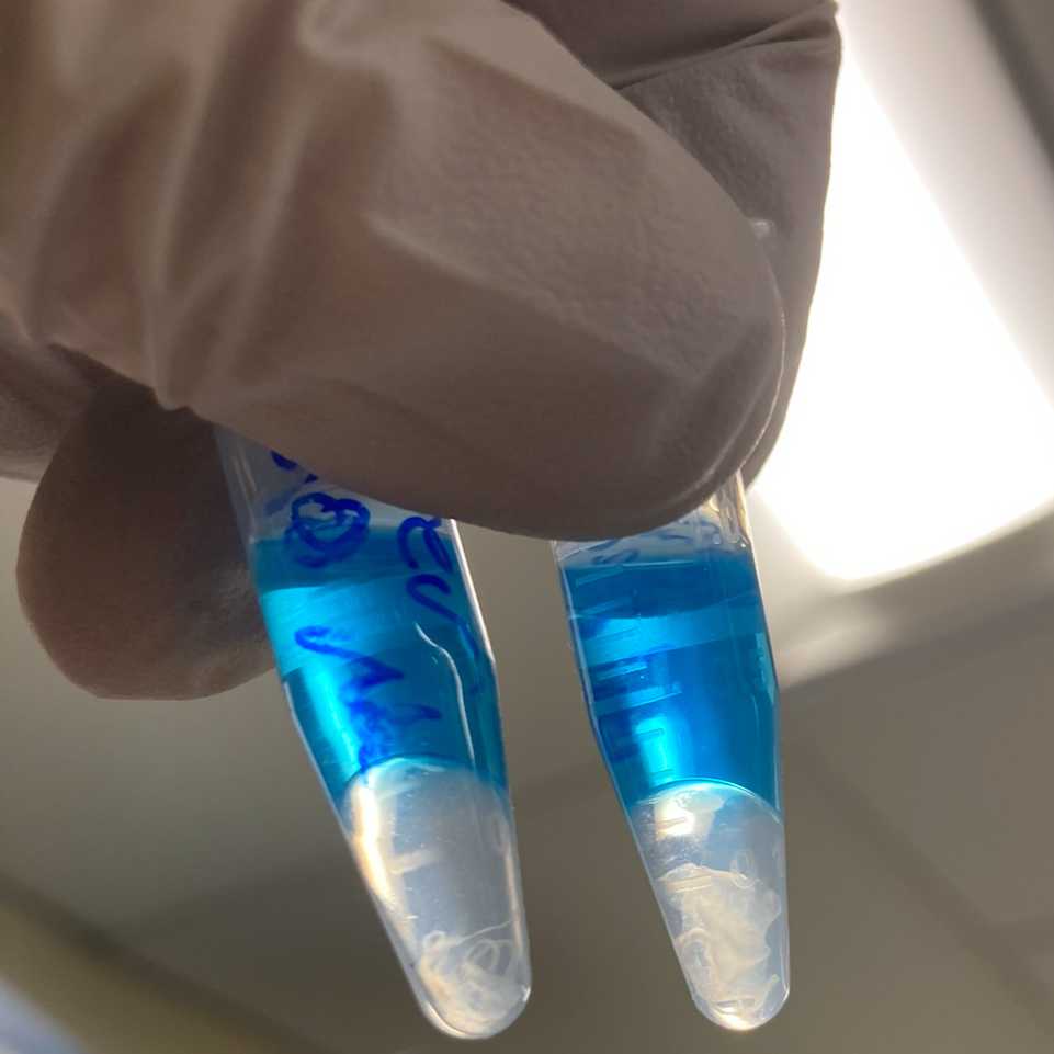
Add 320 µL Buffer RBC to the sample and mix thoroughly.
Add 720 µL ethanol (100%) and mix well by pipetting. Do not centrifuge.
Transfer 700 µL of the sample (including any precipitate) to a pre-labeled spin column placed in a 2 mL collection tube.
Close the lid gently and centrifuge at no less than 8000 x g for 00:00:15 .
15s
Discard the flow-through and reuse the collection tube for subsequent steps.
Safety information
it is critical not to collect RBC containing flow-through in a waste container with bleach or any chlorine-containing compounds, and appropriate safety precautions should be taken when handling these chemicals. Otherwise, guanidine hydrochloride in RBC will release chlorine gas upon reaction with hypochlorite.
Exposure to chlorine gas can lead to severe lung damage or even death.
Process Remaining Sample: Repeat the loading and centrifugation process until the entire sample has passed through the spin column.
Add 500 µL Buffer RPE to the spin column.
Close the lid gently and centrifuge at no less than8000 x g for 00:00:15 .
15s
Discard the flow-through and reuse the collection tube for the next step.
Repeat Washing: Repeat steps 10.7–10.9, but centrifuge for 00:02:00 instead of 15 seconds.
2m
After centrifugation, carefully remove the spin column from the collection tube, ensuring the column does not contact the flow-through to avoid ethanol carryover contamination.
RNA Elution and Drying
Place the spin column in a new 2 mL collection tube.
Open the lid of the spin column and centrifuge at full speed for 00:05:00 .
5m
Discard the collection tube with the flow-through.
To prevent damage to the spin column lids, ensure there is at least one empty position between columns in the centrifuge. Orient the lids in the opposite direction of the rotor's rotation (e.g., if the rotor rotates clockwise, orient the lids counterclockwise).
Note
Proper drying of the spin column membrane is crucial to remove residual ethanol, which could interfere with downstream reactions. Keeping the lids open during centrifugation ensures no ethanol is carried over during RNA elution.
Transfer the spin column to a new 1.5 mL collection tube.
Add 14–30 µL RNase-free water directly onto the spin column membrane. Wait 00:00:30 to prime.
Note
Eluting with smaller volumes of water results in higher RNA concentrations, but may reduce RNA yield. The spin column has a 2 µL dead volume, so elution with 14 µL RNase-free water will yield approximately 12 µL of eluate.
30s
Close the lid gently and centrifuge at full speed for 00:01:00 to elute the RNA.
Save 4 µL for QC and place on ice. If the analysis cannot be performed immediately, store purified and unused RNA at 80 °C for long-term storage.
1m
Analysis and DV200 calculation
Keep RNA samples On ice at all times during the analysis procedure.
For quantitation, use a Qubit 4 Fluorometer to measure RNA concentration to determine the appropriate dilution for loading the instrument and gel type. Qubit RNA HS assay has a quantitatie range from 4 to 200 ng. Qubit RNA HS (High Sensitivity) assay Thermo Fisher ScientificCatalog #Q32852
Note
Pay attention to the quantitative range for : 500-10000 pg/µL for High Sensitivity RNA ScreenTape.
So the minimal concentration will be 0.5 ng/uL.
The DV200, as a %, might be artificially inflated due to input in the non-quantitative range.
Do not use NanoDrop for quantitation due to:
- Inaccuracy: NanoDrop measures sample purity, not the accurate concentration.
- Low Sensitivity: It is unsuitable for samples with low concentration.
The Qubit RNA IQ Assay does not have the necessary sensitivity (>500ng) for small samples.
Equipment
Qubit 4
NAME
Fluorometer
TYPE
Invitrogen
BRAND
Q33238
SKU
DV200 score can be measured using instruments like
Fragment Analyzer (>100 pg with Agilent DNF-472, #DNF-472-0500).
Equipment
TapeStation
NAME
Agilent
BRAND
G2991AA
SKU
LINK
Equipment
Bioanalyzer
NAME
Bioanalyzer
TYPE
Agilent
BRAND
G2991AA
SKU
LINK
Any bioanalyzer will suffice.
SPECIFICATIONS
Equipment
Fragment Analyzer
NAME
capillary based nucleic acid fragment size separation
TYPE
Agilent
BRAND
M5311AA
SKU
LINK
Calculate DV200 following the manufacturer's instructions.
For Fragment Analyzer: https://www.agilent.com/en/promotions/fragment-analyzer-dv200-determination
Assay requirements vary, but as a general guideline, a DV200 below 30% critically impacts performance and represents the minimum acceptable threshold, while a DV200 above 50% is considered adequate.
Protocol references
2. Qiagen, 2021, RNeasy FFPE Handbook, HB-0375-006.
3. Wehmas, L.C., Wood, C.E., Chorley, B.N., Yauk, C.L., Nelson, G.M. and Hester, S.D., 2019. Enhanced quality metrics for assessing RNA derived from archival formalin-fixed paraffin-embedded tissue samples. Toxicological Sciences, 170(2), pp.357-373. https://doi.org/10.1093/toxsci/kfz113
4. Matsubara, T., Soh, J., Morita, M., Uwabo, T., Tomida, S., Fujiwara, T., Kanazawa, S., Toyooka, S. and Hirasawa, A., 2020. DV200 index for assessing RNA integrity in next‐generation sequencing. BioMed research international, 2020(1), p.9349132. https://doi.org/10.1155/2020/9349132
5. Ondracek, R.P., Chen, J., Marosy, B., Szewczyk, S., Medico, L., Mohan, A.S., Nair, P., Pratt, R., Roh, J.M., Khoury, T. and Carpten, J., 2022. Results and lessons from dual extraction of DNA and RNA from formalin-fixed paraffin-embedded breast tumor tissues for a large Cancer epidemiologic study. BMC genomics, 23(1), p.614. https://doi.org/10.1186/s12864-022-08837-6
6. NCI Biorepositories and Biospecimen Research Branch 2024. NCI Biospecimen Evidence-Based Practices (BEBP) - Nucleic Acid Extraction from Formalin-Fixed, Paraffin-Embedded (FFPE) Tissue. protocols.io https://dx.doi.org/10.17504/protocols.io.8epv5r97jg1b/v1
7. Wang, S., 2024. Resolving the bone–optimizing decalcification in spatial transcriptomics and molecular pathology. Journal of Histotechnology, pp.1-10. https://doi.org/10.1080/01478885.2024.2446038
Acknowledgements
Support from Spatial Technologies at Beth Israel Deaconess Medical Center (RRID:SCR_024905).