May 21, 2026
Version 2
Analysis of glucosylceramide from plasma and cerebrospinal fluid V.2
Version 1 is forked from Analysis of glycosphingolipids from human plasma
- David A Priestman1,2,
- Danielle te Vruchte1,
- Kerri-Lee Wallom1,
- Mylene Huebecker1,2,3,4,
- Carla Santos1,2,3,4,
- Reuben Bush1,
- Chia-Chen Celeste Chuang1,2,3,4,
- María Fernández-Suárez1,
- Frances M Platt1,2,3,4
- 1Department of Pharmacology, University of Oxford, Mansfield Road, Oxford, OX1 3QT, United Kingdom;
- 2Aligning Science Across Parkinson’s (ASAP) Collaborative Research Network, Chevy Chase, MD, 20815;
- 3Royal Society Wolfson Research Merit Award Holder;
- 4Wellcome Trust Investigator in Science.


Protocol Citation: David A Priestman, Danielle te Vruchte, Kerri-Lee Wallom, Mylene Huebecker, Carla Santos, Reuben Bush, Chia-Chen Celeste Chuang, María Fernández-Suárez, Frances M Platt 2026. Analysis of glucosylceramide from plasma and cerebrospinal fluid. protocols.io https://dx.doi.org/10.17504/protocols.io.5qpvorrndv4o/v2Version created by Reuben Bush
Manuscript citation:
Analysis of fluorescently labeled glycosphingolipid-derived oligosaccharides following ceramide glycanase digestion and anthranilic acid labeling DCA Neville, V Coquard, DA Priestman, DJM te Vruchte, DJ Sillence, Raymond A Dwek, Frances M Platt, Terry D Butters Analytical biochemistry 331 (2), 275-282, (2004). doi: 10.1016/j.ab.2004.03.051.
License: This is an open access protocol distributed under the terms of the Creative Commons Attribution License, which permits unrestricted use, distribution, and reproduction in any medium, provided the original author and source are credited
Protocol status: Working
We use this protocol and it's working
Created: August 29, 2024
Last Modified: May 21, 2026
Protocol Integer ID: 106678
Keywords: Glycosphingolipids, Glucosyl Ceramide, HPLC, Oligosaccharide Analysis, ASAPCRN, cellular glycosphingolipid, analysis of glucosylceramide, glycosphingolipid, reproducible molar quantities of glucosylceramide, ceramide glycanase enzyme, glucosylceramide, oligosaccharide, glucose, cerebrospinal fluid sample, reproducible quantitation of gsl, labelled chitotriose calibration standard, performance liquid chromatography, fluorescent compound anthranilic acid, chitotriose calibration standard, present in mammalian cell, cerebrospinal fluid interest in the role, cerebrospinal fluid interest, gsl
Funders Acknowledgements:
The Michael J. Fox Foundation for Parkinson’s Research (MJFF) and the Aligning Science Across Parkinson’s (ASAP) Initiative
Grant ID: ASAP-000478
The Michael J. Fox Foundation for Parkinson’s Research (MJFF)
Grant ID: MJFF-01038
Abstract
Interest in the role of cellular glycosphingolipids (GSLs) in health and disease led to us developing a sensitive method to analyse the full complement of GSL structures present in mammalian cells, fluids and tissues. The original qualitative method we developed was published in 2004 and measured the oligosaccharides selectively released from glycosphingolipids using a ceramide glycanase enzyme derived from the medicinal leech. We have now updated and refined this protocol with the focus on achieving sensitive and reproducible quantitation of GSLs in control and patient plasma and cerebrospinal fluid samples. The method uses the fluorescent compound anthranilic acid (2-AA) to label glucose enzymatically released from glucosylceramide prior to analysis using normal-phase high-performance liquid chromatography. The labelling procedure is rapid, selective, and easy to perform. With the inclusion of a 2AA-labelled chitotriose calibration standard, it is possible to obtain accurate and reproducible molar quantities of glucosylceramide.
Guidelines
This protocol requires the use of some hazardous materials. As such, users must be appropriately trained and hazardous materials stored, used, and disposed of in accordance with your institution’s health and safety policies, and approved laboratory policies, risk assessments and codes of practice.
Materials
Acetonitrile 1.00030
Gradient grade for liquid chromatography LiChrosolv® Reag. Ph Eur
Anthranilic acid
A89855 Sigma-Aldrich
Reagent grade, ≥98%
Boric acid
ReagentPlus®, ≥99.5%
B0252 Sigma-Aldrich
Chloroform
Suitable for HPLC, ≥99.8%, amylene stabilized
34854-M Sigma-Aldrich
Discovery® DPA-6S SPE Tube
Gibco™ DPBS, no calcium, no magnesium
14190094
Glycosphingolipid standards
Kinesis SPE Columns: TELOS® C18(EC) 100mg/1ml SPE Columns
Ludger - BioQuant 2AA Labelled Chitotriose Standard
Cat. #: BQ-CAA-CHI-01 Batch: B37I-02
Methanol
34860 Sigma-Aldrich
Suitable for HPLC, ≥99.9%
Cerezyme®: recombinant β-glucocerebrosidase synthesized by Genzyme
Sarstedt 1.5 ml Micro tubes with screw cap and seal
Product reference number 72.692
Sodium acetate trihydrate
S1304 Sigma-Aldrich
Meets USP testing specifications
Sodium cyanoborohydride
Reagent grade, 95%
156159 Sigma-Aldrich
TSKgel® Amide-80 HPLC Column
Safety warnings
This protocol requires the use of some hazardous solvents, reagents and chemicals. Refer to the Safety Data Sheets (SDS) provided by supplier and applicable Control of Substances Harmful to Health (COSHH). The correct personal protective equipment must be worn, and incidents reported in line with your institution’s policy and procedures.
Before start
Check that you have the required reagents, solvents, chemicals, equipment and PPE.
Glycosphingolipid preparation from plasma and cerebrospinal fluid
Use 25 µL of plasma or cerebrospinal fluid for GSL quantification.
Add 175 µL de-ionised water to make the volume up to 200 µL in a 1.5 ml screw-cap tube.
Add 0.8 mL of chloroform/methanol (1:2, v/v) to give (C/M/W 4:8:3 final).
Safety information
Chloroform and methanol are very toxic, and methanol is flammable. Refer to the Safety Data Sheets.
Leave at 4 °C for 16:00:00
16h
Vortex.
Separate into two phases: by adding 0.2 mL PBS and then 0.2 mL chloroform.
Vortex.
Centrifuge at 16,000 x g for 00:10:00 at room temperature.
10m
Remove very carefully the lower phase to a new tube and retain the upper phase.
Dry down the lower phase under a stream of (oxygen-free) nitrogen in heating block (42 °C ).
When dry, re-suspend the lower phase in 20 µL chloroform/methanol (1:3).
Add upper phase to lower phase and vortex.
Pre-equilibrate C18 columns (telos, Kinesis, UK) with 4 x 1 mL methanol and 3 x 1 mL deionised water.
Load lower/upper phase mix onto column, let drip through gravity flow.
Rinse sample tube with 1 mL water, apply to column to wash.
Wash column with 3 x 1 mL water.
Elute GSLs into a new tube with:
2 mL chloroform/methanol (98:2). Push through first 0.5 mL . You can use syringe with adapter that fits into top of column.
2 mL chloroform/methanol (1:3).
1 mL methanol.
Vortex and leave Overnight at 4 °C or carry on to enzymatic digestion.
Glucosylceramide digestion with Cerezyme®
16h
Vortex (5 mL , C18) and dry down samples under a stream of nitrogen in heating block (42 °C ).
When about 150 µL sample remaining, transfer to 1.5 mL screw-cap tube.
Rinse sample tube with 200 µL C:M 2:1, vortex and combine with the rest of the sample in the screw-cap tube.
Rinse sample tube with 200 µL chloroform, vortex and combine with the rest of the sample in the screw-cap tube.
Dry down, under a slow stream of nitrogen in heating block (42 °C ).
Re-suspend in 200 µL C:M 2:1, vortex, dry down under a very slow stream of nitrogen.
Re-suspend in 100 µL C:M 2:1, vortex, dry down under a very slow stream of nitrogen.
Re-suspend in 30 µL C:M 2:1, vortex, dry down under a very slow stream of nitrogen.
Re-suspend in 10 µL C:M 2:1, vortex, dry down under a very slow stream of nitrogen.
Note
NB It is essential that ALL the sample is dried in the bottom of the tube.
Add 25 µL of Cerezyme® in buffer solution to each sample and vortex:
Working solution of enzyme is comprised of:
15 µL Cerezyme® (stored at 4 °C ) and
10 µL buffer solution (stored at 4 °C ) : 0.6 % Triton in 50 millimolar (mM) sodium acetate 5.2 .
Note
Cerezyme®: recombinant β-glucocerebrosidase synthesised by Genzyme.
Incubate at 37 °C for 72:00:00 .
3d
2AA labelling of glucose released from glucosylceramide
1h 30m
Note
Labelling mix: 30 mg/mL 2AA in labelling buffer and 45 mg/mL sodium cyanoborohydride
Labelling buffer: 4% sodium acetate trihydrate and 2% boric acid in methanol
Dissolve 2AA in labelling buffer first with vortexing.
Add 2AA in buffer to sodium cyanoborohydride and vortex.
Safety information
Sodium cyanoborohydride is very toxic. Refer to the Safety Data Sheet.
Add 200 µL labelling mix to the 25 µL sample (digest) in 1.5 mL screw-cap tube.
Incubate in oven at 80 °C for 01:00:00 , vortexing at 00:30:00 .
1h 30m
Allow to cool to Room temperature .
Add 1 mL acetonitrile:water (97:3).
Pre-equilibrate 50 mg Discovery® DPA-6S SPE Tube (supplied by Sigma-Aldrich) with:
1 mL acetonitrile
2 x 1 mL water
2 x 1 mL acetonitrile
Apply samples (1.2 mL ) to equilibrated DPA-6S columns, let drip through gravity flow.
Wash columns with acetonitrile:water (95:5) → Twice add 1 mL into the 1.5 mL tubes to wash and add to columns and then add 2 x 1 mL directly onto the columns
Elute with 1 mL water into new tubes.
Add 60 µL :140 µL (sample:acetonitrile) to HPLC vials and vortex.
For HPLC, inject 50 µL .
DPA-6S eluate is stable stored at -20 °C .
HPLC protocol
1h
The HPLC system consists of a Waters Alliance 2695 separations module and an in-line Waters 2475 multi λ-fluorescence detector set at Ex λ360 nm and Em λ425 nm.
Purified 2AA-labeled oligosaccharides are separated and quantified by normal-phase high-performance liquid chromatography (NP-HPLC) as previously described (Neville et al., 2004, see the reference below).
Note
DCA Neville, V Coquard, DA Priestman, DJM te Vruchte, DJ Sillence, Raymond A Dwek, Frances M Platt, Terry D Butters
Analytical biochemistry 331 (2), 275-282, (2004).
doi: 10.1016/j.ab.2004.03.051.
The solid phase used is a 4.6 × 250 mm TSK gel-Amide 80 column maintained at 30 °C (Anachem, Luton, UK).
The chromatographic flow rate is 0.8 mL/min, and run time was 60 min. The total run time is 00:35:00 .
Gradient conditions for Normal Phase HPLC. All chromatography was controlled and data were collected and processed using Waters Empower software.
35m
The mobile phases are acetonitrile (solvent A), de-ionised water (solvent B) and 100 millimolar (mM) ammonium acetate, 3.85 (solvent C).
In order to calculate molar quantities from integrated peaks in the chromatogram, inject a calibration standard containing 2.5 pmol 2AA-labelled chitotriose (Ludger) with each sample set.
Figures 1-2: HPLC profile and sugar structures for GSLs in cerebrospinal fluid together with the GSL biosynthetic pathway
Figure 1 GSL biosynthesis. Biosynthetic enzymes genes are indicated in the blue grid. Ganglioside names are abbreviated according to Svennerholm [1]. [1] L. Svennerholm, Designation and schematic structure of gangliosides and allied glycosphingolipids, Prog Brain Res 101 (1994) XI-XIV.
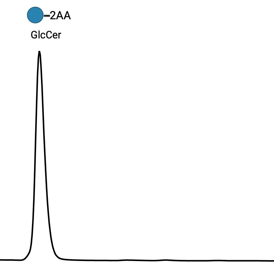
Figure 2 Typical HPLC profile for 2AA-labelled glucose enzymatically released from glucosylceramide.
Note
Figures created with BioRender.com