Feb 02, 2025
An Optimized Porcine Corneal Ex Vivo Model Using Plumbing Rings and Medical Adhesive for Controlled Localized Infection with Topical Treatments
Peer-reviewed method
- Daniel Foulkes1,
- Keri Mclean1,
- Tarunima Sharma1,
- David Fernig1,
- Stephen Kaye1
- 1University of Liverpool
- PLOS ONE Lab Protocols
- Spotlight series


External link: https://doi.org/10.1371/journal.pone.0319911
Protocol Citation: Daniel Foulkes, Keri Mclean, Tarunima Sharma, David Fernig, Stephen Kaye 2025. An Optimized Porcine Corneal Ex Vivo Model Using Plumbing Rings and Medical Adhesive for Controlled Localized Infection with Topical Treatments. protocols.io https://dx.doi.org/10.17504/protocols.io.eq2ly6nkmgx9/v1
Manuscript citation:
Foulkes DM, Mclean K, Sharma T, Fernig DG, Kaye SB (2025) Stable topical application of antimicrobials using plumbing rings in an ex vivo porcine corneal infection model. PLOS One 20(4). doi: 10.1371/journal.pone.0319911
License: This is an open access protocol distributed under the terms of the Creative Commons Attribution License, which permits unrestricted use, distribution, and reproduction in any medium, provided the original author and source are credited
Protocol status: Working
We use this protocol and it's working
Created: December 02, 2024
Last Modified: May 05, 2025
Protocol Integer ID: 113423
Keywords: Ex vivo, infection model, cornea, antimicrobial, porcine, Microbial keratitis, Host-pathogen interactions, Pseudomonas aeruginosa, Localized pathogen inoculation, Plumbing ring model, Antimicrobial treatment assessment, Corneal opacity, versatility of porcine corneal ex vivo model, optimized porcine corneal ex vivo model, porcine corneal ex vivo model, medical adhesive for controlled localized infection, topical treatments infection model, corneal surface, microbial keratitis, controlled localized infection, infection model, medical adhesive, burden of microbial keratitis, evaluation of corneal opacity, pathogen interaction, infection, infection progression, corneal opacity, pathogen, optimized ex vivo protocol, cornea, effective ex vivo model, quantification within the cornea, traditional cell culture system, staphylococcus aureus, adenovirus, precise maintenance of therapeutic agent, ex vivo protocol, reliable assessments of antimicrobial effect, antimicrobial effect, viruses such as herpes simplex, including pseudomo
Funders Acknowledgements:
Fight for Sight
Grant ID: 5175 / 5176
Disclaimer
Spotlight Video
The video below is a supplement with extra context and tips, as part of the protocols.io Spotlight series, featuring conversations with protocol authors.
Abstract
Infection models are essential tools for understanding host-pathogen interactions and developing novel
therapies. Ex vivo infection models offer significant ethical and practical advantages, enabling more accurate and physiologically relevant insights compared to traditional cell culture systems.
The versatility of porcine corneal ex vivo models has made them valuable for studying a range of
pathogens, including Pseudomonas aeruginosa, Staphylococcus aureus, Streptococcus pneumoniae, Candida albicans, Acanthamoeba, and viruses such as Herpes Simplex and adenovirus. Here, we present an
optimized ex vivo protocol that addresses several limitations, using plumbing rings and medical adhesive to enhance experimental accuracy and reproducibility.
The application of a 10 mm plumbing ring to the centre of the cornea allows localized inoculation, preventing the "run-off" of topically added aqueous solutions and ensuring that pathogens remain confined to the site of infection. Additionally, this setup enables precise maintenance of therapeutic agents on the corneal surface, allowing for more reliable assessments of antimicrobial effects.
For analysis, we describe methods for colony-forming unit (CFU) quantification within the cornea, evaluation of corneal opacity, and histological analysis using hematoxylin and eosin staining to assess infection progression over 48 hours. This improved, accessible, and cost-effective ex vivo model provides a standardized platform for evaluating potential treatments, ultimately supporting clinical translation and reducing the burden of microbial keratitis.
Materials
Required Materials
- Whole pig eyes (obtainable from local abattoirs).
- Surgical scissors, forceps and scalpel (size 10) (Fisher scientific).
- Phosphate-buffered saline (PBS), 10% (v/v) iodinated povidone (Ecolab) and ethanol.
- 10 mm Plumbsure Rubber O rings (from Hardware stores such as B&Q).
- LIQUIBAND 0.8 g Exceed topical skin adhesive (Advanced Medical Solutions).
- Luria–Bertani (LB) broth, LB agar (Melford), UltraPure Agarose (Thermo Fisher), DMEM with 10% (v/v) foetal bovine serum (Labtech).
- Sterile 10 cm petri dishes and 12-well plates.
- Histology: Paraformaldehyde, ethanol, xylene.
- Histology: Hematoxylin and eosin (H&E) staining kit (Abcam).
- Glycerol stock of microorganism to be investigated (in this example, the P. aeruginosa strain PA103).
Required instruments
- Biosafety Cabinet (BSC), Class II.
- Tissue homogenizer (such as a Qiagen Tissue ruptor).
- Gel Imaging System: A Bio-Rad ChemiDoc or equivalent.
- Spectrophotometer: equipped with a 12-well plate reader for absorbance measurements.
- Microtome (histology).
Troubleshooting
Preparation of bacterial inoculum
2h 25m
The day before porcine cornea dissection, inoculate 100 mL of LB broth with a pipette scratch of microorganism to be investigated (P. aeruginosa strain PA103 will be discussed in an exemplary protocol) from a glycerol stock (or agar plate, if appropriate). Culture overnight at 37 ºC in a shaker incubator.
10m
Take 2 mL of the PA103 overnight culture and add to 20 mL of fresh LB broth. Allow the subculture to expanded in a shaker incubator (37ºC) until an OD600 of 0.8 is reached.
2h
Take 1 mL of subculture and centrifuge for 5 minutes at 5000 g. Discard the supernatant and resuspend the bacterial pellet in 10 mL of PBS, yielding a PA103 suspension at a concentration of 107 CFU/mL.
Note: When culturing microorganisms other than P. aeruginosa, preliminary methods such as
serial plating should be performed to accurately quantify their concentrations
at various optical densities (OD600).
15m
Preparation of porcine corneas
1h 50m
The morning of slaughter, collect porcine eyes from the
abattoir and transfer to PBS.
Make sure that all regents and equipment used are sterilized (autoclaved where appropriate) before beginning dissections. In a laminar flow hood, use forceps to hold the eye in place and make a small at the edge of the sclera to release ocular pressure and provide an entry point for surgical scissors. Use this procedure for each cornea to be dissected.
5m
Use surgical scissors to cut out the cornea. Maintain 2.5-5 mm of sclera outside the circumference of the cornea.
5m
From the resulting corneoscleral button, remove the uveal tissue using two sets of forceps.
5m
For sterilisation, incubate corneas in 10% (v/v) iodinated povidone for 2 minutes. Afterwards, wash the corneas 3 times in PBS.
5m
In another 10 cm dish, immerse one side of 10 mm plumbing ring onto dispensed LIQUIBAND topical skin adhesive. Use forceps to compress the edges of the ring to the center of the cornea, ensuring a tight seal.
5m
Dissolve agarose (0.5 [w/v]) in DMEM using a water bath heated to 65 °C.
Allow this solution to cool to approximately 37 °C.
1h
Place corneas epithelial side down. Using a pipette, add the cooled agarose DMEN to the endothelial side of corneas to fill the cavity.
10m
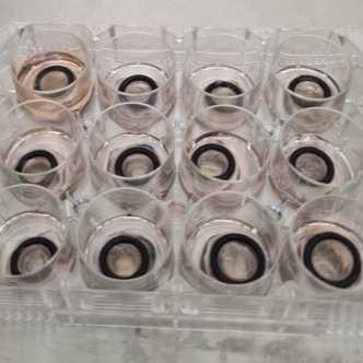
Once the agarose-DMEN has solidified, transfer the corneas into 12-well
plates, epithelial side up.
5m
To induce a controlled epithelial wound on the cornea, add 10 µL 70% ethanol (v/v in PBS) to the center of the corneal surface for 10 seconds. Following ethanol exposure, gently scrape ethanol covered epithelium with the edge of a scalpel to simulate mechanical injury.
5m
Rince the corneas three times with PBS to ensure complete removal of ethanol and debris. Finally, add 1 mL of DMEM to each well, covering the outer edge of the corneal epithelium without submerging the plumbing ring.
5m
Infection of corneas and topical application of treatment(s)
20m
Add 20 µL of the previously prepared bacterial suspension to an Ependorf tube (one Ependorf to be used per cornea). Before addition, ensure that the bacterial suspension is adequately dispersed with a brief vortex.
5m
Make up the final volume to 300 µL with PBS, with topical treatments to be investigated also included.
5m
Add 150 µL of suspension from step 2 to the centre of each cornea, filling the plumbing ring reservoir. In this example, for P. aeruginosa PA103, this equates an inoculum of 1 x 10⁵ colony forming units (CFU) for each cornea.
5m
Transfer the corneas, in 12-well plates, to a 37 °C incubator in 5 % CO2 (v/v). Infection progress should be monitored over the course of several days. Infection progression and extent can vary depending on the species and strain being investigated. It is therefore advised that initial optimization experiments are carried out, inoculating corneas with a range (1 x 10² – 1 x 10⁷) of CFU, observing infection progression at 24 h time points.
5m
Imaging and spectrophotometric analysis of corneal opacity
30m
At assay endpoint, take the corneas from the incubator. Using forceps, carefully remove the plumbing rings and rinse the corneas gently in a beaker of PBS. handle the corneas with care, as infection can cause the epithelial layer to become fragile and prone to detachment. Minimize physical contact with the surface to preserve tissue integrity.
10m
Image the corneas using a Bio-Rad ChemiDoc (or equivalent), selecting the appropriate gel imaging mode (e.g., "Coomassie Stain" or similar) for capturing qualitative data on ulcer size and density.
10m
To assess ulcer density using a spectrophotometer, place the corneas epithelial side up in 6 or 12-well plates and take optical absorbance readings at 400 nm.
10m
For quantitative analysis of ulcer size and density, utilize open-source software such as ImageJ. Use the measurements tool in ImageJ to outline and quantify ulcer areas, applying appropriate thresholding
to assess density and ensure consistent parameters across samples for reliable comparison.
Histological analysis of infected porcine corneas
1d 17h 26m
Incubate the corneas in 4% paraformaldehyde (PFA) for 24 h.
1d
Dehydrate the corneas through a graded series of ethanol (70%, 90% then 100% [v/v in PBS]).
30m
Clear the corneas using xylene and then embedded in paraffin wax.
30m
Slice the corneas into 5 µm-thick sections using a microtome, ensuring consistent thickness for reproducibility.
1h
Mount the sections onto numbered glass slides, ensuring sequential numbering for reliable comparison between samples.
1h
Prepare the samples for staining or labelling by clearing with xylene and hydration through a graded series of ethanol (100%, 95%, 70%, 50% and 0% [v/v in PBS]).
30m
For H&E staining: Immerse slides in hematoxylin stain for 5 minutes and then rinse the slides in running tap water for 1-2 minutes to remove excess stain.
10m
Differentiate in acid alcohol (1% HCl in 70% ethanol) for a few seconds to remove nonspecific staining then rinse in water.
1m
Immerse the slides in 0.1% (v/v) ammonium hydroxide in distilled water for 30 seconds to 1 minute and then rinse with water.
5m
Immerse slides in eosin stain for 30 seconds to 2 minutes and then rinse the slides in water to remove the excess stain.
5m
Dehydrate the samples by immersion in a graded series of ethanol (70%, 95% and 100% [v/v in PBS] and then clear by incubation in xylene (2 minutes).
30m
Add mounting medium to the sample and coverslip.
5m
Allow the samples to dry and then view under a light microscope and take images using a camera or slide scanner.
12h
ImageJ can be effectively used to quantify epithelial thickness and assess stromal swelling. For epithelial measurements, simple linear measurements can be applied directly on calibrated images. Stromal swelling can be quantified by measuring the white space within the stroma. To achieve this, convert H&E-stained images to 8-bit grayscale and define the stroma as the region of interest (ROI). Set threshold values (e.g., 230–255) to isolate white pixels corresponding to stromal spaces. Calculate the percentage area occupied by white pixels within the ROI to quantify stromal swelling accurately. This approach provides a reproducible and standardized method for image-based corneal analysis.
1h