Jun 12, 2026
An integrated transmission electron microscopy workflow for endometrium and isolated endometrial cells
- Katheryn Peterson1,
- Jaydeep Kolape2,
- Daniel Mathew1
- 1Department of Animal Sciences, University of Tennessee, Knoxville, TN, USA;
- 2Advanced Microscopy and Imaging Center, University of Tennessee, Knoxville, TN, USA


Protocol Citation: Katheryn Peterson, Jaydeep Kolape, Daniel Mathew 2026. An integrated transmission electron microscopy workflow for endometrium and isolated endometrial cells. protocols.io https://dx.doi.org/10.17504/protocols.io.5jyl83rddv2w/v1
License: This is an open access protocol distributed under the terms of the Creative Commons Attribution License, which permits unrestricted use, distribution, and reproduction in any medium, provided the original author and source are credited
Protocol status: Working
This protocol has been utilized to prepare bovine endometrial tissue and cultured, primary endometrial cells for transmission electron microscopy.
Created: May 28, 2026
Last Modified: June 12, 2026
Protocol Integer ID: 318152
Keywords: transmission electron microscopy, endometrium, epithelial cells, culture, transmission electron microscopy workflow for endometrium, isolated bovine endometrial epithelial cell, bovine endometrial epithelial cell, endometrial cells for tem, endometrial cells the endometrium, isolated endometrial cell, endometrium in mammal, preparing intact endometrium, bovine endometrium, endometrial remodeling, intact endometrium, endometrium, integrated transmission electron microscopy workflow, transmission electron microscopy, subcellular ultrastructure, cellular morphological change, cultured cells from the same animal, cultured cell, biopsied tissue, postpartum, organelle structure, resolution comparison between biopsied tissue
Funders Acknowledgements:
USDA National Institute of Food and Agriculture.
Grant ID: Hatch Project No. 1022068
Abstract
The endometrium undergoes dynamic morphological changes throughout the menstrual or estrous cycle, pregnancy, postpartum, and disease. Methods to evaluate endometrial remodeling and cellular morphological changes, both within the tissue microenvironment and in isolation (in vitro culture) provide an opportunity to better understand these changes and reproductive disease. Transmission electron microscopy (TEM) offers the resolution needed to examine the subcellular ultrastructure of highly dynamic tissues. Here, we present a unified workflow for preparing intact endometrium and isolated endometrial cells for TEM. The method outlines fixation with glutaraldehyde and osmium tetroxide, graded ethanol dehydration, resin embedding, ultrathin sectioning, and post-staining with uranyl acetate and lead citrate. This approach preserves membrane integrity, organelle structure, and ultrastructural detail, allowing direct high-resolution comparison between biopsied tissue and cultured cells from the same animal if desired. Bovine endometrium and isolated bovine endometrial epithelial cells were utilized in this protocol; however the principles underlying the method are broadly applicable to a variety of tissues and cell types. Overall, this workflow expands the utility of TEM with an emphasis on the study of reproduction and reproductive diseases associated with endometrium in mammals.
Materials
A complete list of materials is provided in Table 1.
Table 1. Tools and reagents needed for this protocol.
| Name of Material | Source | Catalog Number | |
| Chemicals | |||
| Accutase | Sigma | Cat#A6964-500mL | |
| Antibiotic and antimycotic (ABAM) | Gibco | Cat#15-240-062 | |
| DI water | Gibco | Cat#15-230-162 | |
| Dulbecco’s phosphate-buffered saline (DPBS) | Gibco | Cat#14-190-250 | |
| EMBed 812 resin | Electron Microscopy Sciences | Cat#14120 | |
| Ethanol (70%) | VWR | Cat#95041-462 | |
| Glutaraldehyde | Sigma | Cat#G6257-1L | |
| Hank’s Buffered Salt Solution (HBSS) | Gibco | Cat#14175-103 | |
| Methanol | Electron Microscopy Sciences | Cat#18510 | |
| Microscopy-grade ethanol | Electron Microscopy Sciences | Cat#15056 | |
| Lead Citrate | Electron Microscopy Sciences | Cat#17800 | |
| Osmium tetroxide | Sigma | Cat#75632-10ML | |
| Sodium Hydroxide | Sigma | Cat#SX0607N-6 | |
| Uranyl Acetate | Electron Microscopy Sciences | Cat#22400 | |
| Other | |||
| 15 and 50 mL conical tubes | Fisher | Cat#14-959-53A (15 mL); Cat#14-432-22 (50 mL) | |
| BEEM‱ Embedding Capsules, Size 00, 8 mm I.D. | Electron Microscopy Sciences | Cat#70000-B | |
| Copper grids | Electron Microscopy Sciences | Cat#CF200-Cu-50 | |
| Diamond knife | DiATOME | Cat# 15-US | |
| Dissecting trays | Any vendor | N/A | |
| Eyelash tool | Electron Microscopy Sciences | Cat#71182 | |
| Fine forceps | Any vendor | N/A | |
| Fine-tipped curved scissors | Any vendor | N/A | |
| Glass knife | Electron Microscopy Sciences | Cat#12345 | |
| Hemocytometer or automated cell counter | VWR | Cat#15170-173 | |
| Microcentrifuge tubes; 1.5 mL | Fisher Scientific | Cat#05-408-129 | |
| Parafilm M | Genesee Scientific | Cat#16-100 | |
| Pipettor set and tips | Any vendor | N/A | |
| Razor blades | Any vendor | N/A | |
| Scalpel blades | Any vendor | N/A | |
| Serological pipettes | Any vendor | N/A |
A complete list of required equipment is provided in Table 2.
Table 2. Equipment needed for this protocol.
| Name of Equipment | Source | Catalog Number | |
| Centrifuge | Any vendor | N/A | |
| Glass knife maker | LKB | LKB Glass Knife Maker Type 7801B | |
| Incubator with temperature and atmosphere control | Any vendor; capable of 38.8°C; 5.0% CO2 for bovine cell culture | N/A | |
| Oven | Any vendor; capable of 60°C | N/A | |
| Shaking platform or rocker | Any vendor | N/A | |
| Stereomicroscope | Nikon | Cat#SMZ800N | |
| TEM Microscope | JEOL | JEM-1400 | |
| Ultramicrotome | Leica Microsystems | Leica UC7 Ultramicrotome | |
| Vacuum | Any vendor | N/A | |
| Water bath | Any vendor | N/A |
A complete list of required solutions is provided in Table 3. Prepare solutions fresh, on the same day the respective steps are to be completed.
Table 3. Solutions needed for this protocol.
| Name of Solution | Components | Purpose | |
| 3.1 Bovine Uterine Tissue Processing (see Chaney et al., 2020 and Oliver et al., 2023) | |||
| 70% ethanol | Ethanol prepared with sterile water | To drench dissection tray and paper towels | |
| DPBS wash | 500 mL Dulbecco’s phosphate-buffered saline (DPBS) supplemented with 1% antibiotic-antimycotic (ABAM) | To hydrate uterine tissue, use as necessary | |
| HBSS wash | 75 mL Hank’s Buffered Salt Solution (HBSS) supplemented with 1% ABAM, aliquoted into 3 25 mL quantities in 50 mL conical tubes | To collect and wash endometrial strips, final 25 mL volume used in 3.2 | |
| 3.2 Tissue and Cell Collection and Chemical Fixation | |||
| 2.5% glutaraldehyde | Glutaraldehyde prepared in DPBS; volume dependent on number of tissue sections and cell pellets, (1 mL to fix five endometrial sections measuring 1 mm³ and 1 mL for 1 cell pellet). | To serve as a primary tissue and cell fixative | |
| DPBS cell flask wash | DPBS supplemented with 1% ABAM; volume dependent on number of T75 flasks (10 mL per flask) | To wash cell cultures prior to collection | |
| Accutase | Accutase; volume dependent on number of T75 flasks (3 mL per flask) | To enzymatically detach epithelial cells from flasks | |
| DPBS wash | DPBS (no ABAM); volume dependent on number of T75 flasks (6 mL per flask) | To collect and resuspend epithelial cells in | |
| 1% osmium tetroxide | Osmium tetroxide prepared in DPBS; volume dependent on number of tissue sections and cell pellets (1 mL to fix five endometrial sections 1 mL for 1 cell pellet) | To serve as a secondary tissue and cell fixative | |
| 3.3 Dehydration | |||
| DPBS wash | DPBS (no ABAM); volume dependent on number of tissue sections and cell pellets (15 mL total to wash five endometrial 3 times and 15 mL total to wash 1 cell pellet 3 times) | To wash off osmium tetroxide prior to ethanol dehydration | |
| Tissue ethanol series | Microscopy-grade ethanol (EtOH) prepared with DPBS:1 mL 35% EtOH1 mL 50% EtOH1 mL 70% EtOH1 mL 95% EtOH2 mL 100% EtOH | To gradually dehydrate endometrial tissue | |
| Cell ethanol series | Microscopy-grade ethanol (EtOH) prepared with DPBS:1 mL 50% EtOH1 mL 70% EtOH2 mL 90% EtOH3 mL 100% EtOH | To gradually dehydrate endometrial epithelial cells | |
| 3.4 Embedding | |||
| EMBED 812 resin | 9.5 g EMBed, 5.0 g DDSA, 4.0 g NMA, and 0.28g DMP-30. | Use to prepare gradual ethanol : resin embedding steps; prepare each dilution immediately prior to use | |
| 3.5 Sectioning, Staining, and Imaging | |||
| 50% uranyl acetate | Uranyl acetate prepared with methanol; 20 µL per grid | Post-staining solution to increase contrast | |
| 2% lead citrate | 10 mL distilled water0.02 g lead citrate0.1 mL 10N sodium hydroxide | Post-staining solution to increase contrast | |
| Distilled water | Sterile, distilled water; 20 µL per grid | To wash grids between staining steps |
Introduction
Transmission electron microscopy (TEM) is a valuable tool for resolving cellular and subcellular architecture at the nanometer-scale resolution. Unlike light microscopy, TEM offers detailed visualization of intracellular organelles, membrane architecture, junctional complexes, cytoskeletal organization, and vesicular trafficking. Ultrastructural features such as these offer insight into cellular processes, allowing for a better understanding of how cells interact with their surrounding tissue microenvironment or in culture under various conditions. However, TEM workflows are often optimized for a single sample type, such as intact tissue or cultured cells, limiting direct ultrastructural comparisons between an isolated cell in culture and the tissue biopsy from which it is derived.
In eutherian mammals, the endometrium is a structurally dynamic tissue that undergoes continuous structural and functional remodeling across the menstrual or estrous cycle, early pregnancy, placentation, parturition, and postpartum uterine involution. Coordinated uterine function and remodeling depend on interactions among luminal epithelial cells, glandular epithelial cells, stromal fibroblast cells, vascular endothelial cells, and diverse immune populations influenced by ovarian and placental hormones. Throughout the reproductive cycle and pregnancy, these cell populations undergo extensive cellular and subcellular reorganization. Other microscopy-based approaches have identified temporal and molecular contributors of these processes; however, they lack the physical resolution needed to visualize intracellular organization and the nanometer-scale cell-cell interactions that govern uterine biology.
Historically, TEM has been applied to the study of endometrium and placenta to characterize tissue-level organization and cell-cell interactions, providing insight into uterine structure and function in multiple species. Classical studies by F.B.P. Wooding and colleagues, for example, documented the ruminant endometrium and placenta, particularly in sheep, providing detailed descriptions of trophoblast dynamics and placental binucleate cell formation at the feto-maternal interface. While these studies are foundational, protocols that facilitate ultrastructural comparison between endometrial tissue and isolated endometrial cells derived from the same tissue are not available despite widespread use of cell isolation and culture systems in modern reproductive and cell biology research. This limitation constrains mechanistic investigation of endometrial pathologies, such as endometriosis in humans or endometritis in cattle, which rely heavily on isolated cell and culture-based approaches. An approach that enables ultrastructural comparison of endometrial tissue biopsies and corresponding isolated cells before and after experimental treatment provides an impactful, unified framework for evaluating uterine phenotypes, such as those associated with disease and therapeutic responses. Direct comparison of cultured cells with their tissue of origin is also particularly important for validating in vitro models and assessing how isolation, culture environment, or experimental treatments influence cellular architecture and function. For example, disruptions in mitochondrial dynamics, including changes in number, structure, and mitophagy, which influence oxygen metabolism and oxidative stress, have been reported in human endometrial pathologies and aging, emphasizing the need for ultrastructural analysis.
Here, we describe an integrated TEM workflow optimized for both intact endometrial tissue and primary endometrial epithelial cells derived from the same uterus. Individual processing steps are based on established TEM methodologies. The technical novelty of this method lies in its integration and standardization across intact tissue and isolated cell samples, enabling a simplified workflow and direct ultrastructural comparison under unified preparation conditions. Additionally, the protocol supports the preparation of isolated cells in suspension without the need for auxiliary pre-embedding stabilization or localization steps, such as cytospin preparations, agarose or hydrogel encapsulation, BSA/bisacrylamide (BSA-BA)-mediated polymerization, or incorporation of dyes for pre-fixation visualization. These strategies, while occasionally used to immobilize or localize non-adherent cells, introduce additional manipulation steps that may alter cellular morphology and ultrastructural integrity. By eliminating intermediate consolidation and labeling procedures, the present workflow preserves cellular structure while maintaining sample coherence and processing efficiency. This unified protocol therefore provides a standardized approach from tissue dissociation and chemical fixation to imaging of both intact tissue and isolated cells. Practical considerations designed for endometrial tissues include strategies to improve tissue fixation, preserve subcellular features, and maintain cell integrity. Bovine intercaruncular endometrium and primary endometrial epithelial cells were used to develop this protocol; however, it is readily adaptable to other mammalian endometria or epithelial tissues and primary cell populations where comparative ultrastructural analysis between intact tissue and cultured cells is desired. A description of the bovine endometrial cell isolation and culture protocol are described in detail by Chaney et al. (2020) and Oliver et al. (2023). An optimized TEM protocol that enables direct comparison of biopsied, intact endometrial tissue and isolated primary cells provides an impactful tool for studying reproductive biology.
Experimental Design
Bovine uteri and endometrial tissue were sourced from a USDA-inspected animal abattoir (Southeastern Provisions, Bean Station, TN), thus no live animals were used to develop the TEM protocol and Institutional Animal Care and Use Committee (IACUC) approval was not required.
2.1. Materials
A complete list of materials is provided in Table 1.
Table 1. Tools and reagents needed for this protocol.
2.2. Equipment
A complete list of required equipment is provided in Table 2.
Table 2. Equipment needed for this protocol.
2.3. Solutions
A complete list of required solutions is provided in Table 3. Prepare solutions fresh, on the same day the respective steps are to be completed.
Table 3. Solutions needed for this protocol.
Procedure
3.1 Bovine Uterine Tissue Processing (see Chaney et al., 2020 and Oliver et al., 2023)
Timing: 1 h
These steps outline preparation of the bovine uterine horn for the collection of intercaruncular endometrium used to generate intact endometrial sections and isolated endometrial cells for TEM analysis.
1. In the lab, prepare a working area and necessary solutions (Table 3).
1a. Prepare a dissection tray on ice with sterile fine forceps and fine-tipped iris curved scissors readily available in a beaker of 70% ethanol. Line the tray with paper towels and drench with 70% ethanol.
2. Isolate the uterine horn for endometrial collection.
2a. Gently rinse the exterior of the tract with cool tap water to remove residual blood and debris by pinching the dissected end of the reproductive tract (usually near the cervix or uterine body) closed with one hand. Do not allow water to enter the uterus. After washing, pour DPBS wash over the tract.
2b. Dissect the uterine horn from surrounding reproductive tract and broad ligament (including the mesometrium, mesosalphix, and mesovarium) using sterile scissors. The ovaries, oviduct, and all vaginal and cervical tissue should be removed. Keeping the uterine lumen entrance closed, rinse the uterine horn with water and place it on the dissecting tray.
3. Using a sterile scalpel blade, create a longitudinal incision into the uterine horn on the anti-mesometrial side, approximately 2-3 cm above the uterine body, toward the utero-tubal junction, exposing the endometrium.
4. Wash the uterine lumen by pouring approximately 50 mL of DPBS wash medium over the surface of the exposed endometrium. Repeat this process as needed to remove debris and prevent the tissue from drying.
5. Use fine forceps and curved iris scissors to dissect strips of intercaruncular endometrium from the uterine lumen, avoiding the underlying myometrium.
5a. Intercaruncular endometrial strips should be approximately 3 cm long and approximately 1 mm thick to minimize the collection of myometrium.
5b. Place the strips into one of the 50 mL conical tubes containing 25 mL of HBSS. Fill the conical tube with 10 mL of endometrial tissue to ensure enough tissue is collected for fixation and cell dissociation and isolation.
6. Wash endometrial strips by transferring tissue into a new conical tube containing 25 mL of fresh HBSS.
6a. Using sterile forceps, clasp and slowly drag the tissue over the rim of the conical tube before transferring to the new tube. This will help remove unwanted blood.
3.2 Tissue and Cell Collection and Chemical Fixation
Timing: 2 d
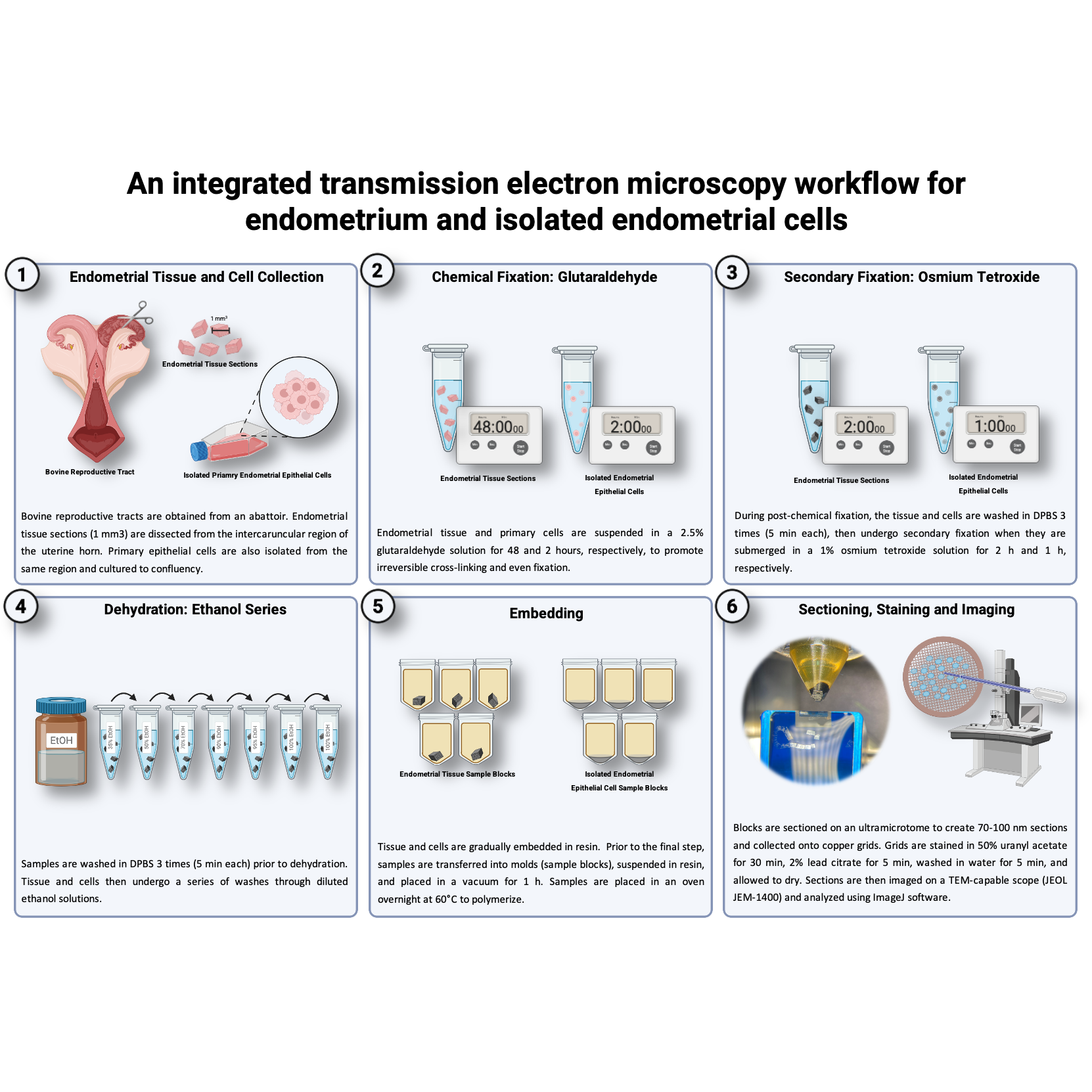
To achieve optimal protein crosslinking and ultrastructural preservation, endometrial tissue and isolated cells will be fixed with glutaraldehyde and osmium tetroxide. Chemical fixation of the endometrial tissue with glutaraldehyde forms irreversible crosslinks between proteins, promoting a slower, more even fixative distribution across the tissue and cells than what is achieved with formaldehyde. While glutaraldehyde-only fixation is sufficient for membrane stabilization, alternative fixation techniques, such as mixed aldehyde formulations (ex. Karnovsky-type glutaraldehyde-paraformaldehyde combinations) may further improve ultrastructural preservation and membrane contrast in future adaptations of this workflow.
7. Remove 1-2 strips of endometrium from the final HBSS wash and place them into a sterile petri dish.
7a. Using a sterile scalpel blade, create tissue sections from the endometrial strips measuring 1 mm3.
Note: The number of sections dissected should be determined based upon experimental goals.
7b. Wash the sections one final time by pouring 25 mL of fresh HBSS into the petri dish. Use sterile forceps to gently dip the sections in and out of the solution.
7c. Place the endometrial sections into 1 mL of a 2.5% glutaraldehyde solution prepared with DPBS (no ABAM) for 48 h at room temperature (R.T.).
Note: The volume of the glutaraldehyde solution should be adjusted to accommodate the number of endometrial sections (ex. 1 mL of the glutaraldehyde solution to fix five endometrial sections measuring 1 mm3). The 2.5% glutaraldehyde solution will also be used to fix isolated endometrial cells. A 48 h fixation period was selected to promote uniform glutaraldehyde penetration in dense endometrial tissue and to minimize tissue gradients across tissue depth. Shorter durations (ie. 12-24 h) may be sufficient for less dense or smaller quantities of tissue, however were not empirically evaluated in this study, but may represent an optimization parameter for users.
Note: If isolation of primary cells is desired, use remaining endometrial strips to isolate cells as described by Chaney et al. 2020 and Oliver et al., 2023, which detail tissue digestion and dissociation, isolation, and culture of primary bovine endometrial fibroblast or epithelial cells (Chaney et al., 2020). This protocol describes fixation of primary cells after 5-7 days of monoculture in T75 flasks. When fixing both intact endometrium and cultured cells from the same uterus, intact tissue must be fixed immediately after dissection, whereas cultured cells are fixed several days later. Extended culture, as described by Chaney et al. (2020) and Oliver (2023), increases epithelial cell purity and yield. Alternatively, using the same digestion and isolation protocols, mixed endometrial cell populations (including uterine glands) may be fixed on the day of tissue collection from the initial digestive filtrate._
8. Once primary endometrial epithelial cells are approximately 80% confluent within a T75 flask, prepare the cultured cells for fixation.
8a. Pour off and discard culture medium. Add 10 mL of DPBS wash medium directly to the culture flask, agitate gently, and pour off and discard the wash solution.
8b. Enzymatically detach the epithelial cells from the T75 flask by applying 3 mL of Accutase for 15 min. Forcefully agitate the flasks by tapping the sides of the flask to encourage cell detachment. Confirm detachment under a microscope before proceeding.
8c. Using a serological pipette, add 6 mL of DPBS to each flask. Transfer the cell solution into a 15 mL conical (or 50 mL conical depending on the number of flasks).
8d. Centrifuge the cells at 700 x g for 7 min at R.T.
8e. Following centrifugation, discard the supernatant from the cell pellet and resuspend the cells in 5 mL of 2.5% glutaraldehyde solution for 2 h.
9. After the 2 d fixation (intact endometrial tissue) or 2 h fixation (primary endometrial epithelial cells) remove the glutaraldehyde solution.
9a. Aspirate and discard the glutaraldehyde solution from the endometrial tissue.
9b. To remove the glutaraldehyde solution from the suspended epithelial cells, centrifuge the cells at 700 x g for 7 min at R.T. Aspirate and discard the supernatant from the cell pellet following centrifugation.
10. Gently wash both the intact endometrial tissue and epithelial cell pellet with 1 mL of DPBS three times, 5 min each, to remove any remaining glutaraldehyde.
Note: After each wash of the cell pellet, centrifuge the cells at 700 x g for 7 min at R.T. before aspirating and discarding the supernatant. Be aware repeated supernatant aspirations will reduce the cell pellet size and number of cells available for TEM evaluation._
Note: After removing the glutaraldehyde solution and washing the intact tissue with DPBS it can be held for multiple days in DPBS. During development of this protocol, fixed tissue sections were held in DPBS at 4°C for five days prior to further processing. Longer storage durations were not evaluated, however prolonged aqueous storage may increase the risk of membrane discontinuities, cytoplasmic extraction, or organelle swelling (Shami et al., 2024, Augusteyn et al., 2008, Mount et al., 1997). Intact tissue can be further processed alongside the fixed primary endometrial epithelial cells isolated from the same uterus.
11. Submerge endometrial tissue for 2 h and cell pellet, for 1 h, in 1 mL of 1% osmium tetroxide solution (1%) at R.T. as a secondary fixative to stabilize unsaturated lipids, cross-linking hydrophobic domains to prevent extraction during subsequent dehydration steps.
3.3 Dehydration
Timing: 3 h
Thorough dehydration is necessary to ensure that water is gradually removed from the sample, replacing it with ethanol as an organic solvent. Excessive water within the tissue can impede resin polymerization and evaporate in the TEM vacuum, affecting interaction between the electron beam and the sample.
12. Following osmium tetroxide fixation, wash the tissue and cell pellet with 5 mL DPBS three times, 5 min each. As described above, after each wash, centrifuge the epithelial cells (700 X g for 7 min) and remove the supernatant.
13. Dehydrate the tissue and epithelial cells by washing with an ethanol series.
13a. Wash the tissue sections (15 min each wash) sequentially in 1 mL of 35%, 50%, 70%, and 95% ethanol before washing the tissue twice in 100% ethanol.
13b. Wash the cell pellet sequentially in 1 mL of 50% followed by 70% ethanol (5 min each), 1 mL of 90% ethanol twice (5 min each), and 1 mL of 100% ethanol three times (10 min each). Centrifuge the cells (700 X g for 7 min) and remove the supernatant between washes.
3.4 Embedding
Timing: 4 d
Embedding the endometrial sections and cell pellet in resin preserves the three-dimensional structure and provides a medium for sectioning. A graded ethanol-to-resin series promotes gradual infiltration of the resin into the samples, while vacuum treatment removes air bubbles for uniform embedding. Final polymerization solidifies the resin to stabilize the tissue and cellular morphology with minimal shrinkage. Epoxide resin was selected because of its medium viscosity, moderate penetration rate, and to avoid excessive shrinkage of membranes and structures.
14. Create labels with small pieces of paper (~6 mm x 3 mm, size to fit along the top portion of the plastics mold) labelled with sample IDs written only in pencil or with computer ink. If only the exterior of the molds is labelled, samples may not be identifiable once removed from molds.
15. Embed endometrial sections and cell pellet with EMBed 812 resin over four total embedding steps, each carried out on a rotary mixer.
15a. Resuspend the endometrial section and cell pellet samples in 1.5 mL of solution across three embedding steps:
• 2 parts (1 mL) ethanol to 1 part (500 µL) resin for 1 h
• 1 part (750 µL) ethanol to 1 part (750 µL) resin overnight
• 1 part (500 µL) ethanol to 2 parts (1 mL) resin for 1 h
15b. After the third embedding step, transfer the endometrial sections and cell pellet samples into labelled molds and suspend them in 500 µL of resin.
16. Place sample molds in a vacuum for 1 h to remove air bubbles.
17. Transfer the sample molds into a 60°C oven to polymerize overnight.
3.5. Sectioning, Staining, and Imaging
Timing: 5 h
Ultra-thin sections of embedded endometrial tissue sections and epithelial cells for TEM imaging are generated by trimming resin-embedded samples with a razor blade and glass knife, then sectioned with a diamond knife to create ultra-thin 70-100 nm sections. Sections are collected onto copper grids and post-stained with uranyl acetate and lead citrate to enhance contrast within the sample to better visualize membranes, organelles, dense protein, and DNA. Final steps include imaging on a TEM-microscope.
18. Remove plastic molds from polymerized samples.
19. Using a dissection- or stereomicroscope and razor blade to remove excess resin at the point of each sample block, trimming the tip of the block into a square pyramid shape. Remove as little sample as possible, while still minimally exposing the sample’s surface.
20. Prepare glass knives from the glass knife strip by using LKB knife maker type 7801B.
21. Cut rough sections with a glass knife to ensure a smooth, even surface at the tip of each block, enabling cutting of continuous, square-shaped sections.
22. Transfer samples to an ultramicrotome and use a diamond knife to trim 70-100 nm sections.
22a. Collect the sample ribbons from the ultramicrotome’s water boat with an eyelash tool, placing them onto copper grids.
23. Prepare a 50% uranyl acetate in methanol solution. Create individual 20 µL drops of the uranyl acetate solution, one per grid. Invert the grid, section-side down, on top of the drop to stain the section.
23a. Using a strip of Parafilm M, add 20 µL drops of the appropriate staining solution or distilled water, one drop per grid. Grids can be transferred between drops using fine forceps. Be sure to invert grids so that the sections are respectively stained or washed. Cover the Parafilm and grids during staining and wash steps with a petri dish or beaker to prevent evaporation.
24. After 30 min, transfer the grids to individual 20 µL drops of distilled water. Wash the grids by allowing them to sit in the water drops for 5 min.
25. Prepare a 2% lead citrate solution by combining 10 mL of distilled water, 0.02 g lead citrate, and 0.1 mL of 10N sodium hydroxide into a 15 mL conical. Shake vigorously until the lead citrate is in solution. As done in previous steps, create individual 20 µL drops of the lead citrate solution to stain the section. Allow them to incubate with the lead citrate solution for 5 min.
26. Wash the sections/grids a final time in new 20 µL drops of distilled water. Following this wash step, allow grids to dry at R.T. overnight.
27. Once the grids are dry they can be loaded into a TEM scope for imaging.
Note: Our laboratory utilized a JEOL JEM-1400 for TEM imaging, however, any scope equipped for TEM is suitable. Images can be further analyzed using the open-source image analysis platform NIH Image J Fiji software to perform quantitative measurements, adjust contrast, segment structures of interest, and conduct various other types of image analysis. Images shown in this manuscript were minimally processed in Image J Fiji to enhance contrast.
Representative Results
This protocol describes a method to generate high-resolution TEM images of endometrium and primary endometrial epithelial cells with well-preserved ultrastructure. Effectively prepared endometrial samples should provide clearly defined tissue architecture, including organized glandular and vascular tissues. Images shown in Figures 1-3 are representative workflow outputs. Image quality can be further improved through laboratory-specific adjustment or optimization of fixation, staining, and imaging parameters. Nuclei are distinguishable with intact membranes and heterochromatin, as well as other organelles such as mitochondria and vacuoles, as well as cytoskeletal or extracellular matrix components (Figure 1). Various cell types, including potential immune cells (Figure 1A), stromal fibroblasts, luminal or glandular epithelia (Figure 1E) may also be present depending on the tissue orientation and sectioning. Cilia, including microtubules, may also be observed (Figure 1F). In primary endometrial epithelial cell sections, successful outcomes are indicated by round, intact cells with continuous plasma membranes, potentially including microvilli, distinct nuclei, and well-preserved organelles (Figure 2). Post-staining with uranyl acetate and lead citrate should result in sufficient contrast between membranes and cytoplasmic contents, allowing detailed visualization of membrane and organelle morphology.
In contrast, poor fixation or dehydration may result in structural artifacts such as ruptured membranes, swollen, or collapsed organelles (Figure 3B). Over-fixation with osmium tetroxide, uranyl acetate, or lead citrate may also lead to an accumulation of artifacts, often presenting as crystallization (Figure 3C) or peppering of random, dark speckles throughout a section (Figure 3D). Incorporation of a basic wash step with deionized water following post-staining may help reduce artifacts related to post-staining. Inadequate staining may result in low-contrast images, making it difficult to distinguish between cellular or organelle membranes. Troubleshooting recommendations are summarized in Table 4.
Figure 1. TEM of intact bovine intercaruncular endometrial tissue and subcellular structures. (A) Potential polymorphic nuclear cell. (B-C) Endometrial stroma cell filaments and organelles such as nuclei, mitochondria, and cytoskeletal components. (D) Stromal tissue showing multiple cells and extracellular matrix components. (E) Endometrial gland. (F) Cross-section of cilia, likely from a glandular epithelial cell (Atkinson et al., 1984, Almeida et al., 1986).
Figure 2. TEM images of primary endometrial epithelial cells. (A-D) Isolated endometrial epithelial cells averaged 15 µm in diameter, with large circular nuclei roughly 6 µm in diameter. Epithelial cell microvilli as well as the nucleus with heterochromatin are visible. Various organelles including mitochondria are apparent. (E) Epithelial cell nuclear envelope, and ribsosomes along the rough endoplasmic reticulum.
Figure 3. Artifacts associated with TEM preparation, ultramicrotomy sectioning, and imaging. (A) Isolated epithelial cell section showing a fold across the center of a cell and knife marks consistent with ultramicrotomy-induced sectioning artifacts. (B) Isolated epithelial cell section with multiple knife marks and membrane disruption, consistent with mechanical sectioning artifacts and dehydration from alcohol-induced damage. (C) Crystallization artifact, likely associated with uranyl acetate precipitation within the tissue section and poor image acquisition parameters resulting in low contrast. (D) Speckling and “peppering” artifacts consistent with lead citrate precipitation during post-staining. Possible over-fixation with osmium tetroxide with apparent speckling and peppering around sectioned microvilli.
Table 4. Troubleshooting Guide
Discussion
This protocol provides a detailed and reproducible method for preparing intact endometrial tissue and endometrial cells from the same uterus for TEM. By optimizing fixation, dehydration, embedding, and staining, this method preserves ultrastructure for high-resolution imaging, facilitating detailed study of the endometrial microenvironment and isolated endometrial cell structures in culture. A major attribute of this technique is its ability to maintain structural features across complex tissues and isolated cells from the same animal integrated into a unified workflow. Conventional TEM protocols are typically optimized independently for intact tissues or cultured cells, resulting in divergent methods for fixation, dehydration, embedding, and staining that ultimately limit direct comparability. In contrast, the workflow described here applies parallel preparation across tissue and cell samples to provide a framework for controlled comparative analysis of ultrastructural features between native endometrial tissue and isolated epithelial cells.
By enabling direct comparison between intact, biopsied tissue and cultured cells from the same source, this protocol allows assessment of how dissociation, in vitro culture, and experimental manipulation influence cellular ultrastructure. Importantly, it also enables evaluation of cellular physiology in isolated cells derived from pathological endometrial biopsies and characterization of ultrastructural response to experimental treatments. Because of the standardized workflow, the protocol supports comparative interpretation of in vivo and in vitro tissues and facilitates validation of cell culture models commonly used in reproductive biology research. To our knowledge, no published TEM protocol is optimized for both intact endometrium and isolated endometrial cells.
Some limitations are inherent to TEM, including the technical expertise required for ultrathin sectioning or complexity involved in sample orientation following osmium fixation. Osmium fixation, while effective at enhancing membrane contrast, uniformly renders the samples black in color, removing the ability to discern between the luminal and deep mucosal regions of the intact endometrial sample at time of sectioning. Achieving an optimal level of moderate membrane contrast may also be difficult, as under- or over-staining samples may impede the resolution of fine structures, unless staining conditions are carefully optimized. Variability in tissue preservation may also occur due to uneven fixative penetration or mechanical damage during ultrathin sectioning (tissue folds or knife marks), particularly for novice users. A troubleshooting guide (Table 3) is provided to support laboratories adopting TEM.
Despite constraints, TEM remains a powerful technique to study complex tissue microenvironments and cell and/or organelle ultrastructure. For reproductive biologists and clinicians studying endometrial function and uterine disease, such as the case of endometriosis or endometritis, this study provides a detailed TEM protocol for the evaluation of biopsied endometrium and isolated endometrial cells.
Conclusion
Overall, this protocol provides a standardized, reproducible method for generating high-quality TEM preparations of intact tissue and isolated cells, with particular applicability to the endometrium. Its use across intact tissue biopsies and cultured cells allows evaluation of the tissue microenvironment and its influence on cellular ultrastructure, while also facilitating the study of how experimental treatments affect cell function. By unifying preparation conditions across biologically distinct sample types, this method facilitates direct ultrastructural comparison between native tissue and corresponding isolated cells. Regarding the endometrium, the approach provides a valuable tool for investigating complex tissue modifications during the reproductive cycle, pregnancy, parturition, and the postpartum period.
Author Contributions: KDP: experimental design, data collection and organization, and manuscript preparation; JK: experimental design and data collection; DJM: acquisition of funding, experimental design, and manuscript preparation.
Funding: This project was supported by Agriculture and Food Research Initiative Competitive Grant no. 2020-67015-31617 and Hatch Project No. 1022068 from the USDA National Institute of Food and Agriculture. It was also supported by the state of Tennessee through the University of Tennessee AgResearch and Department of Animal Science.
The authors declare no conflict of interest.
Protocol references
References
1. Atkinson, B.A.; King, G.J.; Amoroso, E.C. Development of the caruncular and intercaruncular regions in the bovine endometrium. Biol Reprod 1984, 30, 763-774, doi:10.1095/biolreprod30.3.763.
2. Cobb, S.P.; Watson, E.D. Immunohistochemical study of immune cells in the bovine endometrium at different stages of the oestrous cycle. Research in Veterinary Science 1995, 59, 238-241, doi:https://doi.org/10.1016/0034-5288(95)90010-1.
3. Kumro, F.G.; O'Neil, E.V.; Ciernia, L.A.; Moraes, J.G.N.; Spencer, T.E.; Lucy, M.C. Scanning electron microscopy of the surface epithelium of the bovine endometrium. J Dairy Sci 2020, 103, 12083-12090, doi:10.3168/jds.2020-18852.
4. Seo, H.; Melo, G.D.; Oliveira, R.V.; Franco-Johannsen, G.A.; Bazer, F.W.; Pohler, K.G.; Johnson, G.A. Immunohistochemical examination of the uteroplacental interface of cows on days 21, 31, 40, and 67 of gestation. Reproduction 2024, 167, doi:10.1530/rep-23-0444.
5. Wooding, F.B.P. Electron Microscopic Localization of Binucleate Cells in the Sheep Placenta Using Phosphotungstic Acid. Biology of Reproduction 1980, 22, 357-365, doi:10.1093/biolreprod/22.2.357.
6. Wooding, F.B.P.; Hobbs, T.; Morgan, G.; Heap, R.B.; Flint, A.P.F. Cellular dynamics of growth in sheep and goat synepitheliochorial placentomes: an autoradiographic study. Reproduction 1993, 98, 275-283, doi:10.1530/jrf.0.0980275.
7. Klisch, K.; Schraner, E.M. Intraluminal vesicles of binucleate trophoblast cell granules are a possible source of placental exosomes in ruminants. Placenta 2020, 90, 58-61, doi:https://doi.org/10.1016/j.placenta.2019.12.006.
8. Gołąbek-Grenda, A.; Olejnik, A. In vitro modeling of endometriosis and endometriotic microenvironment – Challenges and recent advances. Cellular Signalling 2022, 97, 110375, doi:https://doi.org/10.1016/j.cellsig.2022.110375.
9. Jongsuwanwattana, R.; Sirivaidyapong, S.; Swangchan-Uthai, T. Development of a cell culture system from bovine and bubaline endometrial cells recovered by a minimally invasive technique using a cytobrush. The Veterinary Journal 2025, 312, 106353, doi:https://doi.org/10.1016/j.tvjl.2025.106353.
10. Marino, Y.; Inferrera, F.; Genovese, T.; Cuzzocrea, S.; Fusco, R.; Di Paola, R. Mitochondrial dynamics: Molecular mechanism and implications in endometriosis. Biochimie 2025, 231, 163-175, doi:https://doi.org/10.1016/j.biochi.2025.01.012.
11. Zhang, K.; Zhang, E.; Wu, K.; Cheng, W.; Wei, S. Mitochondrial homeostasis: A key regulator in endometrial physiology and pathology. Drug Discovery Today 2025, 30, 104519, doi:https://doi.org/10.1016/j.drudis.2025.104519.
12. Kobayashi, H.; Nishio, M.; Umetani, M.; Shigetomi, H.; Imanaka, S.; Hashimoto, H. Endometrial Aging and Reproductive Decline: The Central Role of Mitochondrial Dysfunction. Int J Mol Sci 2025, 26, doi:10.3390/ijms26115060.
13. Chaney, H.L.; Grose, L.F.; Charpigny, G.; Behura, S.K.; Sheldon, I.M.; Cronin, J.G.; Lonergan, P.; Spencer, T.E.; Mathew, D.J. Conceptus-induced, interferon tau-dependent gene expression in bovine endometrial epithelial and stromal cells†. Biol Reprod 2020, 104, 669-683, doi:10.1093/biolre/ioaa226.
14. Oliver, M.A.; Peterson, K.D.; Bhandari, S.; Payton, R.R.; Edwards, J.L.; Mathew, D.J. Progesterone-stimulated endometrial cell conditioned media increases in vitro produced bovine embryo blastocyst formation. Animal Reproduction Science 2023, 254, 107264, doi:https://doi.org/10.1016/j.anireprosci.2023.107264.
15. Shami, G.J.; Chen, Z.; Cheng, D.; Wisse, E.; Braet, F. On the long-term storage of tissue for fluorescence and electron microscopy: lessons learned from rat liver samples. Histochem Cell Biol 2024, 163, 12, doi:10.1007/s00418-024-02334-5.
16. Augusteyn, R.C.; Vrensen, G.; Willekens, B. The effect of paraformaldehyde fixation and PBS storage on the water content of the human lens. Mol Vis 2008, 14, 90-94.
17. Mount, S.L.; Schwarz, J.E.; Taatjes, D.J. Prolonged storage of fixative for electron microscopy: effects on tissue preservation for diagnostic specimens. Ultrastruct Pathol 1997, 21, 195-200, doi:10.3109/01913129709021318.
18. Almeida, A.P.; Ayalon, N.; Bartoov, B. Bovine endometrial epithelium ultrastructure 6 and 7 days post-breeding. Animal Reproduction Science 1986, 10, 293-300, doi:https://doi.org/10.1016/0378-4320(86)90004-7.