Aug 05, 2024
Aggregation propensity of α-synuclein seeds in primary neurons
- 1Duke Univeristy
- West lab protocols


External link: https://doi.org/10.1126/sciadv.adq3539
Protocol Citation: Arpine Sokratian 2024. Aggregation propensity of α-synuclein seeds in primary neurons. protocols.io https://dx.doi.org/10.17504/protocols.io.81wgbxe13lpk/v1
Manuscript citation:
Sokratian A, Zhou Y, Tatli M, Burbidge KJ, Xu E, Viverette E, Donzelli S, Duda AM, Yuan Y, Li H, Strader S, Patel N, Shiell L, Malankhanova T, Chen O, Mazzulli JR, Perera L, Stahlberg H, Borgnia M, Bartesaghi A, Lashuel HA, West AB Mouse α-synuclein fibrils are structurally and functionally distinct from human fibrils associated with Lewy body diseases. Science Advances 10(44). doi: 10.1126/sciadv.adq3539
License: This is an open access protocol distributed under the terms of the Creative Commons Attribution License, which permits unrestricted use, distribution, and reproduction in any medium, provided the original author and source are credited
Protocol status: Working
We use this protocol and it's working
Created: January 23, 2024
Last Modified: August 05, 2024
Protocol Integer ID: 93933
Keywords: ASAPCRN, synuclein seeds in primary neuron, synuclein fibril, synuclein seed, synuclein, treated neuron, monomer protein, primary neuron, protein, neuron, primary hippocampal culture, antibody, immunostaining analysis, fibril
Funders Acknowledgements:
ASAP
Grant ID: 020527
Abstract
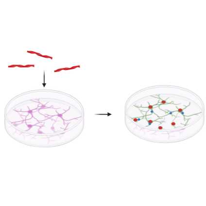
This protocol describes the details of the treatment of primary hippocampal culture with α-synuclein fibrils or monomer protein. It also includes details of the collection of cell lysates for immunoblotting or ELISA measurements. Another part of the protocol describes immunostaining analysis of treated neurons to measure the seeding propensity of α-synuclein fibrils visualized with phospho-S129 α-synuclein antibodies.
Safety warnings
Hazard Identification and Risk of Exposure to the
Hazards:
Inhalation or spread through food or drink that contain fibrils aerosols or fibrils.
Protective gloves, safety glasses and lab coat must always be used when handling anything that possibly could contain α-synuclein fibrils. Food or drink is strictly prohibited in any environment where α-syn fibrils are used.
Routes of Transmission: Prior to assigning containment requirements, it is imperative to
understand the routes of transmission.
Some issues to address:
- What are the exposure routes/risks of most concern:
Inhalation or spread through food or drink that contain fibril aerosols or fibirls accordingly. Fibrils possibly might reach the brain regions through the olfactory epithelium; Risk of accidental needlestick/droplet splash while handling fibrils for in vitro or in vivo work.
- What are the consequences of exposure (potential illness, etc)
Fibrils may be considered as infectious material. Minimum to no hazard is expected from α-syn protein. There is no evidence that transmission of fibrils can lead to development Parkinson’s disease. However, taking into account prion-like properties of α-syn fibrils should therefore be handled cautiously and wisely. Strictly recommended using disposable materials and Personal Protective Equipment (PPE) such as gloves, face mask, etc.
PRECAUTIONS:
Laboratory work where high concentration of fibrils (more than 300 uM) is needed must comply with biosafety level 2 (BSL2) containment as described in the current edition of the CDC/NIH’s
Biosafety in the Microbiological and Biomedical Laboratories: http://www.cdc.gov/od/ohs/biosfty/bmbl5/bmbl5toc.htm
Sharps safety precautions:
The use of sharps (glass pipettes, glass slides and cover slips, scalpels and
lancets) should be eliminated, when possible. Appropriate precautions should be
taken to avoid percutaneous injuries. These items should be disposed of
immediately in a puncture-resistant sharps container. Bending, recapping or
clipping of needles is prohibited. As described in CDC’s sharps safety website:
https://www.cdc.gov/sharpssafety/index.html
Procedural Methods
and Materials:
- Laboratory work where high concentration of fibrils (more than 300 uM) is needed must comply with biosafety level 2 (BSL2) containment. This means all aerosol generating procedures must be performed within the biosafety cabinet.
- All the fibrils work involves using PPE, aerosol-tight centrifuges, water bath sonicator in a closed cabinet, homogenization of frozen brain samples using probe-tip sonicator under the hood (collection of protein fractions in BSL2 cabinets), chromatography equipment in a closed-door fridge, sealed plates, safe lock microfuge tubes (or tubes wrapped/sealed with parafilm), and use of filtered tips for pipettes. All personnel must strictly adhere to these procedures.
- Use of proper PPE as stated in the section below. Use of available N95 respirators is voluntary (same for the use of available sleeve protectors). Follow safety precautions for sharps (for e.g., to avoid accidental needle sticks) while working with PFFs in the lab and for doing in vivo work.
Personal Protective
Equipment (PPE): Appropriate PPE includes gloves, lab coat and safety glasses,
face mask (voluntary N95 respirator use and sleeve protectors), face / bench
top splash shield for specific procedures as stated above.
Methods to minimize personal exposure: Strictly adhere to sharps safety precautions using needles or any material that can potentially cause wounds. Use disposable supplies where possible. Use the minimal amounts of α-fibrils needed for an experiment. Keep fibrils in closed tubes. 10% of SDS solution in water must be used for decontaminating work areas. Do not use NaOH or Sodium Hypochlorite or ethanol. Do not leave samples containing fibrils unattended at the bench.
Methods to prevent the release of fibrils/protect workers from aerosols,
splashes, splatters: protective gloves and clothing always be always be worn when handling frozen vials. High concentration of fibrils(>1mg/mL) always be handled under Biosafety cabinet and containment caps will be used while centrifugation. Centrifuge cups will be opened inside a biosafety cabinet. Face shield or benchtop splash shield will be used when working at the open bench.
Specimen transport
and removal of material(s) from the laboratory: Transported in secondary
container (plastic/Styrofoam) in a closed box. The closed box is carried in a
bag.
Standard
microbiological methods: hand washing after removal of gloves and before
leaving the work area, no mouth pipetting, strictly no food or drink in
refrigerators where material is stored, no eating in work area.
Cleaning &
Disinfection: Work area must be
cleaned with 10% SDS in water. Wipes used must be immediately disposed into
biohazard waste container. Any piece of equipment or supplies that possibly
have been exposed to fibrils must be wiped with 10% of SDS.
Waste Generation and Disposal Methods: The solutions that contain α-syn fibrils must be
decontaminated with 10% of SDS in water for 30 minutes and be thrown as a
biohazard waste in a sealed container/bag (use a minimal volume of fibrils needed
for an experiment, do not generate large volumes of fibril-containing liquids).
Use small biohazard bags to collect tips and consumables of experiment
performed, appropriately tie neck of bag in single knot and place in into
secondary biohazard waste container.
Spill and Accident Response Procedure: Describe all emergency procedures including spill clean-up. Describedisinfectants and
environmental decontamination. (ex., Outside of a BSC: If spill is a
respiratory hazard, evacuate 30 minutes to allow aerosols to settle. Place absorbent towels over the spill, apply freshly
prepared 10% SDS solution to entire area of spill starting on the outer edges
and working inward, pick up sharp items with mechanical device (not hands),
place all clean-up materials in a biohazard bag)
Prepare primary hippocampal neurons according to protocol
at DIV7 treat the culture with α-synuclein fibrils/seeds or monomeric protein at concentration 0.64 nM or 64 pM relative to estimated molecular weight measured as calculated size by DLS acquisitions and protein weight denatured in 6M GuHCL and calculated via Nanodrop.
At DIV21 for 14 days of incubation or at DIV14 for 7 days of incubation culture media should be removed - aspirate the media
Here are options to use the prepared cultures for the western-blot analysis or ELISA (step-case) or for the immunofluorescence
Immunnoblotting of the primary culture7 steps
Add 200 ul of lysis buffer (1% triton X-100 in PBS + prot/phos inhibitors),
Re-suspend the culture, check on a scope if there is any remaining attached cells. Collected the cell lysate into the protein-low-binding eppendorf tube.
Sonicate using prob-tip for 10 sec, 10% power
Vortex 10 sec each tube. Spin at 10,000g for 20 min at 4C. Transfer the supernatant into another eppendoft tube (200uL)
Transfer 40 ul of lysed cells to the tube containing 50 ul of sample buffer containing 10%DTT prepared for a western blot analysis
Remaining lysates store at -80 with another aliquot of 10 ul for BCA analysis.
Spin down a tube of normal donkey serum (20k xg for 10 min, 4C). Remove supernatant, do not use pellet.
Rinse with 3x with TBS. Use plastic bulb dropper and be careful. Prefer to remove w/ 200uL pipette, not vaccum. DO NOT let cells dry out, only do a row at a time.
Block/ Permeabilize with 300 uL 0.1% saponin, 3% Normal Donkey serum in TBS 30 min RT [KEEP COVERED]
During block, prepare primary antibodies:
Add primary antibody at desired concentration with a supplement of 0.02% saponin, 1% donkey serum, in TBS. Spin down the tube (20k xg for 10 min, 4C). Remove supernatant, do not use pellet
MJFR14 (EPY, 1:4,000), Tau5 (1:2,000), NeuN (1:2,000)
Remove block from wells.
Add primary antibody solutions to well. (300uL)
Overnight at 4C. Gentle rocking (no orbital shaker). Cover in Foil and make sure no loss of liquid.
Rinse with 1x with 300uL of TBS
Prepare secondary antibodies.
Use anti-rabbit AlexaFlour 647, anti-mouse AlexaFlour 488, anti-chicken AlexaFlour 555 at 1:1000 concentration, supplemented with 0.02% saponin, 0.1% normal goat serum in PBS. Spin down the tube (20k xg for 10 min, 4C). Remove supernatant, do not use pellet. Add Hoescht 1:10,000 to staining buffer
Rinse two times with TBS
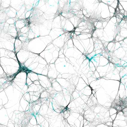
Add TBS imaging buffer, take pics on Keyence.
Imaging: let the plate come up to RT, wipe bottom of plate with 70% ethanol before imaging
Take x25 20x pictures per well.
Images were obtained using Keyence BZ-X810 and Zeiss880 and coded for a blinded approach to analyze using CellProfiler and imageJ.