May 10, 2024
Agarose Gel Electrophoresis for quality control of DNA aptamers in biosensor assays
- Geisianny AM Moreira1,
- Diana Vanegas1,
- Eric S McLamore1
- 1Clemson University
- SNAPS research group


Protocol Citation: Geisianny AM Moreira, Diana Vanegas, Eric S McLamore 2024. Agarose Gel Electrophoresis for quality control of DNA aptamers in biosensor assays. protocols.io https://dx.doi.org/10.17504/protocols.io.bydcps2w
License: This is an open access protocol distributed under the terms of the Creative Commons Attribution License, which permits unrestricted use, distribution, and reproduction in any medium, provided the original author and source are credited
Protocol status: Working
We use this protocol and it's working
Created: September 19, 2021
Last Modified: May 10, 2024
Protocol Integer ID: 53380
Keywords: electrophoresis, DNA aptamer, agarose gel, biosensor, quality control, dna aptamers in biosensor assay, dna aptamer, size of dna aptamer, agarose gel electrophoresis for quality control, aptamer loading on lig electrode, agarose gel electrophoresi, biosensor assay, procedure for electrophoresi, aptamer loading, electrophoresi, agarose gel, dna, lig electrode
Abstract
This protocol describes the procedure for electrophoresis in agarose gel to check the quality, purity, and size of DNA aptamers. The general objective is that this is the first and fundamental step to start aptamer loading on LIG electrodes.The complete process requires approximately 2 hours and 15 minutes to complete.
Materials
- Electrophoresis system
- Gel casting set (casting tray, well combs)
- Agarose (powder)
- 10X TAE buffer (Tris-acetate-EDTA)
- Gels Stain
- Loading dye
- DNA Ladder - low range
- Pipette (0.5 to 10 µl range)
- Sterile pipette tips (0.5 to 10 µl range)
- Parafilm
- DI water
- Microwave
- Ethanol (70% or greater)
- Mobile phone with camera for imaging gels
Safety warnings
Should be proper attire PPE (lab coat, gloves, glasses, tie hair back, face mask/shield, etc.) during all steps.
Before start
Sanitize the entire workstation (bench) with ethanol (70% or greater) before starting.
Prepare the agarose gel (Timing: 30 minutes)
- Assemble the gel casting
- Prepare the 1X TAE working solution. To make 1X TAE from 10X TAE stock solution, dilute 100 ml of the stock solution into 900 mL of DI water.
- Prepare 3% agarose gel
- Adjust the mass of agarose in a given buffer volume (1X TAE) to make gels in 3% concentration. For example, for the short casting tray, 40 mL is enough. If the casting tray is more extensive, you will need a larger buffer volume, so the amount of agarose will change.
- Short casting tray: to make a 3% agarose gel in 40 mL 1X TAE, measure 1,2 g of agarose.
30m
- Mix agarose powder with 40 mL 1XTAE in a microwavable flask.
- Microwave in pulses until the agarose be dissolved entirely. Microwave for 30 sec, stop and swirl, and then continue towards a boil. Keep an eye on it; the solution tends to boil over. The agarose will take a while to dissolve because the concentration is high. Shake gently to avoid bubbles. Use a bottle with a much larger volume capacity than used (e.g., for 40 mL of gel, use a 200 mL bottle). HOT! Be careful stirring, and eruptive boiling can occur.
- After the agarose has completely dissolved, let the solution cool for 5 minutes. Add 2 µl of the Gel stain to the agarose solution. Shake gently to dilute the stain. The protocol for using SmartGlow gel stain is 5 µl per 100 ml of agarose. If the volume is smaller or larger, adjust the amount needed with a simple rule of three.
- Pour the agarose into a gel tray, and then put the well comb in place. Pour slowly to avoid bubbles. Let sit at room temperature for 15-20 minutes until it has completely solidified.
Loading samples and running gel (Timing: 1 hour and 40 minutes)
- Use a piece of parafilm to prepare the samples before applying them to the gel.
- Prepare the samples for a final volume of 6 µl, as follows: 3 µl DI water + 1 µl loading dye + 2 µl sample.
- Once solidified, place the agarose gel into the electrophoresis unit.
- Add 5 µl Gel Stain in 200 mL 1X TAE running buffer. Fill the gel box with 1X TAE until the gel is covered.
1h 30m
- Carefully load a DNA ladder into the first lane of the gel. Carefully load your samples into the additional wells of the gel. Be careful in this step not to tear or pierce the gel and avoid bubbles during the application of the samples. Place the very top of the tip of the pipette into the buffer just above the well. Very slowly and steadily, push the sample out and watch as the sample fills the well.
- Run the gel at 35 V for 99 minutes.
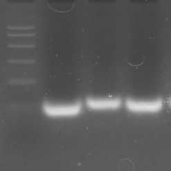
- After the electrophoresis is complete, view and document the gel using a blue light illuminator (coupled to the electrophoresis system). Use a mobile phone with camera for imaging gels.
- Using the DNA ladder in the first lane as a guide, you can infer the size of the DNA in your sample lanes. Samples of DNA aptamers are considered good quality when they have a single band, as the predicted size, and without smearing.
Clean up work area (Timing: 5 minutes)
- Dispose of the gel in biohazardous waste.
- Place the 1X TAE running buffer in an appropriate bottle and can be reused up to 3 times. After maximum use, dispose of in chemical hazard containers.
- Wash the casting tray and well combs. Keep in the proper place.
- Clean the bench with ethanol.
5m