Nov 24, 2020
A simple, non-invasive approach to detect vagal nerve response patterns that predict a positive treatment response to gastric electrical stimulation therapy for gastroparesis
- Matthew Ward1,2,
- Bartek Rajwa3,
- John M Wo2,
- Anita Gupta2,
- John Furness4,5,
- Terry Powley6,
- Thomas V Nowak2
- 1Weldon School of Biomedical Engineering, Purdue University, West Lafayette, IN, USA;
- 2Indiana University School of Medicine, Indianapolis, IN, USA;
- 3Bindley Bioscience Center, Purdue University, West Lafayette, IN, USA;
- 4Department of Anatomy and Neuroscience, University of Melbourne, Parkville, Australia;
- 5Florey Institute of Neuroscience and Mental Health, Parkville, Australia;
- 6Dept. of Psychological Sciences, Purdue University, West Lafayette, IN, USA
- SPARCTech. support email: [email protected]


Protocol Citation: Matthew Ward, Bartek Rajwa, John M Wo, Anita Gupta, John Furness, Terry Powley, Thomas V Nowak 2020. A simple, non-invasive approach to detect vagal nerve response patterns that predict a positive treatment response to gastric electrical stimulation therapy for gastroparesis. protocols.io https://dx.doi.org/10.17504/protocols.io.bax7ifrn
Manuscript citation:
Ward M.P., Rajwa B., Wo J.M., Gupta A., Furness J.B., Powley T.L., and T.V. Nowak. An emerging method to noninvasively measure and identify vagal response markers to enable bioelectronic control of gastroparesis symptoms with gastric electrical stimulation. Submitted to Journal of Neuroscience Methods 336, pp. 1-13, Feb 2020. DOI: https://doi.org/10.1016/j.jneumeth.2020.108631
License: This is an open access protocol distributed under the terms of the Creative Commons Attribution License, which permits unrestricted use, distribution, and reproduction in any medium, provided the original author and source are credited
Protocol status: In development
We are still optimizing this protocol
Created: January 04, 2020
Last Modified: November 24, 2020
Protocol Integer ID: 31455
Keywords: Gastroparesis, Gastric Electrical Stimulation, Vagus Nerve, Compound Nerve Action Potential, Neurostimulation, Bioelectronics, vagal nerve involvement in ges therapy, gastric electrical stimulation, positive treatment response to gastric electrical stimulation therapy, gastric electrical stimulation therapy, vagal nerve response pattern, vagal nerve involvement, vagal sensory afferent, bioelectronic control of gastroparesis symptom, vagal activity with noninvasive electrode, nerve response, gastroparesi, vagal response, gastroparesis protocol title, increased vagal response, idiopathic gastroparesi, vagal response marker, vagal activity, vagal mechanism, transcutaneous recording of cervical vagal activity, ges in conscious subject, right cervical vagal nerve, gastroparesis symptom, cutaneous vagal cnap analysis, right vagal fiber response, cervical vagal activity, electrical stimulation, left vagal abactivation, stimulation, refractory nausea, ges therapy, vomiting, resistant nausea, activity of different ner
Abstract
Protocol Title: A simple, non-invasive approach to detect vagal nerve response patterns that predict a positive treatment response to gastric electrical stimulation therapy for gastroparesis [1]
Background: Gastric electrical stimulation (GES) can be a life-changing, device-based treatment option for drug-resistant nausea and vomiting associated with diabetic or idiopathic gastroparesis (GP).Despite over two decades of clinical use, the mechanism of action remains unclear.We hypothesize a vagal mechanism, whereby GES activates vagal sensory afferents that project to the nucleus of the solitary tract and onward to influence brain structures that mediate the biological processes leading to nausea and vomiting (e.g., area postrema). Here, we describe a noninvasive method to investigate vagal nerve involvement in GES therapy in human subjects.We have developed transcutaneous recording of cervical vagal activity that is synchronized with GES in conscious subjects, along with methods of discriminating the activity of different nerve fiber groups that are activated.We are refining the system of recording and analysis so that it can be applied in the clinic.
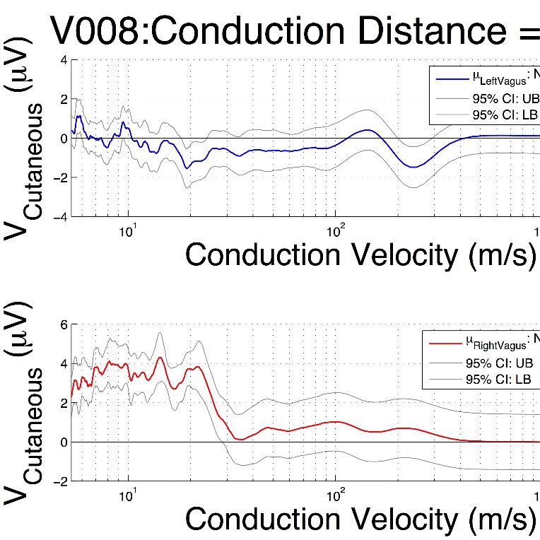
New Method: Sixty-six adults receiving GES therapy (Medtronic Enterra I/II) to treat refractory nausea and vomiting secondary to diabetic, idiopathic, or postoperative GP were enrolled in the study (IRB #:1206008988). Subjects completed a GCSI symptom survey upon enrollment. The left and right cutaneous vagal electroneurograms (vENG) were collected from the skin surface over the left and right cervical vagal nerves, respectively, without changing the prescribed GES parameters. The mean response to GES was computed for each subject and classified according to the Letter System for nerve fiber classification. Symptom scores were compared between groups of subjects with or without significant Ab, Ag, Ad, or B fiber responses, defined as volleys in the mean response to GES whose peak amplitude is significantly different from 0 V ata=0.05. Nerve responses were correlated with GP symptoms.
Results: Of the 66 subjects, 28 had diabetic GP (type 1:9; type 2:19), 35 had idiopathic GP, and 3 had postsurgical GP. Stimulus pulse current and charge did not predict treatment efficacy, but did predict a significant increase in total symptom score in type 1 diabetics as GES stimulus charge per pulse increased (p < 0.01), representing a notable side effect and providing a method to identify it. In contrast, the number of significant left and right vagal fiber responses that were recorded was directly related to patient symptom improvement.Increased vagal responses correlated with significant decreases in total symptom score (p < 0.05). Taken together, our analysis suggests that it is possible to extract meaningful information pertaining to vagal activity with noninvasive electrodes and that specific fiber response profiles predict improvements in specific symptoms of gastroparesis.
Conclusions: Cutaneous vagal CNAP analysis is a useful technique to unmask relationships among GES parameters, vagal recruitment, efficacy and side-effect management. Our results suggest that CNAP-guided GES optimization will provide the most benefit to patients with idiopathic and type 1 diabetic GP, especially when tuned for left vagal Ag and right vagal Ad/B fiber responses. The possible side effects associated with left vagal Abactivation in type 1 diabetics underscore the need to consider disease etiology and fiber recruitment profiles in the patient and parameter selection process.
Funding Acknowledgment
This work is supported, in part, by NIH SPARC OT2OD028183 and NIH SPARC OT2OD023847.
Publication/Abstract Reference:
[1] Ward M.P., Rajwa B., Wo J.M., Gupta A., Furness J.B., Powley T.L., and T.V. Nowak. An emerging method to noninvasively measure and identify vagal response markers to enable bioelectronic control of gastroparesis symptoms with gastric electrical stimulation. Journal of Neuroscience Methods 336, pp. 1-13, Feb 2020. PMID:32087238
Guidelines
The methods outlined within this protocol were developed for human subjects under an Institutional Review Board (IRB) approved protocol at the Digestive and Liver Disorders (DALD) Center at the Indiana University School of Medicine (Indianapolis, IN) (IRB #: 1206008988). Anyone who wishes to use these methods should obtain IRB approval from their institution before beginning.
Materials
1. Gastroparesis Cardinal Symptom Index (GCSI) Survey/Symptoms Interview Form
2. Medtronic Enterra Gastric Electrical Stimulation System Programming Log
3. Alcohol Wipes and Skin Prep Gel
4. Conductive Electrolyte Gel
5. Electrocardiogram Pad Electrodes (7-9 electrodes per subject)
6. ADInstruments PowerLab System
7. Clean towels to remove any gel
8. GES System Interrogation Unit/Programmer
9. IRB-approved Protocol and Informed Consent Form
10. Computer running Matlab R2015a or newer with the Signal Processing Toolbox
Safety warnings
While these methods do not contain any invasive procedures, they are experimental. Therefore, care must be taken by any investigator who performs these methods to first understand the nature of all electrode materials and any risks that the steps outlined in the protocol may pose to subjects.
Before start
Gain IRB approval for this protocol at your institution before enrolling any subjects or performing any of the procedures outlined within this protocol.
Preparation
Review the nature of the study and the informed consent document with the prospective subject. Be sure to answer any questions that s/he may have. Move to Step 2 if the subject understands the nature of the study and agrees to enroll into the study by signing the informed consent document.
Symptom Survey and GES Device Interrogation
1. Have the subject complete the Gastroparesis Cardinal Symptom Index (GCSI) survey [14].
2. Interrogate the gastric electrical stimulation device to determine the current stimulus parameter settings, stimulating electrode impedance and stimulating electrode configuration (e.g., bipolar stimulation or monopolar stimulation using the implanted pulse generator housing as the return electrode). Record these parameters/settings on a separate document (this document should be kept with the GCSI symptom survey data).
Skin Preparation and Recording Electrode Placement
1. Instruct the subject to lay down on their back, resting their head and neck on pillows so that the neck is not strained. Proper neck support will reduce signal contamination from muscle activity (e.g., from the sternocleidomastoids).
2. Clean the skin surface overlying the left and right cervical vagus nerve with alcohol swabs and allow it to dry.
Recording Protocol
1. Following the conventional cutaneous vagal recording protocol, use the ADInstruments PowerLab System and cutaneous ECG electrodes to record the Lead II ECG (for SKNA recordings), the electrogastrogram, and the electroneurograms (ENG) from the skin surface overlying the left and right mid-cervical vagus nerves (be sure to apply a small amount of conductive electrolyte gel to each pad electrode before placement) with the GES device:
A) turned ON for at least 8 minutes (without adjusting the parameter settings)
B) with the GES device turned OFF for at least 2 minutes
2. Mark the ECG electrode locations and the COMMON/GROUND electrode location on the figure below
3. Measure and record the straight line distance from the GES stimulating electrodes to the closest recording electrode on the left cervical vagus nerve
4. Complete the checklist below to ensure that each step has been completed
Conduction distance (in cm): ________________
Vagal electrode locations marked on figure? (YES / NO)
Collected 8+ min of data with GES device ON? (YES / NO)
Collected 2+ min of data with GES device OFF? (YES / NO)
ElecPlacementImage.png
Fig. 2. Electrode Placement Guide
Cleanup
1. After recordings have been collected, carefully remove the electrodes from the subject and gently wipe away any residual electrolyte gel with a clean cloth.
2. De-identify and store the raw data on a secure computer or server as per your IRB guidelines.