Nov 26, 2025
A simple and fast single step method to isolate and stain the CD27 B cells (plasma cells) with quantum dots magnetic beads antibody conjugate for fluorescent microscopy
- Sudhir Bhatia1,
- Gudrun Baersch1
- 1Genekam Biotechnology AG


Protocol Citation: Sudhir Bhatia, Gudrun Baersch 2025. A simple and fast single step method to isolate and stain the CD27 B cells (plasma cells) with quantum dots magnetic beads antibody conjugate for fluorescent microscopy. protocols.io https://dx.doi.org/10.17504/protocols.io.q26g7n4o9lwz/v1
Manuscript citation:
Bhatia, S. (2025). A Simple and Fast Single-Step Method to Isolate and Stain CD27 B Cells (Plasma Cells) Using Quantum Dot Magnetic Bead Antibody Conjugates for Fluorescent Microscopy. Medical Science and Discovery, 12(9), 284–287. https://doi.org/10.36472/msd.v12i9.1315
License: This is an open access protocol distributed under the terms of the Creative Commons Attribution License, which permits unrestricted use, distribution, and reproduction in any medium, provided the original author and source are credited
Protocol status: Working
We use this protocol and it's working
Created: August 04, 2025
Last Modified: November 26, 2025
Protocol Integer ID: 224031
Keywords: CD27 B cells, plasma cells, quantum dots magnetic beads antibody conjugate, immune response , human antibody , microscopy, nanomedicine, quantum dot magnetic bead antibody, magnetic beads antibody conjugate, methods cd27 cells from mononuclear cell culture, methods cd27 cell, magnetic bead antibody, results cd27 cell, quantum dot magnetic bead, antibody, specific fluorescence under the microscope, fluorescence microscope, quantum dot, mononuclear cell, plasma cell, magnetic bead, specific fluorescence, fluorescent microscopy background today, cell, mononuclear cell culture, fluorescence, other cell, using quantum dot, cd27, new possibilities for immunotherapy, conventional dyes such as fitc, conventional dye, long time before the cell, immunotherapy
Disclaimer
Genekam Biotechnology AG
Duissernstr. 65a
47058 Duisburg
Germany
Abstract
Background Today, mononuclear cells are stained in a two-step process: in the
first step, the cells are isolated, and in the second step, they are stained.
This two-step process takes a long time before the cells can be observed under
a fluorescence microscope. In addition, quantum dots have further advantages
over conventional dyes such as FITC, Cy3, Cy5, etc., as they do not fade and emit
fluorescence in a narrow range. We decided to develop a new method in which
the cells can be isolated and stained in a single step using quantum dot magnetic bead
antibody conjugates for CD27-positive B cells.
Material and Methods CD27 cells from mononuclear cell cultures were stained and
isolated simultaneously in magnetic racks using quantum dot magnetic bead antibody
conjugates with two different wavelengths (510 and 700 nm). They were then observed
under a fluorescence microscope.
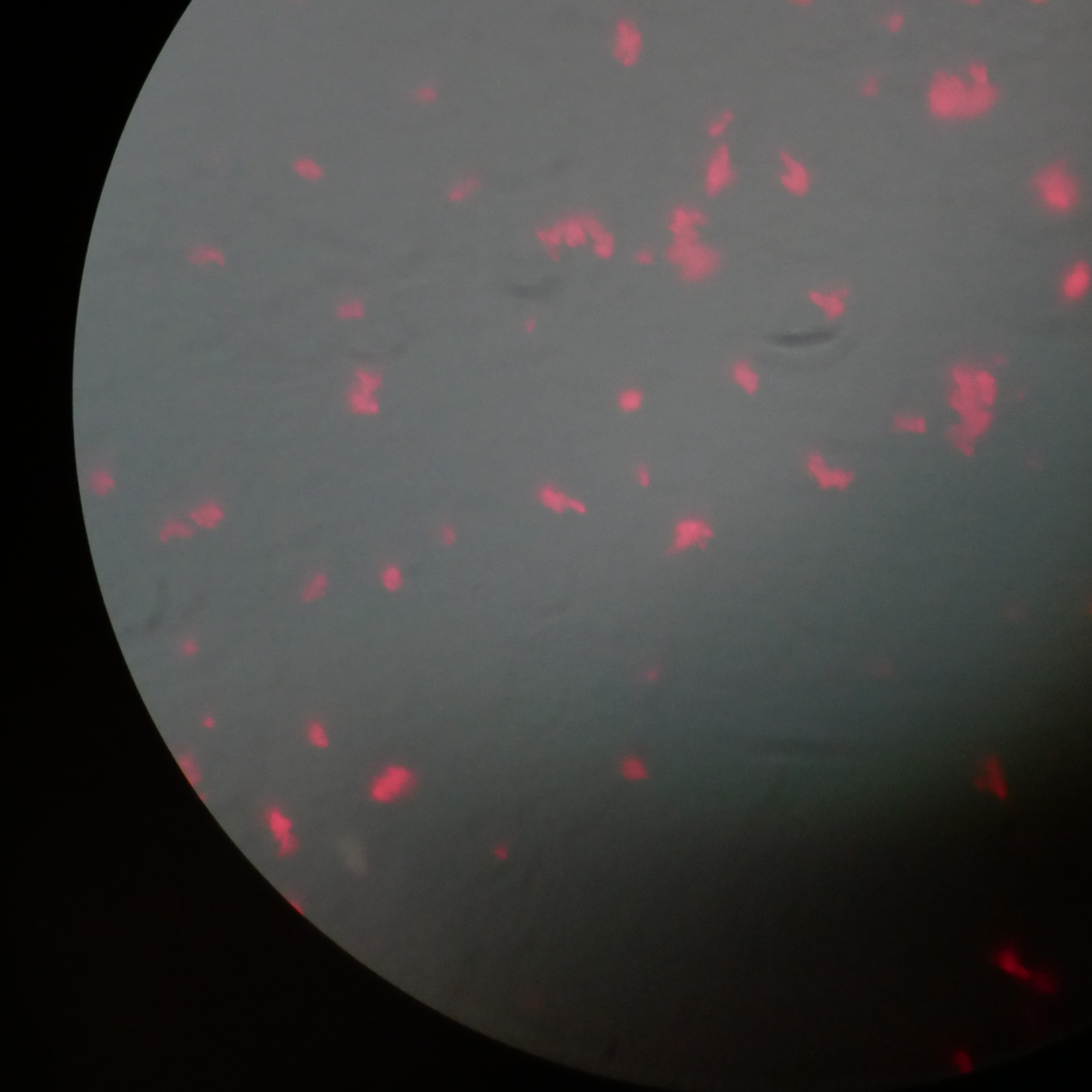
Results CD27 cells were successfully isolated and stained within 30 minutes, emitting
a specific fluorescence under the microscope so that only the specific CD27 B cells were
observed and no other cells were present. Under the normal microscope, the magnetic beads
adhered to their surfaces.
Conclusions This may be the first report of the successful development of a rapid one-step
method for isolating and staining CD27 B cells from mononuclear cells using quantum dot
magnetic bead antibody conjugates within 30 minutes, compared to the two-step method,
which takes more than an hour. This method opens up new possibilities for immunotherapies and
clinical applications.
Image Attribution
Quantum dot magnetic bead conjugate Genekam
Materials
Quantum dots magnetic beads antibody conjugate (MICROBOSS Nanomedicine GmbH, Germany)
Magnetic racks (Genekam Biotechnology AG, Germany)
PBS (Washing buffer, Lonza, USA)
Fluorescent microscope (Zeiss, Germany)
50 ml sterile tubes
Microscopic slides (Thermofischer, Germany)
5 ml tubes
Safety warnings
1. Please use a high-quality fluorescence microscope, as inferior and cheap microscopes do not deliver results and are therefore a waste of time.
2. Please keep away your mobile telephone and camera from the magnetic rack as the magnet can damage your display.
3. Work under laminar flow.
Ethics statement
Since human blood samples are being used, approval from an ethics committee may therefore be required. Please check this point.
Before start
Please read the protocol properly before start of the experiment.
Protocal A: There are two protocols: A and B. Use one protocol.
Centrifuge 5 ml of cell culture of human mononuclear cells at 18000 rpm and wash twice with PBS. Collect the pellet.
Add 2 μl of quantum dot magnetic bead antibody conjugates (510 nm and 700 nm) on the pellet and incubate it in the dark for 10 minutes with intermittent mixing.
Wash the cells with 5 ml PBS while placing on a magnetic rack for 3 minutes. Remove the fluid while keeping pellet in the tube.
Suspend the pellet in 100 μl PBS; apply 1 μl to a slide. Let it dry and fix in ethanol for 5 minutes, and mount it the slide.
Observe the CD27 poisitive cells using a Zeiss fluorescence microscope with UV, blue, and green filters at various magnifications, with and without oil immersion. Attempts using a lower-cost Biobase microscope were unsuccessful. Do not waste your time using low quality microscope.
Alternative Protocol B
Inocubate the 5 ml of cell culture with 2 μl of quantum dot magnetic bead antibody conjugate for 10 minutes.
Suspend the cells in 5 ml on a magnetic rack, discard the supernatant and wash pellet twice with 5 ml PBS.
Apply 2 μl of the pellet after suspending it in 100 µl PBS to a slide. Let it dry, fix it, and mount it.
Observe the CD27 positive cells under a Zeiss fluorescence microscope with UV, blue, and green filters at various magnifications, with and without oil immersion. Attempts using a lower-cost Biobase microscope were unsuccessful.
Protocol references
Bhatia, S. (2025). A Simple and Fast Single-Step Method to Isolate and Stain CD27 B Cells (Plasma Cells) Using Quantum Dot Magnetic Bead Antibody Conjugates for Fluorescent Microscopy. Medical Science and Discovery, 12(9), 284–287. https://doi.org/10.36472/msd.v12i9.1315
Acknowledgements
Ms Kornelia Niklis for her Laboratory work support.