Apr 24, 2024
Version 2
A protocol for tissue clearing and three-dimensional imaging of human sigmoid mucosal biopsies V.2
- Pu-Qing Yuan1,
- Tao Li1,
- Yvette Taché1
- 1University of California at Los Angeles (UCLA)
- SPARC
- A single cell RNA sequencing protocol for the pig colon


Protocol Citation: Pu-Qing Yuan, Tao Li, Yvette Taché 2024. A protocol for tissue clearing and three-dimensional imaging of human sigmoid mucosal biopsies . protocols.io https://dx.doi.org/10.17504/protocols.io.6qpvr8dkblmk/v2Version created by Pu-Qing Yuan
License: This is an open access protocol distributed under the terms of the Creative Commons Attribution License, which permits unrestricted use, distribution, and reproduction in any medium, provided the original author and source are credited
Protocol status: Working
We use this protocol and it's working
Created: April 24, 2024
Last Modified: April 24, 2024
Protocol Integer ID: 98739
Keywords: Tissue clearing, 3D imaging, human sigmoid mucosal biopsy, nerve fibers , 3d imaging of human sigmoid mucosal biopsy, dimensional imaging of human sigmoid mucosal biopsy, entire human sigmoid mucosal biopsy specimen, nerve fibers in the sigmoid colonic mucosa, piece of entire human sigmoid mucosal biopsy specimen, sigmoid colonic mucosa, human colonic enteric nervous system, phenotyping of human colonic enteric nervous system, original clarity tissue clearing technique, enteric glial cell, biopsy sample, protocol for tissue clearing, cleared biopsy sample, tissue clearing, distributions of mast cell, clear spatial views of nerve innervation, configurations of mast cell, mast cell, nerve fiber, immunofluorescence, 3d imaging, dimensional imaging, immunofluorescence protocol, nerve innervation
Funders Acknowledgements:
NIH/SPARC
Grant ID: 1OT2OD024899-01
Abstract
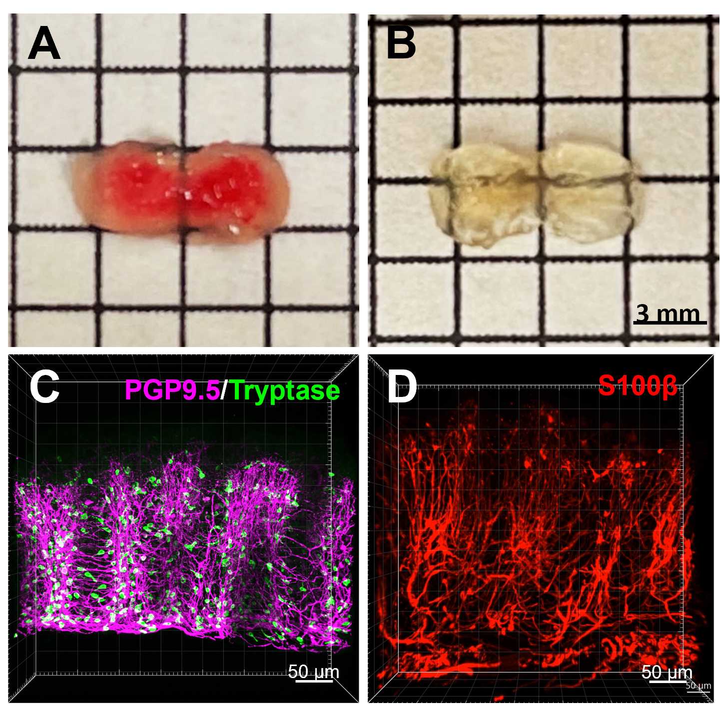
This protocol was developed for tissue clearing and 3D imaging of human sigmoid mucosal biopsies by adapting and modifying the original CLARITY tissue clearing technique/immunofluorescence protocols that we established for 3D imaging and phenotyping of human colonic enteric nervous system. By using this new protocol, a piece of entire human sigmoid mucosal biopsy specimen with the volume about 6x3x1-2 mm (length x wide x thickness) can be completely cleared within 3-4 days. The 3D images and videos generated from cleared biopsy samples showed clear spatial views of nerve innervation, distributions of mast cells and enteric glial cells, and configurations of mast cells with nerve fibers in the sigmoid colonic mucosa.
Attachments

a protocol for CLARI...
39.8MB