Feb 20, 2026
A block staining method using ethanolic phosphotungstic acid for the visualisation of collagens in the TEM
- Astrid Obermayer1,
- Stefan Hainzl2,
- Ulrich Koller2,
- David Lang3,
- Bernd Lamprecht3,
- Walter Stoiber1
- 1Department of Environment and Biodiversity, EM Core Facility, University of Salzburg, Austria;
- 2EB House Austria, Research Program for Molecular Therapy of Genodermatoses, Department of Dermatology and Allergology, University Hospital of the Paracelsus Medical University, 5020 Salzburg, Austria;
- 3Department of Pulmonology, Kepler University Hospital, Johannes Kepler University, Linz, Austria


Protocol Citation: Astrid Obermayer, Stefan Hainzl, Ulrich Koller, David Lang, Bernd Lamprecht, Walter Stoiber 2026. A block staining method using ethanolic phosphotungstic acid for the visualisation of collagens in the TEM. protocols.io https://dx.doi.org/10.17504/protocols.io.rm7vz9m9xgx1/v1
License: This is an open access protocol distributed under the terms of the Creative Commons Attribution License, which permits unrestricted use, distribution, and reproduction in any medium, provided the original author and source are credited
Protocol status: Working
We use this protocol and it's working
Created: July 02, 2025
Last Modified: February 20, 2026
Protocol Integer ID: 221574
Keywords: transmission electron microscopy, phosphotungstic acid, collagen, anchoring fibrils, elastic fibres, block staining, visualisation of small fibrillar collagen, fibrillar collagen, small fibrillar collagen, small collagen vii anchoring fibril, visualization of collagen, collagens in the tem conventional tem imaging, ethanolic phosphotungstic acid for the visualisation, standard staining method, staining procedure, staining method, using ethanolic phosphotungstic acid, collagen, ethanolic phosphotungstic acid, preparation of ethanolic phosphotungstic acid, fibril, keratin fibre, used stain, human skin, hemidesmosomal plaque, accentuating keratin fibre, tem conventional tem imaging, biological sample
Abstract
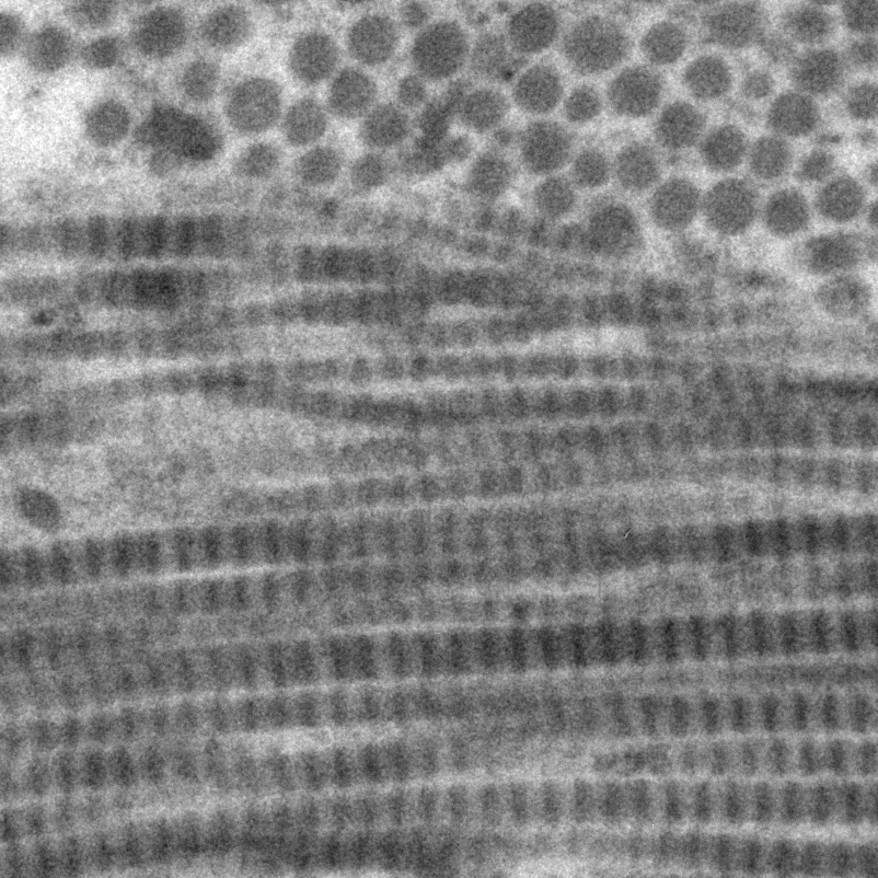
Conventional TEM imaging of biological samples requires contrast enhancement by staining with heavy metal salts. The most widely used stains are uranyl acetate or its non-radioactive lanthanoid replacements, and lead citrate. However, particularly for the visualisation of small fibrillar collagens, these substances proved of limited use. We therefore developed a preparation of ethanolic phosphotungstic acid (E-PTA) as an improvement to overcome this deficiency. Were able to establish a highly effective and time saving block staining procedure that can be integrated in the dehydration steps. The method reliably visualizes fibrillar collagens, prominently including the small collagen VII anchoring fibrils of the human skin, and various other extracellular matrix components. Collagen I/III fibrils are conspicuous in transverse and longitudinal section, accurately showing the characteristic banding patten in the latter. The new E-PTA based block staining method also clearly depicts all relevant intracellular structures, particularly accentuating keratin fibres and
desmosomal and hemidesmosomal plaques. We therefore conclude that beyond the visualization of collagen this method is also a fast, inexpensive and versatile non-radioactive alternative to standard staining methods.
Image Attribution
Astrid Obermayer, University of Salzburg, Austria
Guidelines
This protocol describes the workflow of animal and human tissue sample fixation with aldehydes and osmium tetroxide, followed by an innovative block staining procedure with ethanolic PTA integrated into the dehydration steps, and subsequent epoxy resin embedding. Although originally aimed at the visualization of collagen fibrils, the block staining procedure turned out a much more versatile method. Accordingly, it is possible to adapt fixation steps or incubation times to the specific requirements of the particular type of sample and/or the established procedures of your lab.
The materials section provides a suggested list of chemicals, but the exact lots and vendors of the components listed are not critical. However, it should be made sure that non-denatured ethanol and EM-grade fixatives are used.
If a resin different to the one listed is used, infiltration times must be adapted according to manufacturer’s instructions.
Cacodylate buffer can be substituted with PBS, but it should be kept in mind that the fixation quality of structures such as intracellular membranes benefits from cacodylate buffer containing calcium ions.
It is best to use glassware as specimen vials, as glass is resistant to propylene-oxide. If you prefer to use plastic ware, please test its suitability beforehand.
Choose reagent volumes according to sample size and use at least 20x the sample volume to ensure that the sample is fully immersed in and surrounded by the liquid.
Materials
- Dimethylarsinic acid sodium salt trihydrate, molecular weight: 214.03 g/mol, CAS-nr: 6131-99-3, EG-nr.: 204-708-2, Carl Roth 5169.1
- Osmium tetroxide, (4% solution), molecular weight: 254.23 g/mol, CAS-nr: 20816-12-0, EG-nr. 244-058-7, Sigma-Aldrich 251755
- EM-grade glutaraldehyde (1,5 pentanedial) 25 % aqueous solution, molecular weight: 100.12 g/mol, CAS-nr: 111-30-8, EG-nr. 203-856-5, Carl Roth 4157.1
- Paraformaldehyde, molecular weight: 30,03 g/mol, CAS-nr: 30525-89-4, EG-nr. 608-494-5, Carl Roth 0964.1
- Calcium chloride, molecular weight: 110.98 g/mol, CAS-nr: 10043-52-4, EG-nr.233-140-8, Sigma-Aldrich C5670
- Phosphotungstic acid molecular weight: 2880,17 + x H2O g/mol, CAS-nr: 12501-23-4, EG-nr. 603-020-3, Carl Roth 2635.1
- 96% Ethanol undenatured, molecular weight 46,07 g/mol, CAS nr. 64-17-5, EG-nr. 200-578-6, Carl Roth P075.4
- 100% Ethanol undenatured, molecular weight 46,07 g/mol CAS nr. 64-17-5 EG-nr. 200-578-6, Carl Roth 9065.4
- 1,2-Propylene-oxide, molecular weight: 58.08 g/mol, CAS-nr. 75-56-9, EG-nr. 200-879-2, Sigma Aldrich 8.07027
- Agar Low Viscosity Resin (ALVR) Kit, Agar Scientific, AGR1078
Solutions:
10% PFA stock solution 100 mL
10 g paraformaldehyde
Fill to 100 mL with aqua bidest, dissolve by heating to approx. 60°C (do not boil!) and adjust pH with NaOH until the solution appears clear.
Store at 4C° up to one month, or at -20°C for several months.
CCB buffer (100 mM cacodylate buffer containing 2mM CaCl2) 1 L
21.403 g dimethylarsinic acid sodium salt trihydrate (M=214,03g/mol) and
0.222 g CaCl2 (M=110,98g/mol)
Fill to 1 L with aqua bidest. and stir until fully dissolved.
Adjust to 7.4
2% PFA, 2.5% GA fixative 50 mL
10 mL 10% PFA stock solution
5 mL 25% glutaraldehyde aqueous solution, EM grade
35 mL 35 ml CCB
Best prepared fresh before use, store at 4°C up to one month
1% OsO4 solution 10 mL
2.5 ml 4% OsO4 solution
7.5 ml CCB
Prepare fresh immediately before use. Very toxic, handle with care!
Ethanol dilutions 96 mL each
70% ethanol: 70 mL 96% ethanol, fill to 96 mL with aqua bidest. (26 mL ).
80% ethanol: 80 mL 96% ethanol, fill to 96 mL with aqua bidest. (16 mL ).
90% ethanol: 90 mL 96% ethanol, fill to 96 mL 96 ml with aqua bidest. (6 mL ).
1% ethanolic phosphotungstic acid 10 mL
0.1 g phosphotungstic acid
Fill to 10 mL with 70% ethanol
Prepare fresh immediately before use
Safety warnings
Perform all steps under a fume hood and wear protective gear.
Osmium tetroxide is highly reactive, severely toxic and corrosive. Handle with extreme care, avoid any contact with the solution or the fumes. Collect all solutions and the buffer of at least the first two washing steps for special waste disposal according to manufacturer’s instructions and local rules.
Glutaraldehyde and paraformaldehyde are toxic, corrosive and environmental hazards. Avoid any contact with the solutions and the fumes. Collect all solutions and the buffer of at least the first two washing steps for special waste disposal according to manufacturer’s instructions and local rules.
Cacodylic acid is toxic and an environmental hazard. Avoid any contact with the solution and the fumes.
Collect all solutions for special waste disposal according to manufacturer’s instructions and local rules.
Before start
This protocol comprises working steps for 4 lab days, so make sure you plan them accordingly before you start. As some process step times can be altered depending on sample type and other requirements, the
number of required days may change.
After the last washing step following fixation with OsO4, samples may be interim stored in 70% ethanol at 4°C over night or for up to approximately 2 weeks, e.g. enabling to collect samples from several experiments. Caution: Do not store in higher concentrations of ethanol, as this might lead to severe shrinking artefacts. Alternatively, interim storage in buffer is also possible, however, this does not provide against microbial contamination as the washing steps are usually not performed under sterile conditions.
Primary fixation
1d 1h
Place samples in a solution of 2% Paraformaldehyde and 2.5% Glutaraldehyde in 100mM cacodylate buffer containing 2mM CaCl2 (CCB) 4 °C Overnight
1d
Wash in CCB (1/4)Room temperature 00:15:00
15m
Wash in CCB (2/4)Room temperature 00:15:00
15m
Wash in CCB (3/4)Room temperature 00:15:00
15m
Wash in CCB (4/4)Room temperature 00:15:00
15m
Secondary fixation
4h
Fix samples in a freshly prepared solution of 1% OsO4 in CCBRoom temperature
03:00:00
Safety information
Highly toxic! Handle with great care.
3h
Wash in CCB (1/4)Room temperature 00:15:00
15m
Wash in CCB (2/4)Room temperature 00:15:00
15m
Wash in CCB (3/4)Room temperature 00:15:00
15m
Wash in CCB (4/4)Room temperature 00:15:00
15m
If applicable store samples in 70% ethanol at 4°C over night or until further use (up to approx. 2 weeks) 4 °C
Block staining
1h
Incubate samples in a freshly prepared solution of 1% PTA in 70% ethanol Room temperature 01:00:00
1h
Dehydration
2h 15m
80% ethanolRoom temperature 00:30:00
30m
90% ethanolRoom temperature 00:30:00
30m
96% ethanolRoom temperature 00:30:00
30m
100% ethanol (1/3)Room temperature 00:15:00
15m
100% ethanol (2/3)Room temperature 00:15:00
15m
100% ethanol (3/3)Room temperature 00:15:00
15m
5. Infiltration
3h 45m
Propylenoxide (1/3)Room temperature 00:15:00
15m
Propylenoxide (2/3)Room temperature 00:15:00
15m
Propylenoxide (3/3)Room temperature 00:15:00
15m
1:1 mixture of propylenoxide:ALVR resinRoom temperature 01:00:00
1h
1:2 mixture of propylenoxide:ALVR resinRoom temperature 02:00:00
2h
ALVR resin Room temperature Overnight
Embedding
1d
Place in fresh resin in suitable embedding moulds
Cure 60 °C 24:00:00
1d
Protocol references
Bulmer D. Observations on histological methods involving the use of phosphotungstic and phosphomolybdie acids, with particular reference to staining with phosphotungstic acid /haematoxylin. J Cell Sci. 1962;s3-103: 311–323. doi:10.1242/jcs.s3-103.63.311
Burry RW, Lasher RS. A quantitative electron microscopic study of synapse formation in dispersed cell cultures of rat cerebellum stained either by Os-UL or by E-PTA. Brain Res. 1978;147: 1–15. doi:10.1016/0006-8993(78)90768-0
Doughty MJ. Options for determination of 2-D distribution of collagen fibrils in transmission electron micrographs — Application to the mammalian corneal stroma. Microsc Res Tech. 2011;74: 184–195. doi:10.1002/jemt.20890
Doughty MJ. Assessment of collagen fibril spacing in relation to selected region of interest (ROI) on electron micrographs—application to the mammalian corneal stroma. Microsc Res Tech. 2012;75: 474–483. doi:10.1002/jemt.21080
Fry KR, Spira AW. An ethanolic phosphotungstic acid (EPTA) analysis of photoreceptor and synaptic ultrastructure in the guinea pig retina. J Histochem Cytochem. 1980;28: 142–148. doi:10.1177/28.2.6153396
Gluenz E, Shaw MK, Gull K. Structural asymmetry and discrete nucleic acid subdomains in the Trypanosoma brucei kinetoplast. Mol Microbiol. 2007;64: 1529–1539. doi:10.1111/j.1365-2958.2007.05749.x
Terner JY. Phosphotungstic acid-hematoxylin, reactivity in vitro. J Histochem Cytochem Off J Histochem Soc. 1966;14: 345–351. doi:10.1177/14.4.345
Acknowledgements
The authors are grateful to Angela Pittner and Sonja Bruckner for excellent technical support.