Nov 20, 2023
811.1 CODEXV1 Staining and Imaging Protocol
- 1University of Rochester Medical Center
- Human BioMolecular Atlas Program (HuBMAP) Method Development Community
- LungMap2 Consortium


Protocol Citation: Jeffrey Purkerson, Gloria S Pryhuber, heidie_huyck 2023. 811.1 CODEXV1 Staining and Imaging Protocol. protocols.io https://dx.doi.org/10.17504/protocols.io.261ged5pov47/v1
Manuscript citation:
Dylag AM, Misra RS, Bandyopadhyay G, Poole C, Huyck HL, Jehrio MG, Haak J, Deutsch GH, Dvorak C, Olson HM, Paurus V, Katzman PJ, Woo J, Purkerson JM, Adkins JN, Mariani TJ, Clair GC, Pryhuber GS. New insights into the natural history of
bronchopulmonary dysplasia from proteomics and multiplexed immunohistochemistry. Am J Physiol Lung Cell Mol Physiol. 2023;325(4):L419-l33. Epub 20230725. doi: 10.1152/ajplung.00130.2023. PubMed PMID: 37489262.
License: This is an open access protocol distributed under the terms of the Creative Commons Attribution License, which permits unrestricted use, distribution, and reproduction in any medium, provided the original author and source are credited
Protocol status: Working
We use this protocol and it's working
Created: August 25, 2023
Last Modified: November 20, 2023
Protocol Integer ID: 86978
Keywords: lung, multiplexed immunofluorescence, FFPE, tissue sections, Phenocycler, multiplexed immunofluorescent staining of lung tissue, imaging of ffpe lung tissue section, multiplexed immunofluorescence staining, multiplexed immunofluorescent staining, ffpe lung tissue section, labeling of lung tissue section, lung tissue section, immunofluorescence, lung tissue, custom antibody conjugation, multiplexed imaging with the keyence bz, codexv1 staining, multiplexed imaging, imaging protocol, staining technology, imaging protocol this protocol, antibody, x810 microscope, imaging
Funders Acknowledgements:
NHLBI (LungMAP) URMC
Grant ID: U01HL148861
NIH Human BioMolecular Atlas Program (HuBMAP)
Grant ID: U54HL165443
Abstract
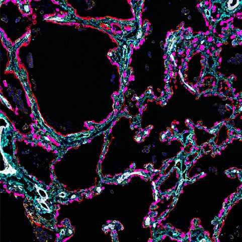
This protocol describes multiplexed immunofluorescent staining and imaging of FFPE lung tissue sections utilizing the Phenocycler-Keyence 800 platform (Akoya Biosciences). The approach is based on the CODEX® multiplexed immunofluorescence staining technology developed by Garry Nolan and colleagues (1). The protocol is closely aligned with the CODEX® User Manual Rev C provided by Akoya (2) and includes sections that describe A) coating of coverslips with Poly-L-Lysine, B) Lung section preparation C) Labeling of lung tissue sections with antibody-barcode conjugates, D) Multiplexed Imaging with the Keyence BZ-X810 microscope, E) Custom antibody conjugation. Thus, the protocol is designed to provide information regarding specific reagents (i.e., antibodies), conditions (i.e., dilutions), and procedures used in multiplexed immunofluorescent staining of lung tissue. Images produced utilizing this protocol are published in reference 3.
Attachments

Guidelines
An overview of Multiplexed Imaging using barcode conjugated antibodies can be found in the attached CODEX® User Manual Rev C provided, with permission, from Akoya. Note, multiplexed immunofluorescent imaging with Phenyocycler-Keyence 800 has been supplanted by the Phenocycler-Fusion platform.
Materials
Key equipment, reagents, buffers, and supplies can be found in the attached CODEX® User Manual Rev C (Akoya Biosciences).
1. Poly-L-Lysine powder; P5899 (Sigma-Aldrich, St. Louis, MO).
2. Antigen Unmasking Solution, Citric Acid Based (H-3300, Vector Lab, Burlingame, CA).
3. CODEX Staining kit P/N 7000008, contains Hydration; Staining; and Storage buffer; N, G, J, and S blocker; and final fixative reagent (Akoya Biosciences, Marlborough, MA).
4. Paraformaldehyde (20% solution); P/N: 15713-S EM Grade (Electron Microscopy Sciences, Hatfield, PA)
5. 10X CODEX Buffer P/N 232119
6. dPBS; 17-512Q (Lonza, Walkerville, MD).
7. DMSO, ACS Reagent Grade (≥99.9%); 472301-1L Sigma-Aldrich.
8. Humidified antibody incubation chamber, room temperature.
Safety warnings
Procedures involving xylenes and paraformaldehyde should be performed in a chemical fume hood. Nitrile gloves are not impermeable to xylenes therefore change gloves promptly upon contact with xylenes.
Use heat resistant gloves to handle glassware after antigen retrieval.
Ethics statement
The protocol does not utilize laboratory animals.
Before start
Ensure all reagents, supplies, equipment needed to complete the protocol are available in sufficient quantities to complete the procedure.
Coat glass coverslips with poly-L-Lysine
Glass coverslips (22mm x 22 mm, #1.5, Thermo Fisher, Waltham) are acid-washed and coated with Poly-L-Lysine.
Acid-wash glass coverslip in glass beaker or petri dish containing 1 N HCl and place in an oven set to 50-60 C Overnight with occasional agitation.
Wash coverslip 4X with ddH20
Rinse coverslips in 100% ethanol and dry them between sheets of Whatman filter paper. Store coverslips in a clean container.
Prepare 1% Poly-L-Lysine stock solution by diluting 100 mg of Poly-L-Lysine powder into 10 mL ddH2O. Prepare a working solution by diluting Poly-L-Lysine to 0.1% in ddH20.
Place coverslips in a cover glass staining rack and immerse in a beaker containing 0.1% Poly-L-Lysine (Fischer Scientific, Waltham, MA) and incubate. Overnight , minimum 12:00:00 Room temperature
12h
Wash coverslips via immersion in a series of 6 X 50 ml beakers containing ddH20; 3-5 dips in each beaker.
Store lysine-coated coverslips in a cool dry place and use within 2 months.
Lung section preparation
Bake Sections to promote tissue adherence to coverslips.
3h
FFPE Lung sections are prepared as described in dx.doi.org/10.17504/protocols.io.kxygxejwdv8j/v2 except that tissue sections are placed on poly-L-lysine coated glass coverslip from previous section. Standard section thickness is 4-5 micron.
Heat tissue sections on coverslips in a dry oven 60 °C Overnight .
Labeling of lung tissue sections with antibody-barcode conjugates
Deparaffination and Rehydration
Incubate coverslips placed in a coverslip staining rack in Coppin Jars containing xylene (3X) followed by a descending ethanol series and finally by molecular biology grade distilled water (2X)
Xylene #1- 00:05:00
5m
Xylene #2 00:05:00
5m
Xylene #3 00:05:00
5m
100% Ethanol #1 00:05:00
5m
100% Ethanol #2 00:05:00
5m
90% Ethanol 00:05:00
5m
70% Ethanol 00:05:00
5m
50% Ethanol 00:05:00
5m
30% Ethanol 00:05:00
5m
ddH2000:05:00
5m
ddH2000:05:00
5m
Antigen Retrieval6
Heat-Induced Epitope Retrieval (HIER) reverses protein crosslinking in FFPE tissue
Dilute Antigen Unmasking Solution, Citric Acid Based (H-3300, Vector Lab, Burlingame, CA), AR6 buffer to 1/100 in ddH20 in 50 ml beaker. Immerse coverslips in buffer in staining rack. Wrap the jar containing the buffer and coverslips in aluminum foil.
Place beaker on rack in the InstantPot® pressure cooker
Heat on high pressure setting 00:20:00
20m
After releasing pressure remove beaker, partially unwrap foil without uncovering beaker, and allow coverslips to cool for a minimum of 01:00:00 . Attempting to rinse/wash without allowing slides to cool may reduce tissue adherence.
1h
Prepare Antibody Buffer
Prepare Blocking buffer no earlier than 1 h before staining (i.e. while slide are cooling in AR6 buffer and/or incubating in Staining buffer see below) and keep on ice. Staining buffer and reagents were obtained from Akoya Biosciences (CODEX Staining kit P/N 7000008, Akoya Bioscience).
Component 2 Tissue Sections
Staining Buffer 362 µL
N Blocker 9.5 µL
G Blocker 9.5 µL
J Blocker 9.5 µL
S Blocker 9.5 µL
Total Volume 400 µL
Pipette volume of blocking buffer corresponding to total buffer volume minus volume of antibodies to be added to blocking buffer (See Table below). Add antibodies to the blocking buffer as per table, including calculation for number of tissue sections to be stained.
The Blocking buffer volume must be >60% of total antibody buffer volume for effective blocking. If needed, reduce staining buffer volume (µL) to achieve 60% blocking buffer volume in Ab solution.
| Antibody | Vendor | Cat# | Barcode | Dilution | ul/tissue section | |
| Ki67 | Akoya | 4250019 | BX047 | 1:200 | 1 | |
| HLADR | Akoya | 4550118 | BX033 | 1:200 | 1 | |
| CD68 | Akoya | 4550113 | BX015 | 1:200 | 1 | |
| CD45 | Akoya | 4550121 | BX021 | 1:200 | 1 | |
| CD4 | Akoya | 4550112 | BX003 | 1:200 | 1 | |
| CD3e | Akoya | 4550119 | BX045 | 1:200 | 1 | |
| CD31 | Akoya | 4450017 | BX001 | 1:200 | 1 | |
| CD20 | Akoya | 4450018 | BX007 | 1:200 | 1 | |
| PanCK | Akoya | 4450020 | BX019 | 1:200 | 1 | |
| KRT14 | Akoya | 4450031 | BX002 | 1:200 | 1 | |
| ECAD | Akoya | 4250021 | BX014 | 1:200 | 1 | |
| Vimentin* | Biolegend | 677802 | BX046 | 1:200 | 1 | |
| SFTPC* | Invitrogen | PA5-71842 | BX020 | 1:200 | 1 | |
| LYVE1* | R&D Systems | AF2089 | BX025 | 1:40 | 5 | |
| Col1A1* | Abcam | ab88147 | BX054 | 1:200 | 1 | |
| CD1c* | Novus | ab156708 | BX016 | 1:200 | 1 | |
| ACTA2* | Abcam | ab240654 | BX052 | 1:2000 | 0.1 | |
| CXCL4* | Peprotech | 500-P05 | BX004 | 1:50 | 4 | |
| Total Ab Vol | 24.1 |
Whenever possible barcodes and reporters are assigned to specific antibodies based on predicted antigen abundance and relative channel sensitivity in accordance with the CODEX® User Manual Rev C (Akoya Biosciences, Marlborough, MA). * Denotes custom-conjugated antibody. Refer to custom-conjugation method section at the end of the protocol.
Labeling of lung tissue sections with antibody-barcode conjugates
Wash Coverslip and Incubate with Ab Solution.
Aliquot 4 ml Hydration buffer (Akoya Biosciences) and 4 ml Staining buffer (Akoya biosciences) into a 6 well plate. 2 wells/coverslip Hydration buffer and 1 well/coverslip Staining buffer/
With flat nose-forceps carefully remove coverslips from cooled AR6 buffer and transfer to wells of the 6 well-plate in the following sequence:
Hydration buffer 00:02:00
Hydration buffer 00:02:00
Staining buffer 00:20:00 -00:30:00 max.
54m
Carefully dab edge of coverslip on a Kimwipe to remove excess buffer and then place in humidity chamber and pipette 190 μL Ab solution onto cover tissue section.
Incubate coverslips in a humidity chamber for 03:00:00 Room temperature
3h
Post Stain Wash-Fixation
Tissue slides are briefly washed in staining buffer followed by sequential fixation with paraformaldehyde, ice-cold methanol, and final fixation solution.
Into a 6 well plate, aliquot 4 ml Staining buffer (2 wells/coverslip) and 1.6% paraformaldehyde freshly diluted (0.8ml 20% paraformaldehyde + 9.2 mL in Storage Buffer) (1 well/coverslip).
With flat nose-forceps transfer coverslip to Staining buffer well #1 00:02:00
2m
With flat nose-forceps transfer coverslip to Staining buffer well #1 00:02:00
2m
With flat nose-forceps transfer coverslip to 1.6% paraformaldehyde 00:10:00
10m
Rinse coverslip sequentially 3X in 50 ml conicals containing 1xPBS (3 dips each).
Incubate coverslip on ice in a well containing 5 ml pre-chilled (-20 °C methanol00:05:00
5m
Rinse coverslip sequentially 3X in 50 ml conicals containing 1xPBS (3 dips each).
Fixative Reagent (Akoya) tubes are single use only, do not re-freeze, finger thaw, only one tube (20ul) placed in 1 ml PBS per 5 slides.
With flat-nose forceps carefully dab coverslip on a Kimwipe to remove excess buffer and then place in humidity chamber. Pipette 190 μL diluted fixative reagent onto slide to cover tissue section 00:20:00 .
20m
Rinse coverslip sequentially 3X in 50 ml conicals containing 1xPBS (3 dips each).
Photobleaching and Storage
On the evening prior to imaging immerse the coverslip in 2-3 ml Storage buffer (Akoya Biosciences) in a 6-well plate and photobleach by illumination with a 200 mA, 15 watts, 1600 lumens bulb 4 °C Overnight .
Coverslips may be stored for up to 5 days in a 6-well plate containing Storage buffer.
Multiplexed Imaging with the Keyence BZ-X810 microscope
Detailed instructions for setting up CODEX® experiment runs utilizing the Keyence BZ-X810 microscope can be found in the Keyence 800 QRC RevE documentation provided by Akoya Biosciences.
Set up the Keyence BZ-X810 inverted epifluorescence microscope with : A) Keyence Pan Fluorite 4X/0.13 NA and Nikon® 20x Plan Fluorite 0.75 NA objectives, B) four filter cubes including CH1: DAPI (Ex 350 Em 461), CH2: Cy3/R ATT0550 (Ex545 Em 605), CH3: Cy5 AF633(Ex 632 Em 647), CH4: Cy7 AF750 (Ex 743 Em 767), and interface with a Phenocycler® fluidics system (Akoya Biosciences).
Mount Coverslip a Keyence stage insert and stain with DAPI (14.3 µM) for 3-5 min to facilitate identification of a region of interest (ROI).
Once an ROI is identified with the 4X objective, image 10-30 Z planes
within the ROI with the 20X objective over 8 cycles with the indicated reporters
and exposure times for respective channels.
| Cycle # | Cy3-Reporter | Cy5-Reporter | Cy7-Reporter | |
| 1 | Blank | Blank | Blank | |
| 2 | E-Cad – BX014 | CD4 – BX003 | CD20 – BX007 | |
| 3 | COL1A1 – BX054 | CD68 – BX015 | CD31 – BX001 | |
| 4 | Ki67 – BX047 | CD45 – BX021 | CXCL4 – BX004 | |
| 5 | KRT14 – BX002 | HLA-DR – BX033 | VIM – BX046 | |
| 6 | ACTA2 – BX052 | CD1c – BX042 | LYVE1 – BX025 | |
| 7 | SFTPC – BX020 | CD3e – BX045 | PanCK – BX019 | |
| 8 | Blank | Blank | Blank | |
| Exposure* (ms) | 250 | 335 | 500 |
*DAPI channel exposure ranged from 4-8 ms.
Image Processing (i.e. background subtraction, deconvolution, extended depth of field, shading correction, full stitching and diagnostic output) of Z-plane tile stacks was performed with Codex Processor 1.8.3.14 software (Akoya Biosciences).
Custom Conjugation
For antibodies containing sodium azide (NaN3) (0.05-0.1%) or Trehalose (5%) buffer exchange is performed utilizing Zeba Spin Desalting columns 7K MWCO (89890, 2ml, Thermoscience) equilibrated in PBS in accordance with the manufacturer's recommendations.
Success of Antibody-Barcode chemical conjugation is determined by resolving unconjugation and conjugated Ab's on BioRAD‱s MiniProtean TGX Gel 4-15% Bis-Tris Protein Gels in accordance with Guidelines in the Phenocycler-Fusion User Guide (Akoya Biosciences®, Malborough, MA).
Expected result
Barcode conjugated Ig heavy and light chains exhibit an Electrophoretic Mobility Shift (EMS) relative to the corresponding unconjugated Ab.
Figure 1. A representative SDS-PAGE Gel providing quality assurance of Ab barcode conjugation.
Protocol references
1. Black, S., Phillips, D., Hickey, J.W. et al. CODEX multiplexed tissue imaging with DNA-conjugated antibodies. Nat Protoc 16, 3802–3835 (2021). https://doi.org/10.1038/s41596-021-00556-8.
3. Dylag AM, Misra RS, Bandyopadhyay G, Poole C, Huyck HL, Jehrio MG, Haak J, Deutsch GH, Dvorak C, Olson HM, Paurus V, Katzman PJ, Woo J, Purkerson JM, Adkins JN, Mariani TJ, Clair GC, Pryhuber GS. New insights into the natural history of bronchopulmonary dysplasia from proteomics and multiplexed immunohistochemistry. Am J Physiol Lung Cell Mol Physiol. 2023 Oct 1;325(4):L419-L433. doi: 10.1152/ajplung.00130.2023. Epub 2023 Jul 25. PMID: 37489262.