Oct 28, 2020
1 Preparation of Mica Substrate
- Philip J. Haynes1,2,3,
- Kavit H. S. Main1,4,
- Alice Pyne5
- 1London Centre for Nanotechnology, University College London, London WC1H 0AH, UK;
- 2Molecular Science Research Hub, Department of Chemistry, Imperial College London, W12 0BZ, UK;
- 3Department of Physics and Astronomy, University College London, London, WC1E 6BT, UK;
- 4UCL Cancer Institute, University College London, London, WC1E 6DD, UK;
- 5Department of Materials Science, Sir Robert Hadfield Building, University of Sheffield, S1 3JD


Protocol Citation: Philip J. Haynes, Kavit H. S. Main, Alice Pyne 2020. 1 Preparation of Mica Substrate. protocols.io https://dx.doi.org/10.17504/protocols.io.bnb2maqe
License: This is an open access protocol distributed under the terms of the Creative Commons Attribution License, which permits unrestricted use, distribution, and reproduction in any medium, provided the original author and source are credited
Protocol status: Working
We use this protocol and it's working
Created: October 12, 2020
Last Modified: October 28, 2020
Protocol Integer ID: 43098
Keywords: Atomic force microscopy, AFM, DNA, Supercoiling, Double helix, DNA-protein binding, atomic force microscopy of dna, atomic force microscopy, microscopy technique, mica substrate, preparation of mica substrate, bound dna, sample surface at nanometre resolution, protein interaction, tertiary structure of surface, molecular complex, substructure of single molecule, interactions with protein, protein, dna, molecular complexes in an aqueous environment, nanometre resolution, single molecule, tertiary structure, sample surface, mica, sharp probe,
Abstract
This is part 1 of the "Atomic Force Microscopy of DNA and DNA-Protein Interactions" collection of protocols.
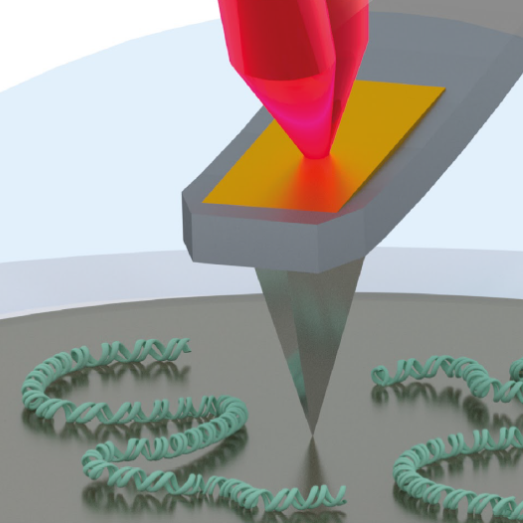
Collection Abstract: Atomic force microscopy (AFM) is a microscopy technique that uses a sharp probe to trace a sample surface at nanometre resolution. For biological applications, one of its key advantages is its ability to visualize substructure of single molecules and molecular complexes in an aqueous environment. Here, we describe the application of AFM to determine the secondary and tertiary structure of surface-bound DNA, and it’s interactions with proteins.
Troubleshooting
Safety warnings
For hazard information and safety warnings, please refer to the SDS (Safety Data Sheet).
Preparation of Mica Substrate
Cut the adhesive PTFE into circles of the same size as the steel sample discs (15 mm), using either a punch or scalpel (see Note 7).
Peel off the backing of the PTFE cut-out and adhere to the steel disc.
Mix the Araldite® 2-part epoxy resin 50:50 on a disposable surface e.g. weighing boat. Using a pipette tip transfer a small amount of the mixed epoxy to the centre of the teflon (use tip as a capillary to help bring up the glue).
Cleave the 6 mm mica disc on one side with scotch tape. With the cleaved mica disc facing down, immediately place on top of the epoxy droplet and press flat.
Leave the glue to dry and cure Overnight .
Once cured, cleave the mica using Scotch tape to reveal an atomically flat clean substrate (see Note 8).